

Free-breathing cardiovascular magnetic resonance flow quantification can be an alternative to standard breath-holding approach
- PMID: 40542034
- PMCID: PMC12181356
- DOI: 10.1038/s41598-025-06126-2
Free-breathing cardiovascular magnetic resonance flow quantification can be an alternative to standard breath-holding approach
Abstract
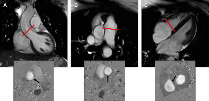
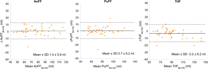
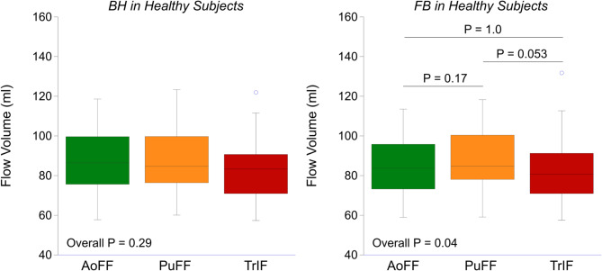
Cardiovascular magnetic resonance (CMR) evaluation of valvular heart disease is an important diagnostic tool when echocardiography is inconclusive. Phase contrast flow quantification is usually performed during breath hold (BH), which can be challenging in patients suffering from dyspnea and heart failure. The purpose of the present study is to compare a free-breathing (FB) with the conventional BH approach for flow quantification in the aortic, pulmonary and tricuspid valves in 20 healthy subjects (HS) and 25 patients with tricuspid regurgitation (TR). Aortic (AoFF) and pulmonary forward flow volume (PuFF), and tricuspid inflow volume (TrIF) were evaluated. Mean, standard deviation (SD) and limits of agreement (LoA) were calculated. There were good agreements between phase contrast flow volumes obtained by FB and BH approach. Mean difference ± SD / LoA for AoFF during BH versus FB were 1 ± 6 / -10 to 13 ml. The corresponding for PuFF were 1 ± 6 / -11 to 13 ml, and for TrIF - 3 ± 6 / -15 to 9 ml, respectively. Thus, free-breathing CMR flow acquisition can be an important alternative in the assessment of stroke volume, valvular regurgitant volume and be useful in all patients with difficulties to hold their breath.
Keywords: Breath-hold; Cardiovascular magnetic resonance; Free-breathing; Phase contrast flow quantification; Valvular heart disease.
© 2025. The Author(s).
Conflict of interest statement
Declarations. Competing interests: The authors declare no competing interests.
Figures
Similar articles
-
Deep-learning reconstruction for noise reduction in respiratory-triggered single-shot phase sensitive inversion recovery myocardial delayed enhancement cardiac magnetic resonance.Magn Reson Imaging. 2025 Oct;122:110460. doi: 10.1016/j.mri.2025.110460. Epub 2025 Jul 14. Magn Reson Imaging. 2025. PMID: 40669733
-
Comparison of the effectiveness of inhaler devices in asthma and chronic obstructive airways disease: a systematic review of the literature.Health Technol Assess. 2001;5(26):1-149. doi: 10.3310/hta5260. Health Technol Assess. 2001. PMID: 11701099
-
MRI-based quantification of whole-organ renal metabolic rate of oxygen during free-breathing.Magn Reson Med. 2025 Oct;94(4):1529-1545. doi: 10.1002/mrm.30583. Epub 2025 May 25. Magn Reson Med. 2025. PMID: 40415411 Free PMC article.
-
Abnormal coronary vascular response in patients with long COVID syndrome - a case-control study using oxygenation-sensitive cardiovascular magnetic resonance.J Cardiovasc Magn Reson. 2025 Summer;27(1):101890. doi: 10.1016/j.jocmr.2025.101890. Epub 2025 Apr 2. J Cardiovasc Magn Reson. 2025. PMID: 40185235 Free PMC article.
-
Pushing/bearing down methods for the second stage of labour.Cochrane Database Syst Rev. 2017 Mar 26;3(3):CD009124. doi: 10.1002/14651858.CD009124.pub3. Cochrane Database Syst Rev. 2017. PMID: 28349526 Free PMC article.
References
-
- Iung, B. et al. Contemporary presentation and management of valvular heart disease: the eurobservational research programme valvular heart disease II survey. Circulation140, 1156–1169 (2019). - PubMed
-
- Vahanian, A. et al. 2021 ESC/EACTS guidelines for the management of valvular heart disease. Eur. J. Cardiothorac. Surg.60, 727–800 (2021). - PubMed
-
- Zoghbi, W. A. et al. Recommendations for noninvasive evaluation of native valvular regurgitation: A report from the American society of echocardiography developed in collaboration with the society for cardiovascular magnetic resonance. J. Am. Soc. Echocardiography: Official Publication Am. Soc. Echocardiography. 30, 303–371 (2017). - PubMed
-
- Chaturvedi, A. et al. Quantitating aortic regurgitation by cardiovascular magnetic resonance: significant variations due to slice location and breath holding. Eur. Radiol.26, 3180–3189 (2016). - PubMed
-
- Kilner, P. J., Gatehouse, P. D. & Firmin, D. N. Flow measurement by magnetic resonance: a unique asset worth optimising. J. Cardiovasc. Magn. Resonance: Official J. Soc. Cardiovasc. Magn. Reson.9, 723–728 (2007). - PubMed
MeSH terms
LinkOut - more resources
Full Text Sources
Medical
