. 2025 Jun 20;15(1):20223.
doi: 10.1038/s41598-025-06813-0.

Impacts of commercial bile acids on growth performance, immune responses and expression genes of lipid metabolism in Nile tilapia fingerlings Oreochromis niloticus

Affiliations

Impacts of commercial bile acids on growth performance, immune responses and expression genes of lipid metabolism in Nile tilapia fingerlings Oreochromis niloticus

Yasser Marzouk et al. Sci Rep. .

Abstract

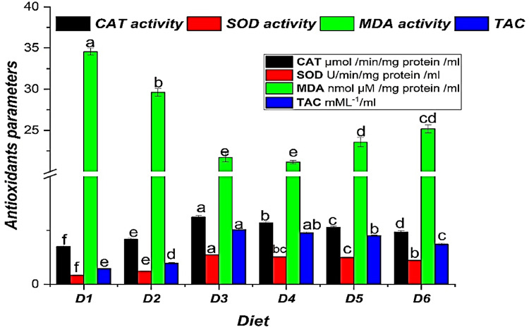
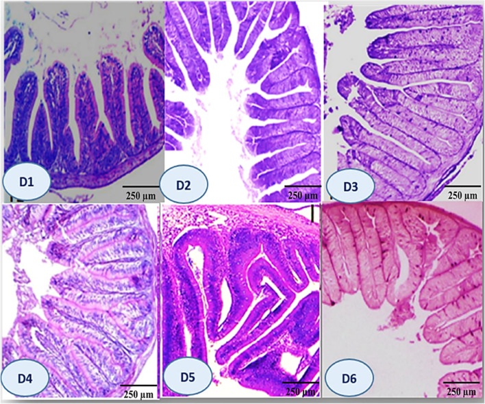
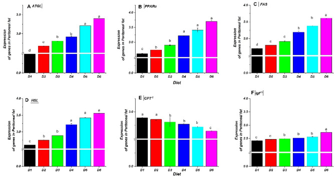
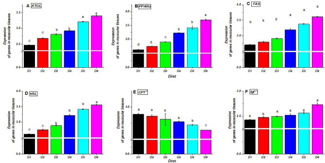
The current investigation evaluated the impact of the dietary addition of commercial bile acids (BAs) on growth, nutrient assimilation, immunity, antioxidant status, intestinal and hepatic histomorphometry, and gene expression of lipid metabolism in Nile tilapia (Oreochromis niloticus). In a study conducted for seventy days, 180 healthy fingerlings weighing 9 ± 0.5 g were divided into 18 hapas measuring 0.7 × 0.7 × 1.0 m. The fish were fed on six meals enriched with varied amounts of BAs: 0.0 (D1), 0.1 (D2), 0.2 (D3), 0.3 (D4), 0.4 (D5), and 0.5 (D6) g/kg diet. Nile tilapia fed the D3 diet exhibited significantly enhanced growth performance, with a specific growth rate of 1.89%/day and had the greatest feed conversion ratio (1.25), protein productive value, and energy utilization (33.28%). Fish fed the D3 exhibited significantly the highest crude protein content (64.50%). Energy content varied significantly, with D1 showing the lowest value (533.34 Kcal/100 g) and D3 the highest (604.27 Kcal/100 g). D3 improved biochemical indicators, immunological parameters, and digestive enzymes of O. niloticus. Histological analysis revealed notable liver and intestinal integrity enhancements among fish receiving BA-enriched diets, especially D3. Additionally, gene expression related to lipid metabolism in liver, peritoneal fat, and muscle tissues was upregulated in the treatment groups, especially 0.2 g/kg BAs compared to the control group. Results illustrate significant modulation of lipid metabolism gene expression parameters (Adipose triglyceride lipase; ATGL, Hormone-sensitive lipase; HSL, Peroxisome proliferator-activated receptor α; PPARα, Fatty acid synthase; FAS) by BAs treatments and were upregulated in BA-fed groups (D2-D6). Conversely, Carnitine palmitoyl transferase 1; CPT-1and Insulin-like growth factor-II; Igf-II expression declined, particularly when the BAs dose was increased. Accordingly, dietary 0.2 g/kg BAs supplementation positively influences on physiological, biochemical parameters, and lipid metabolic of Nile tilapia, making it a promising feed additive for aquaculture.

Keywords: Antioxidants; Digestive enzyme; Intestinal and liver morphology; Intestinal microbiota.


Conflict of interest statement

Declarations. Competing interests: The authors declare no competing interests.

Figures

Fig. 1
Fig. 1
Hematological parameters of Nile tilapia (O. niloticus) fingerlings fed diets supplemented with graded levels of commercial bile acids (BAs). Grouped bars represent the mean values (n = 9) ± standard error (SE) for Red Blood Cell count (RBC, 10⁶/mm³), White Blood Cell count (WBC, 10³/mm³), Hemoglobin concentration (Hb, g/dl), and Packed Cell Volume (PCV, %). Within each parameter group (indicated by color), bars topped with different letters show statistically significant differences between dietary groups (P ≤ 0.05).
Fig. 2
Fig. 2
Antioxidant parameters in Nile tilapia (O. niloticus) fingerlings fed diets supplemented with graded levels of commercial bile acids (BAs). Bars represent the mean (n = 9) ± standard error (SE) for Catalase activity (CAT; µmol/min/mg protein/ml), Superoxide Dismutase activity (SOD; U/min/mg protein/ml), Malondialdehyde level (MDA; nmol µM/mg protein/ml), and Total Antioxidant Capacity (TAC; mML−1/ml). Within each measured parameter, bars topped with different letters indicate statistically significant differences between dietary groups (P ≤ 0.05).
Fig. 3
Fig. 3
Following 70 days of feeding with different concentrations of commercial bile acids (BA), photomicrographs of the Nile tilapia’s (O. niloticus) gastrointestinal tract showed substantial histological changes amongst all treatments (H&E staining, 250× magnification): D1 (Control) had a typical unaltered intestinal wall including well-organized villi, a small count of goblet cells in the mucosa, a well-defined columnar epithelium, submucosa, and standard amounts of absorptive vacuoles. D2: A small number of goblet cells and slightly formed villi were shown, with a modest height rise. D3 demonstrated notable improvements, including augmented villus height, breadth, and crypt depth, enhanced mucosal folds, increased goblet cells in the lamina propria, and heightened levels of absorptive vacuoles. D4: Showed notable improvement in villus length and arrangement, with well-maintained villous architecture and no epithelial degeneration. D5 demonstrated a moderate increase in villus length and structure, preserving near-normal architecture and epithelial integrity. D6 exhibited standard villus height, characterized by significant villus fusion, broad villus tips, decreased goblet cells, mild lymphocytic infiltration, and limited mucosal folds. Figure 4 B: D1 (control diet without BAs); diets 2, 3, 4, 5, and 6 enriched with commercial BAs 0.10, 0.20, 0.30, 0.40, and 0.50 g/kg diet, respectively, n=3.
Fig. 4
Fig. 4
Morphometric analysis of the gastrointestinal tract in Nile tilapia (O. niloticus) fingerlings fed diets supplemented with graded levels of commercial bile acids (BAs). (A) Mean (± standard error, SE; n=3) crypt depth, villus width, and villus height in millimeters; within each measured parameter, bars topped with different letters indicate statistically significant differences between dietary groups (P≤ 0.05). (B) Qualitative representation of goblet cell abundance, indicated by '+' symbols (+, ++, +++, ++++). For statistical comparisons (P<0.05) between dietary groups for each parameter shown in the figure. Diets included a control (D1, 0 g/kg BA) and treatments supplemented with 0.10 (D2), 0.20 (D3), 0.30 (D4), 0.40 (D5), and 0.50 (D6) g/kg BA.
Fig. 5
Fig. 5
The liver microscopic structure of Nile tilapia feeding with different levels of commercial BA shows round polygonal hepatocytes organized in cord-like formations, constrained on one side by hepatic capillaries or sinusoids. At the core of each cord are narrow bile canaliculi located next to the hepatocytes, along with the pancreatic portion that encircles the afferent vein (D1 control). D2: shows subcapsular leukocytic infiltration and marked lytic and cavitation within the hepatic tissues. D3: showing slight cloudy swelling of hepatocytes and few pyknotic nuclei. D4: demonstrates focal to diffuse vacuolar degeneration of hepatocytes. The vacuoles exhibited severity and diffuseness, accompanied by focal leucocytic infiltration. D5 exhibits diffuse deterioration of hepatocytes, focal necrosis, leukocytic infiltration, and fat cell deposits in the central vein. D6 demonstrates severe deterioration with focal necrosis characterized by pyknotic nuclei, fat cell deposits, alterations in the typical liver architecture, and parenchymal fibrosis with fat deposits (H&E X 200). D1 (control diet without BAs); diets 2, 3, 4, 5, and 6 enriched with commercial BAs 0.10, 0.20, 0.30, 0.40, and 0.50 g/kg diet, respectively, n=3.
Fig. 6
Fig. 6
Relative expression of fatty acid metabolism-related genes in the liver tissues of Nile tilapia (O. niloticus) fingerlings fed diets supplemented with graded levels of commercial bile acids (BAs). The relative mRNA expression for (A) ATGL, (B) HSL, (C) PPARα, (D) CPT−1, (E) FAS, and (F) Igf−II. Values are presented as mean ± standard error (SE), n = 3 replicates per diet. Within each panel, bars with different letters indicate statistically significant differences (P ≤ 0.05). Gene abbreviations: ATGL, adipose triglyceride lipase; PPARα, peroxisome proliferator-activated receptor α; CPT−1, carnitine palmitoyl transferase-1; FAS, fatty acid synthase; HSL, hormone-sensitive lipase; Igf−II, insulin-like growth factor-II. Diets included a control (D1, 0 g/kg BA) and treatments supplemented with 0.10 (D2), 0.20 (D3), 0.30 (D4), 0.40 (D5), and 0.50 (D6) g/kg BA.
Fig. 7
Fig. 7
Relative expression of fatty acid metabolism-related genes in the peritoneal fat of Nile tilapia (O. niloticus) fingerlings fed diets supplemented with graded levels of commercial bile acids (BAs). The relative mRNA expression for (A) ATGL, (B) HSL, (C) PPARα, (D) CPT−1, (E) FAS, and (F) Igf−II. Values are presented as mean ± standard error (SE), n = 3 replicates per diet. Within each panel, bars with different letters indicate statistically significant differences (P ≤ 0.05). Gene abbreviations: ATGL, adipose triglyceride lipase; PPARα, peroxisome proliferator-activated receptor α; CPT−1, carnitine palmitoyl transferase- 1; FAS, fatty acid synthase; HSL, hormone-sensitive lipase; Igf−II, insulin-like growth factor-II. Diets included a control (D1, 0 g/kg BA) and treatments supplemented with 0.10 (D2), 0.20 (D3), 0.30 (D4), 0.40 (D5), and 0.50 (D6) g/kg BA.
Fig. 8
Fig. 8
Relative expression of fatty acid metabolism-related genes in the muscular tissues of Nile tilapia (O. niloticus) fingerlings fed diets supplemented with graded levels of commercial bile acids (BAs). The relative mRNA expression for (A) ATGL, (B) PPARα, (C) FAS, (D) HSL, (E) CPT−1, and (F) Igf−II. Values are presented as mean ± standard error (SE), n = 3 replicates per diet. Within each panel, bars with different letters indicate statistically significant differences (P ≤ 0.05). Gene abbreviations: ATGL, adipose triglyceride lipase; PPARα, peroxisome proliferator-activated receptor α; CPT−1, carnitine palmitoyl transferase- 1; FAS, fatty acid synthase; HSL, hormone-sensitive lipase; Igf−II, insulin-like growth factor-II. Diets included a control (D1, 0 g/kg BA) and treatments supplemented with 0.10 (D2), 0.20 (D3), 0.30 (D4), 0.40 (D5) and 0.50 (D6) g/kg BA.

Similar articles

References

    1. FAO. FAO yearbook of fishery and aquaculture statistics. Rome. (2024).
    1. FAO. Food and Agriculture Organization of the United Nations: The state of world fisheries and aquaculture. Rome, Italy. Retrieved from (2022). https://www.fao.org/3/cc0461en/cc0461en.pdf.
    1. El-Ouny, Y. M. et al. Effect of fishmeal replacement with dried red wigglers (Eisenia fetida) worm meal on growth and feed utilization, production efficiency, and serum biochemistry in nile tilapia (Oreochromis niloticus) fingerlings. Aquaculture Rep.29, 101518 (2023).
    1. Abdel-Latif, H. M. R. et al. Growth Performance, Antioxidant Activities, and Immunological Responses of hapa-reared Thinlip Mullet (Liza ramada) Juveniles Fed on Diets Supplemented with spirulina (Arthrospira platensis)130p. 359–367 (Fish & Shellfish Immunology, 2022). - PubMed
    1. Khalil, H. S. & Nasr-Allah, A. Comparative study on the effect of raceway and In-Pond raceway systems on different nile tilapia, Oreochromis niloticus strains fed diets replacing soybean meal by poultry byproduct meal on: water quality, growth performance and production efficiency. Aquacult. Int.33 (4), 258 (2025).

MeSH terms

Substances

LinkOut - more resources