. 2025 Jun 20;16(1):312.
doi: 10.1186/s13287-025-04433-z.

Human induced neural progenitor cells generated from three-dimensional aggregate-based culture significantly improve post-stroke recovery in tMCAO mice

Affiliations

Human induced neural progenitor cells generated from three-dimensional aggregate-based culture significantly improve post-stroke recovery in tMCAO mice

Zeqin Fu et al. Stem Cell Res Ther. .

Abstract

Background: Despite the high prevalence of cerebral ischemic stroke, effective clinical treatments remain limited. With the development of regenerative medicine, induced neural progenitor cells (iNPCs) demonstrate ideal potential and good availability for autologous transplantation therapy. However, current differentiation protocols for iNPCs still have room for improvement in terms of purity, reproducibility, scalability and differentiation potential.

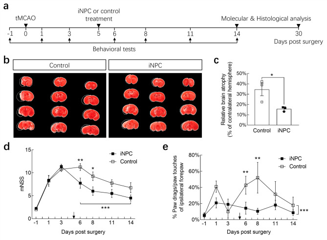
Methods: We aimed to develop a scalable, stable, and efficient 3D aggregate-based method for iNPC production in suspension culture, avoiding detrimental cell dissociation and replating processes. We evaluated the therapeutic potential of iNPCs in the chronic phase of a transient middle cerebral artery occlusion (tMCAO) mouse model and explored iNPC subpopulations via single-cell RNA sequencing to elucidate their pleiotropic therapeutic potentials.

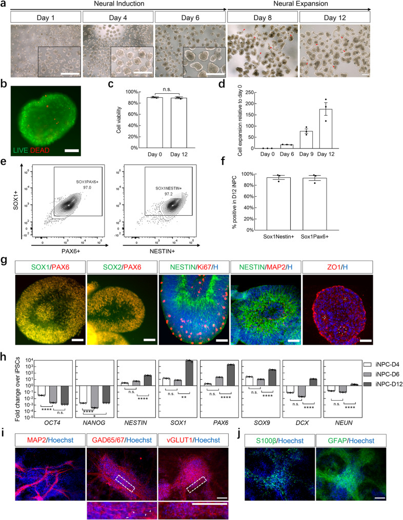
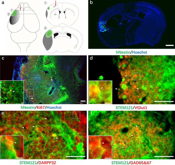
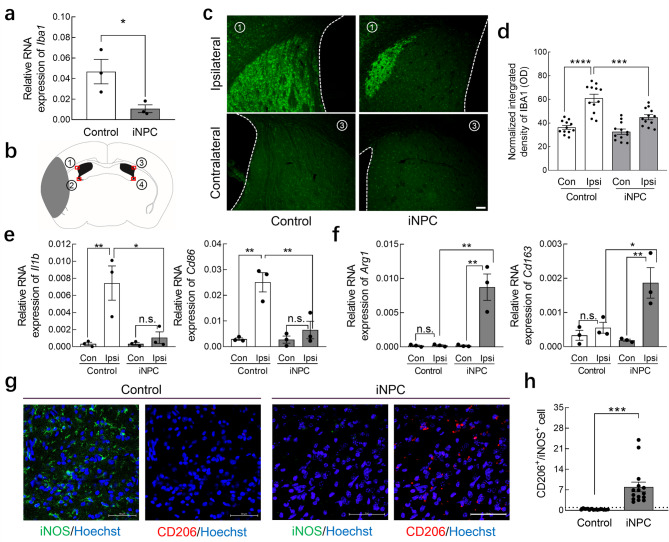
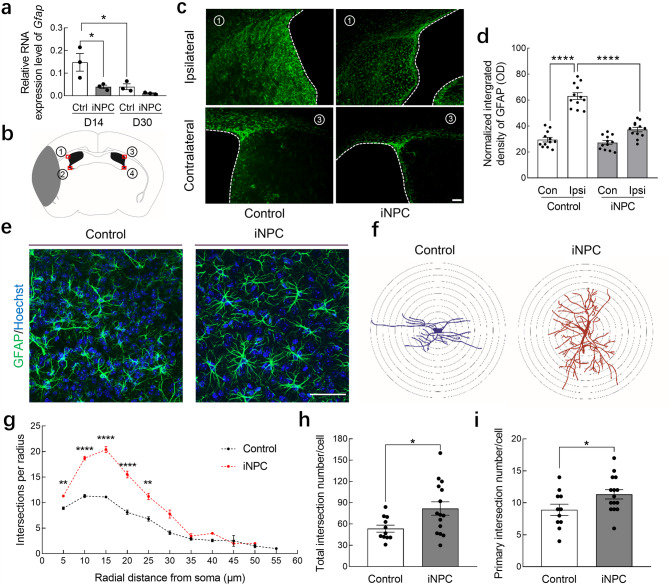
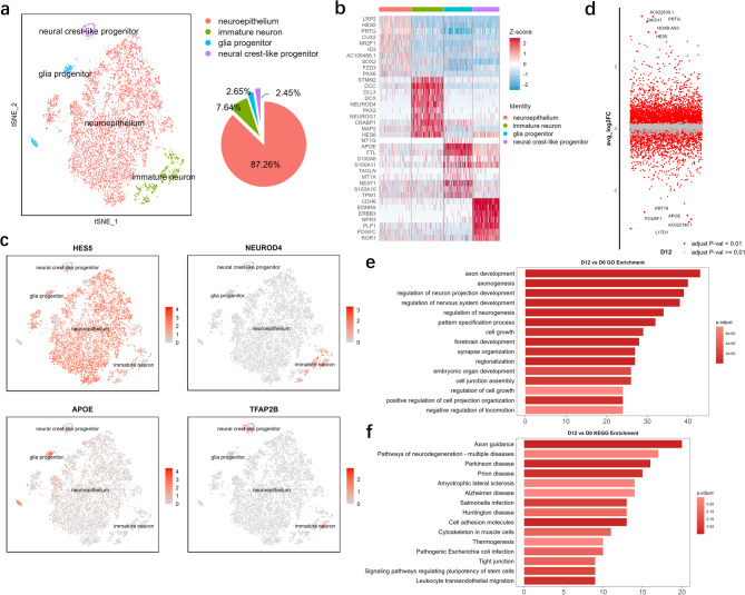
Results: iNPCs generated from three iPSC lines displayed high NPC marker expression and an average 176-fold cell expansion over the 12-day culture period. These iNPCs could spontaneously differentiate into both neurons and glial cells in vitro. In the tMCAO model, transplanted iNPCs remodeled the microenvironment by alleviating neuroinflammation, inhibiting chronic microgliosis and astrogliosis, promoting M2 polarization of microglia, and preserving astrocytic morphology in the ischemic penumbra. Mechanistically, iNPCs can be divided into four subpopulations, with neuroepithelia being the most abundant and capable of rapidly replenishing damaged cells and mitigating microenvironmental deterioration.

Conclusions: We developed a simple and efficient 3D aggregate-based method for iNPC differentiation. These iNPCs showed excellent potential for post-stroke recovery and represent a valuable tool for clinical translation.

Keywords: 3D aggregates; Chronic ischemic stroke; Induced neural progenitor cells; Post-stroke recovery; Stem cell transplantation.


Conflict of interest statement

Declarations. Ethics approval and consent to participate: Human PBMC samples were collected and studied at Shenzhen Beike Biotechnology Co., Ltd with written informed consent from all participants from which the iPSCs and iNPCs have been derived. The research ethics and animal experiments were reviewed and approved by the Experimental Ethics Committee of the Institutional Review Boards at Shenzhen Beike Biotechnology Co., Ltd. The reference IRB number is BK-SL-20230419-03, titled ‘Large-scale preparation and application of induced neural progenitor cells and their exosomes’, approved on Apr.19th 2023. Consent for publication: Not applicable. Competing interests: This study was sponsored by the Shenzhen Beike Biotechnology Co., Ltd. The authors declare that the core technology in this research has been patented under: CN117448272A and CN116396936A.

Figures

Fig. 1
Fig. 1
Efficient generation and characterization of iPSC-derived NPCs via a 3D aggregate-based method. (a) Schematic overview and representative bright-field images of iPSC-derived NPC aggregates. The enlarged figure on day 1 showed aggregates with uniform structure, and the enlarged figure on day 4, day 6 showed a typical apicobasal polarized morphology. Red arrowheads on day 8, day 12 indicated newly formed aggregates shedding from the original ones. (b) Viability of a day 12 iNPC aggregate. Green: AO; Red: PI. (c) Cell viability on day 0 and day 12 during iNPC generation. (d) Cell expansion ratio on day 0, day 6, day 9 and day 12 during iNPC generation. (e) Representative flow cytometry results on day 12 iNPCs. (f) Average percentage of SOX1/PAX6 + and SOX1/NESTIN + cells in day 12 iNPCs. (g) Representative immunostaining images of NPC markers (NESTIN, SOX1, SOX2 and PAX6), proliferation marker (Ki67), mature neuronal marker (MAP2) and apical surface marker (ZO-1) from day 12 iNPC aggregates. White arrows indicated the centers of inner rosettes in a neural aggregate. (h) Gene expression of pluripotency marker (NANOG, OCT4), NPC marker (NESTIN, SOX1, PAX6), gliogenic marker (SOX9), proneuronal marker (DCX) and mature neuronal marker (NEUN) in day 4, day 6 and day 12 samples during iNPC generation, measured by RT-PCR. (i-j) Representative immunostaining images of the (i) neurogenic markers (MAP2, GAD65&67, vGLUT1) and (j) gliogenic markers (S100β, GFAP) from iNPCs subjected to a 14-day in vitro differentiation culture. Dotted rectangles marked the position of enlarged area below. White arrowheads demonstrated potential spine structures. Scale bar in (a): 500 μm; scale bar in (b, i-j): 100 μm; scale bar in (g): 50 μm. n = 3 for (c-d, f). n.s. represented no significance by Student’s t test in (c). * represented p < 0.05, ** represented p < 0.01, **** represented p < 0.0001, and n.s. represented no significance by one-way ANOVA followed by Tukey’s post hoc test in (h)
Fig. 2
Fig. 2
Effects of transplanted iNPCs on post-stroke recovery in tMCAO mice. (a) Summary of the experimental timeframes. (b) TTC staining of coronal brain sections from iNPC and control group on day 14 after tMCAO surgery. Brain infarcts were marked by dotted lines. (c) Quantification of relative brain atrophy. (d) mNSS of iNPC and the control group. n = 8. (e) Cylinder test result of the ipsilateral forepaw in iNPC and the control group. n = 3 for (b-c), n = 8 for (d-e). * represented p < 0.05 by Student’s t test in (c), * represented p < 0.05, ** represented p < 0.01 and *** represented p < 0.001 by two-way ANOVA followed by Tukey’s post hoc test in (d-e)
Fig. 3
Fig. 3
Transplanted hiNPCs migrated and differentiated in the ischemic injured site. (a) Horizontal and coronal brain diagrams. The injection site was indicated by the red arrows and green dots in M1 cortex and striatum, green color indicated the transplanted iNPC disseminated areas, the grey area indicated the ischemic lesion site. (b) hiNPCs identified with hNestin in a coronal brain section. (c) Proliferating hiNPCs identified with hNestin and Ki67. (d-f) Differentiated hiNPCs identified with human cytoplasmic protein specific antibody (STEM121) and the glutamatergic marker (d) vGlut1, dopaminergic marker (e) DARPP32 or GABAergic marker (f) GAD65&67. The white arrowheads in (c-f) indicated signal colocalization. The small figures on the bottom left corner in (c-f) were the magnification of the dashed-line squares. CPu: caudate putamen. Scale bar in (b): 1 mm; scale bar in (c-f): 100 μm; scale bar in the enlarged figures of (c-f): 10 μm
Fig. 4
Fig. 4
Transplanted hiNPCs alleviated microgliosis and promoted M2 polarization on day 30 after cerebral ischemia. (a, e-f) Quantitative RT-PCR analysis of (a) microglia marker (Iba1), (d) M1 microglia markers (Il1b, Cd86), and (e) M2 microglia markers (Arg1, Cd163) mRNA expression level relative to Gapdh in the ipsilateral or contralateral hemispheres from control and iNPC-treated mice. (b) Coronal brain diagrams. Red boxes indicated the sites of immunostaining figures taken in (c) and for statistical analysis, the grey area indicated the ischemic lesion site. (c) Representative figures of Iba1 (Green) immunostaining in the ipsilateral and contralateral side of control and iNPC treated group. White dotted lines marked the border of the ventricle. (d) Statistical analysis on normalized integrated density of Iba1 signals surrounding the ipsilateral or contralateral ventricles of control or iNPC-treated mice in (c). (g) Representative figures of immunostaining with antibodies against iNOS (green) and CD206 (red) in the penumbra region, nuclei were counterstained with Hoechst (blue). (h) Statistical analysis of CD206+/iNOS + ratio in (g). Scale bar in (c): 100 μm; scale bar in (g): 50 μm. n = 3 for (a, e-f), n = 12 for (d), and n = 15 from 3 mice for (h). * represented p < 0.05, ** represented p < 0.01, and n.s. represented no significance by one-way ANOVA followed by Tukey’s post hoc test in (e-f). * represented p < 0.05 and *** represented p < 0.001 by Student’s t test in (a, h). Con: contralateral, Ipsi: ipsilateral
Fig. 5
Fig. 5
Transplanted iNPCs inhibited reactive astrogliosis but preserved astrocytic processes in the ischemic penumbra zone. (a) Quantitative RT-PCR analysis of reactive astrocytic marker Gfap in control and iNPC-treated mice. (b) Coronal brain diagrams. Red boxes indicated the sites of immunostaining figures taken in (c) and for statistical analysis, the grey area indicated the ischemic lesion site. (c) Representative figures of Gfap (Green) immunostaining in the ipsilateral and contralateral side of control and iNPC-treated group. White dotted lines marked the border of the ventricle. (d) Statistical analysis on normalized integrated density of Gfap signals surrounding the ipsilateral or contralateral ventricles of control or iNPC-treated mice in (c). (e) Representative immunostaining of Gfap in the penumbra zone of ipsilateral hemisphere. (f) Sholl analysis for the measurement of astrocytic branching. Represented astrocyte morphologies from control and iNPC-treated group were outlined from Gfap staining. Concentric rings were placed 5 μm apart around the soma. (g) Astrocytic branches shown by intersection numbers per radius against radial distance from soma. (h-i) Summary of (h) total intersection number and (i) primary intersection number in the reactive astrocytes from control and iNPC-treated group. Scale bar in (c): 100 μm, scale bar in (e): 50 μm. n = 3 for (a), n = 11 for control and n = 15 for iNPC from 3 mice for (g-i). * represented p < 0.05, and **** represented p < 0.0001 by one-way ANOVA followed by Tukey’s post hoc test in (a, d). ** represented p < 0.01, and **** represented p < 0.0001 by two-way ANOVA followed by Tukey’s post hoc test in (g). * represented p < 0.05 by Student’s t test in (h-i)
Fig. 6
Fig. 6
Differential changes and functional enrichment in hiNPCs. (a) t-SNE plots of the scRNA-seq data of iNPCs (D12) and sector graph illustrating the percentage of each cluster in iNPCs. (b) Heatmap of 10 DEGs in each cluster. (c) t-SNE plots showing the expression levels of marker genes in each cluster of iNPCs. (d) Differential expression gene analysis showing iPSCs and iNPCs. (e-f) GO and KEGG enrichment analyses of iNPCs

Similar articles

References

    1. McKinley KL, Longaker MT, Naik S. Emerging frontiers in regenerative medicine. Science. 2023;380(6647):796–8. - PMC - PubMed
    1. Lam M, Sanosaka T, Lundin A, Imaizumi K, Karlsson D, et al. Single-cell study of neural stem cells derived from human iPSCs reveals distinct progenitor populations with neurogenic and gliogenic potential. Genes Cells. 2019;24(12):836–47. - PMC - PubMed
    1. Song B, Cha Y, Ko S, Jeon J, Lee N, Seo H, et al. Human autologous iPSC-derived dopaminergic progenitors restore motor function in parkinson’s disease models. J Clin Invest. 2020;130(2):904–20. - PMC - PubMed
    1. Kanemura Y, Yamamoto A, Katsuma A, Fukusumi H, Shofuda T, Kanematsu D, et al. Human-Induced pluripotent stem Cell-Derived neural progenitor cells showed neuronal differentiation, neurite extension, and formation of synaptic structures in rodent ischemic stroke brains. Cells. 2024;13(8):671. - PMC - PubMed
    1. Imai R, Tamura R, Yo M, Sato M, Fukumura M, Takahara K, et al. Neuroprotective effects of Genome-Edited human iPS Cell-Derived neural stem/progenitor cells on traumatic brain injury. Stem Cells. 2023;41(6):603–16. - PMC - PubMed

LinkOut - more resources