. 2025 Jun 20;56(1):121.
doi: 10.1186/s13567-025-01556-2.

LGALS3BP/90K suppresses porcine reproductive and respiratory syndrome virus replication by enhancing GP3 degradation and stimulating innate immunity

Affiliations

LGALS3BP/90K suppresses porcine reproductive and respiratory syndrome virus replication by enhancing GP3 degradation and stimulating innate immunity

Qingyu Fan et al. Vet Res. .

Abstract

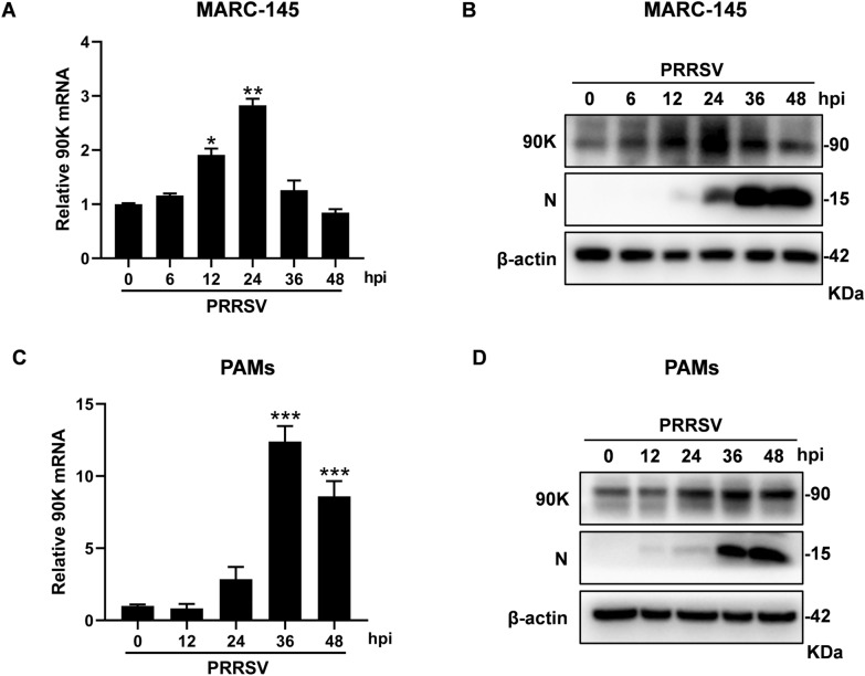
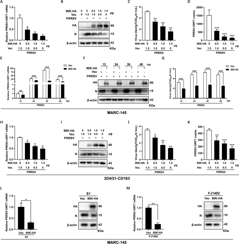
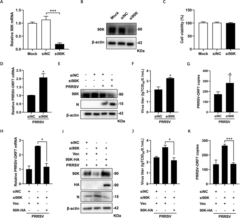
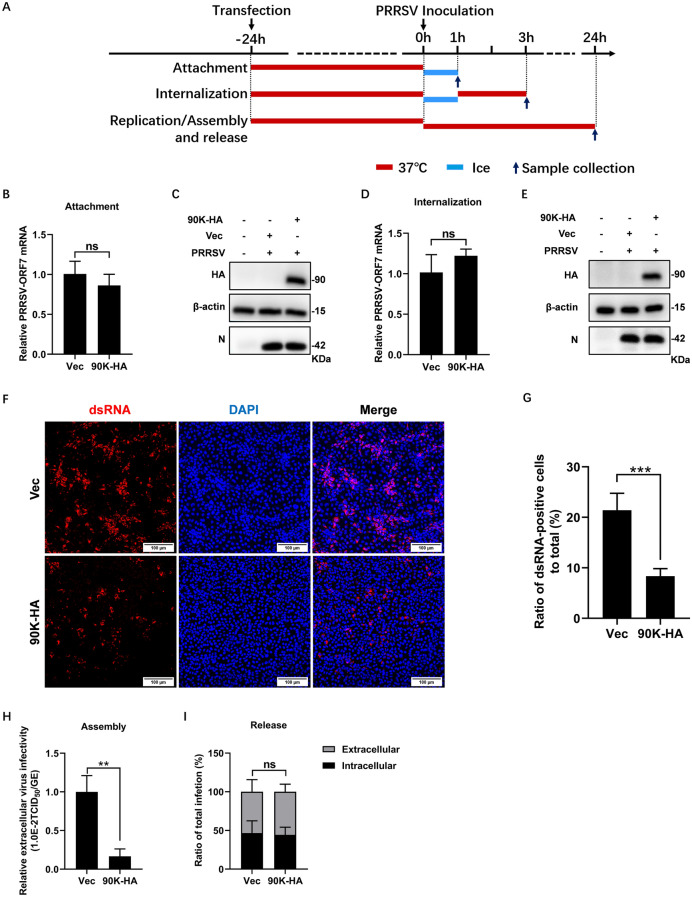
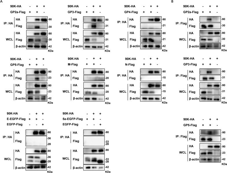
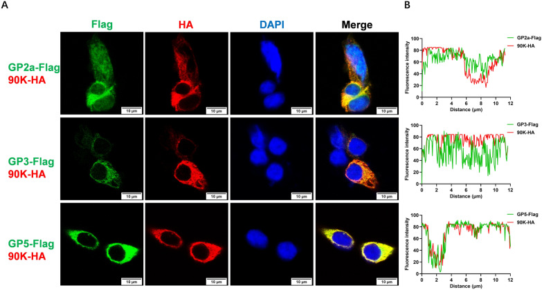
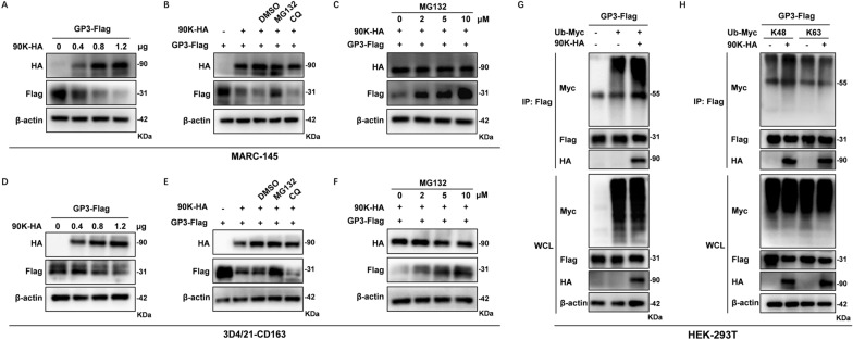
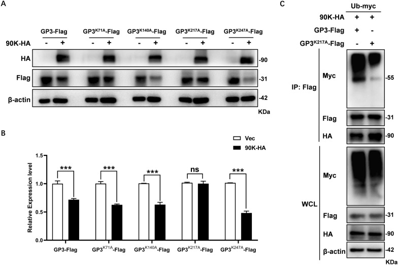
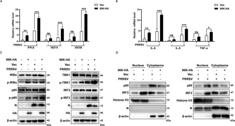
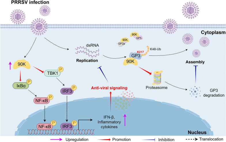
Porcine reproductive and respiratory syndrome virus (PRRSV) seriously affects the world pig industry, and existing vaccines are not enough to protect against PRRSV efficiently. Identifying host factors that inhibit PRRSV infection may provide potential targets for antiviral therapies. Galectin 3 binding protein (LGALS3BP), also referred to as 90K, is a multifunctional protein that plays significant roles in viral infections, but its role in PRRSV infection is not clearly defined. In this study, we found that 90K expression increased during PRRSV infection. Additionally, 90K inhibits PRRSV replication by restricting both viral RNA synthesis and viral assembly. Further studies showed that 90K can interact with various PRRSV structural proteins, such as GP2, GP3, and GP5. Moreover, 90K targets K217 in GP3 for K48-linked ubiquitination, facilitating GP3 degradation through the proteasomal pathway. Meanwhile, we found that 90K induces the expression of type I interferon (IFN) and inflammatory cytokines by activating NF-κB and IRF3. Overall, our findings suggest that 90K plays a negative role in PRRSV replication by inducing PRRSV-GP3 degradation and activating innate immune responses.

Keywords: LGALS3BP/90K; PRRSV; innate immunity; proteasomal degradation; viral replication.


Conflict of interest statement

Declarations. Competing interests: The authors declare that they have no competing interests.

Figures

Figure 1
Figure 1
The expression of 90K can be induced in cells infected with PRRSV. MARC-145 cells or PAMs were infected with PRRSV BB0907 (MOI = 0.1) for indicated hours. A and C RNA was extracted from cells and 90K mRNA levels were measured by qRT-PCR. These values were normalized to the levels of β-actin mRNA. The bars represent the means ± SD from three independent experiments. (*, p < 0.05; **, p < 0.01; ***, p < 0.001). B and D Protein expression levels of endogenous 90K and PRRSV-N were detected by western blot (WB). The antibodies used are listed in the adjacent column. β-actin was used as the internal loading control.
Figure 2
Figure 2
Overexpression of 90K inhibits PRRSV replication. AD MARC-145 cells were transfected with different doses of HA-tagged 90K expressing plasmid (90K-HA) (0, 0.5, 1, or 1.5 μg), along with an empty vector (Vec) adjusted accordingly. At 24 h post-transfection (hpt), the cells were infected with PRRSV BB0907 (MOI = 1.0). A Cells were collected at 24 hpi. The mRNA levels of PRRSV-ORF7 were detected by qRT-PCR. B Protein expression levels of 90K-HA and PRRSV-N were detected by WB. C Supernatants were harvested for determining viral titers by TCID50 assay. D PRRSV-ORF7 copies in the supernatant were also determined by qRT-PCR. EG MARC-145 cells were transfected with 1 µg of either 90K-HA or Vec. At 24 hpt, cells were infected with PRRSV BB0907 (MOI = 1.0) for indicated hours. Cells and supernatants were collected to access PRRSV replication levels by qRT-PCR (E), WB (F), and TCID50 assay (G). HK Parallel dose–response experiments were conducted using 3D4/21-CD163 cells. PRRSV-N expression levels were detected by qRT-PCR (H) and WB (I). Viral titers and PRRSV-ORF7 copies in the supernatant were determined by TCID50 assay (J) and qRT-PCR (K). L and M MARC-145 cells were transfected with either 90K-HA or Vec and subsequently infected with PRRSV strains S1 or FJ1402 (MOI = 1.0). At 24 hpi, cells were harvested for qRT-PCR and WB analysis. The bars represent the means ± SD from three independent experiments. (*, p < 0.05; **, p < 0.01, ***, p < 0.001).
Figure 3
Figure 3
Knockdown of 90K promotes PRRSV replication. AC MARC-145 cells were transfected with 90K-specific siRNA (si90K) or control siRNA (siNC). Cells were collected after 36 h (h) to access endogenous 90K expression using qRT-PCR (A) and WB (B). C The cytotoxicity of siRNAs on MARC-145 cells was assessed using a Cell Counting Kit-8 (CCK-8). DG MARC-145 cells were transfected with either si90K or siNC. After 36 h, the cells were infected with PRRSV BB0907 (MOI = 0.1) for 24 h. Levels of PRRSV-ORF7 mRNA and PRRSV-N protein were measured by qRT-PCR (D) and WB (E), while viral titers and PRRSV-ORF7 copies in the supernatant were assessed using the TCID50 assay (F) and qRT-PCR (G). HK MARC-145 cells were transfected with either si90K or siNC. After 24 h, the cells were transfected with either 90K-HA or Vec for 24 h, and then infected with PRRSV BB0907 (MOI = 0.1) for another 24 h. Levels of PRRSV-ORF7 mRNA and PRRSV-N protein were measured by qRT-PCR (H) and WB (I), while viral titers and viral RNA in the supernatant were assessed using the TCID50 assay (J) and qRT-PCR (K). The bars represent means ± SD from three independent experiments. (*, p < 0.05; **, p < 0.01; ***, p < 0.001).
Figure 4
Figure 4
90K inhibits PRRSV RNA replication and virion assembly. A Overview of the experiment designed for examining PRRSV attachment, internalization, replication, assembly, and release. B and C MARC-145 cells were transfected with 90K-HA or Vec. At 24 hpt, the cells were inoculated with PRRSV BB0907 (MOI = 10) and kept on ice for 1 h. Cells were then washed three times with ice-cold PBS. The expression levels of PRRSV-N were quantified using qRT-PCR (B) and WB (C). D and E Transfected cells were inoculated with PRRSV BB0907 (MOI = 10) and cultured on ice for 1 h, and were then shifted to 37 °C. After 2 h, cells were washed three times with ice-cold citrate buffer. PRRSV-N expression levels were quantified by qRT-PCR (D) and WB (E). F Transfected cells were inoculated with PRRSV BB0907 (MOI = 1). At 24 hpi, cells were fixed and inoculated with anti-dsRNA antibody (red). Nuclei were stained with DAPI (blue). Scale bars, 100 μm. G Random fields of view were recorded and the ratio of dsRNA positive cells were quantified with ImageJ. H Transfected cells were infected with PRRSV BB0907 (MOI = 1) for 24 h. Viral assembly efficiency was represented as the ratio of TCID50/mL to the total number of PRRSV genome equivalents (GE). I The efficiency of virus release was determined as the ratio of intra- and extracellular infectivity relative to the total infectivity. The bars represent means ± SD from three independent experiments. (**, p < 0.01; ***, p < 0.001; ns, not significant).
Figure 5
Figure 5
90K interacts with multiple PRRSV structural proteins. A HEK-293 T cells were co-transfected with plasmids that express HA-tagged 90K and Flag-tagged PRRSV structural proteins. After 48 h, the cell lysates were immunoprecipitated with anti-HA antibodies. Whole-cell lysate (WCL) and immunoprecipitation (IP) complexes were analyzed using immunoblotting with anti-HA and anti-Flag antibodies. B HEK-293 T cells were co-transfected with plasmids for Flag-tagged PRRSV structural proteins and HA-tagged 90K. At 48 hpt, cells were lysed and immunoprecipitated with anti-Flag antibodies. WCL and IP complexes were used for immunoblotting analysis with anti-Flag and anti-HA antibodies.
Figure 6
Figure 6
90K colocalize with PRRSV-GP2a, GP3 and GP5 in the cytoplasm. A HEK-293 T cells were co-transfected with 90K-HA and plasmids encoding Flag-tagged PRRSV-GP2, GP3 or GP5. At 24 hpt, the cells were fixed and analyzed using indirect immunofluorescence to identify Flag-tagged structural proteins (green) and HA-tagged 90K (red). The nuclei were stained with DAPI (blue). Scale bars, 10 μm. B The graphs illustrate the use of ImageJ software to conduct line scan analysis, showing the relative positions of the two markers.
Figure 7
Figure 7
90K degrades PRRSV-GP3 through a proteasome-dependent pathway. A and D MARC-145 (A) or 3D4/21-CD163 (D) cells were co-transfected with plasmid encoding Flag-tagged GP3 (GP3-Flag) and increasing doses of 90K-HA (0, 0.4, 0.8, or 1.2 μg). The expression levels of PRRSV-GP3 were measured using WB at 24 hpt. B and E MARC-145 (B) or 3D4/21-CD163 (E) cells were co-transfected with GP3-Flag and 90K-HA. At 14 hpt, cells were treated with either dimethyl sulfoxide (DMSO), MG132 (10 μM), or CQ (160 μM) for 10 h. GP3-Flag expression levels were detected by WB. C and F MARC-145 (C) or 3D4/21-CD163 (F) cells were co-transfected with GP3-Flag and 90K-HA and treated with increasing doses of MG132 (0, 2, 5, or 10 μM). The cell lysates were analyzed by WB using the specified antibodies. G HEK-293 T cells were co-transfected with GP3-Flag, Ub-Myc, and 90K-HA or Vec. At 36 hpt, cells were collected for Co-IP assay with an anti-Flag antibody. This was followed by WB analysis with indicated antibodies. H HEK-293 T cells were co-transfected with plasmids encoding GP3-HA, 90K-HA, and Myc-tagged Ub mutants (Ub-K48 or Ub-K63). At 24 hpt, Co-IP assay was performed as described above and then analyzed by WB.
Figure 8
Figure 8
90K targets the K217 site of GP3 for ubiquitination. A HEK-293 T cells were co-transfected with expression plasmids containing 90K-HA and either GP3-Flag or its mutants (GP3K71A, GP3K140A, GP3K217A, or GP3K247A). At 24 hpt, the cells were collected for WB. B At least three independent replicates were analyzed by densitometry to determine the relative protein expression levels. The bars represent the means ± SD from three independent experiments. (***, p < 0.001; ns, not significant). C HEK-293 T cells were co-transfected with plasmids encoding 90K-HA, Ub-Myc and either Flag-tagged GP3 or its mutant GP3K217A. At 24 hpt, cells were collected for Co-IP with an anti-Flag antibody. The WCL and IP complexes were analyzed by immunoblotting with indicated antibodies.
Figure 9
Figure 9
90K induces cytokine expression by activating NF-κB and IRF3. MARC-145 cells were co-transfected with either 90K-HA or Vec for 24 h. Afterward, the cells were either infected or not infected with PRRSV (MOI = 1). A and B At 36 h, cells were collected for qRT-PCR to measure the expression levels of indicated cytokines.  C Cell lysates were analyzed by WB to determine the expression levels of indicated signal molecules. D Cytosolic and nuclear extracts were prepared and subjected to WB analysis to detect the translocation of NF-κB and IRF3. Histone-H3 and β-actin were used as markers for nuclear and cytosolic fraction, respectively. The bars represent the means ± SD of three independent experiments. (*, p < 0.1; **, p < 0.01; ***, p < 0.001; ns, not significant).
Figure 10
Figure 10
Model of the mechanism by which 90K suppresses PRRSV replication. 90K can be induced during PRRSV infection. 90K restricts PRRSV RNA replication and assembly. Mechanistically, 90K interacts with several PRRSV structural proteins, such as GP2a, GP3, and GP5. Additionally, it targets the K217 site of GP3 for K48-linked ubiquitination, which leads to its degradation through a proteasome-dependent pathway. Furthermore, this protein upregulates the expression of p-p65 and p-IRF3, facilitating the translocation of p65 and IRF3 from the cytoplasm to the nucleus, which promotes the production of IFNs and inflammatory cytokines. By Figdraw.

Similar articles

References

    1. Artini M, Natoli C, Tinari N, Costanzo A, Marinelli R, Balsano C, Porcari P, Angelucci D, D’Egidio M, Levrero M, Iacobelli S (1996) Elevated serum levels of 90K/MAC-2 BP predict unresponsiveness to alpha-interferon therapy in chronic HCV hepatitis patients. J Hepatol 25:212–217 - PubMed
    1. Boeters M, Garcia-Morante B, van Schaik G, Segales J, Rushton J, Steeneveld W (2023) The economic impact of endemic respiratory disease in pigs and related interventions—a systematic review. Porcine Health Manag 9:45 - PMC - PubMed
    1. Bosquillon DJL, Akbil B, Mhlekude B, Leyens J, Postmus D, Harnisch G, Jansen J, Schmidt ML, Aigner A, Pott F, Chua RL, Krist L, Gentile R, Muhlemann B, Jones TC, Niemeyer D, Fricke J, Keil T, Pischon T, Janke J, Conrad C, Iacobelli S, Drosten C, Corman VM, Ralser M, Eils R, Kurth F, Sander L, Goffinet C (2023) 90K/LGALS3BP expression is upregulated in COVID-19 but may not restrict SARS-CoV-2 infection. Clin Exp Med 23:3689–3700 - PMC - PubMed
    1. Creagh EM, O’Neill LA (2006) TLRs, NLRs and RLRs: a trinity of pathogen sensors that co-operate in innate immunity. Trends Immunol 27:352–357 - PubMed
    1. de Vries AA, Post SM, Raamsman MJ, Horzinek MC, Rottier PJ (1995) The two major envelope proteins of equine arteritis virus associate into disulfide-linked heterodimers. J Virol 69:4668–4674 - PMC - PubMed

MeSH terms

LinkOut - more resources