. 2025 Jan-Dec:39:3946320251351083.
doi: 10.1177/03946320251351083. Epub 2025 Jun 21.

Blocking leukotriene receptors improve experimentally induced gastric ulcers in rats by inhibiting inflammation and apoptosis

Affiliations

Blocking leukotriene receptors improve experimentally induced gastric ulcers in rats by inhibiting inflammation and apoptosis

Hanan M Hassan et al. Int J Immunopathol Pharmacol. 2025 Jan-Dec.

Abstract

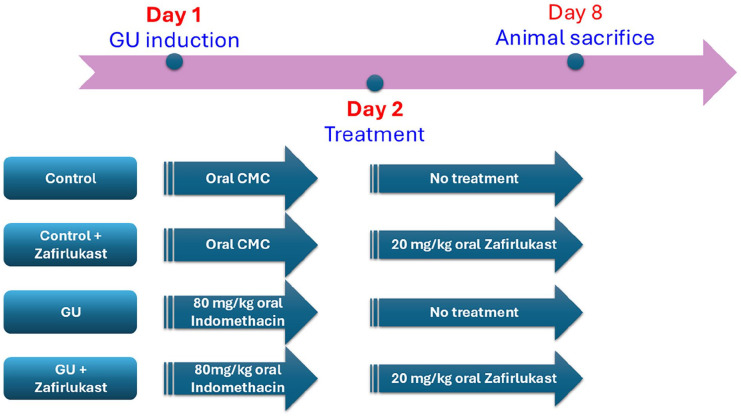
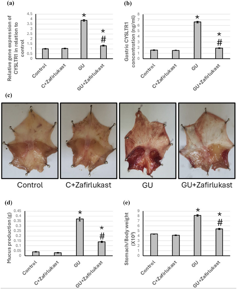
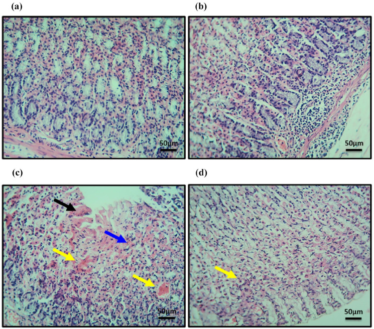
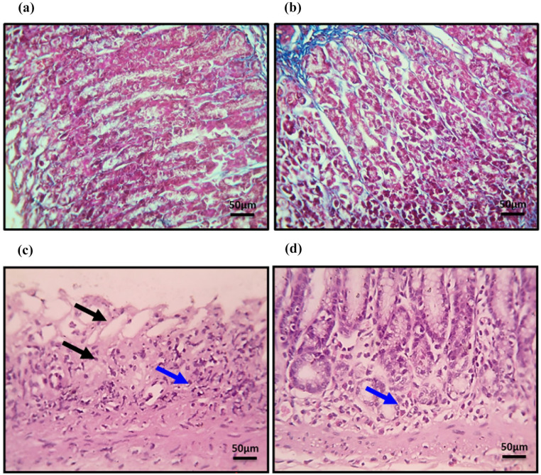
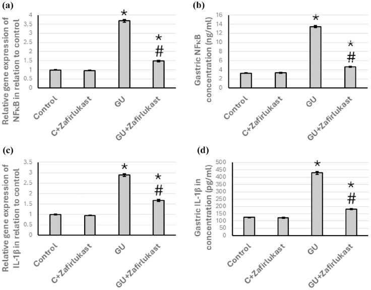
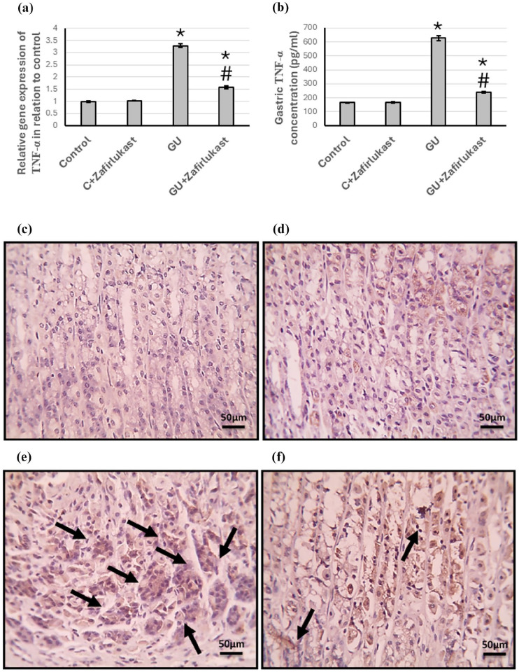
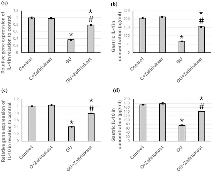
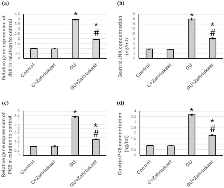
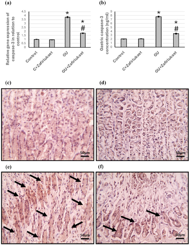
To investigate whether obstructing the cysteinyl leukotriene receptor-1 (CYSLTR1) with zafirlukast diminishes experimentally induced gastric ulcer (GU) in rats by modulating inflammation and apoptosis. Gastric ulcers affect approximately 10% of the global population and can lead to serious complications such as gastrointestinal perforation and bleeding. Leukotrienes are proinflammatory compounds, and cysteinyl leukotrienes, such as LTC4, LTD4, and LTE4, that are potent proinflammatory mediators. Rats were orally administered a single oral dose of 80 mg/kg of indomethacin to induce GU. The rats were administered an oral dose of 20 mg/kg Zafirlukast. Gastric tissues were collected for macrostructural and microstructural analyses. A portion of gastric tissue was used to assess the genetic expression and protein levels of CYSLTR1, NFκB, TNF-α, IL-1β/4/10, JNK, PKB, and caspase-3. The gastric sections were subjected to hematoxylin/eosin and Masson trichrome staining and immunohistochemical staining with anti-TNF-α and anti-caspase-3 antibodies. Zafirlukast blocked the expression of CYSLTR1. Analysis of micro-images of GU rats revealed damage to surface cells and glandular epithelial cells caused by inflammatory cell infiltration, which was mitigated by Zafirlukast. Additionally, Zafirlukast treatment significantly reduced NFκB, TNF-α, IL-1β, JNK, PKB, and caspase-3 while increasing IL-4 and IL-10. Zafirlukast successfully reduced experimentally induced gastric ulcers in rats. Its mechanism of action includes inhibition of CYSLTR1, diminishing the inflammatory pathway. This is demonstrated by a decrease in the levels of NFκB, TNF-α, and IL-1β, along with an increase in the levels of IL-4 and IL-10. Additionally, Zafirlukast exerted anti-apoptotic effects by downregulating the expression of JNK, PKB, and caspase-3.

Keywords: Jun N-terminal kinase (JNK); cysteinyl leukotriene receptor 1 (CYSLTR1); nuclear factor (NF)κB; protein kinase B (PKB); tumor necrosis factor-α (TNF-α).


Conflict of interest statement

Declaration of conflicting interestsThe author(s) declared no potential conflicts of interest with respect to the research, authorship, and/or publication of this article.

Figures

Figure 1.
Figure 1.
Timeline of the animal treatment in the study.
Figure 2.
Figure 2.
Effect of GU and 20 mg/kg Zafirlukast on gene expression of CYSLTR1 (a) and its gastric protein levels (b). Representative images of the whole stomach from different treated groups (c). The images of control rats exhibited a normal appearance without any observable abnormalities. The image of GU’s stomach exhibited mucosal hemorrhagic lesions and areas of ulceration. In contrast, the stomach from the GU treated with Zafirlukast showed a substantial reduction in hemorrhage and ulcer areas, indicating a positive impact of the treatment. Finally, statistical analysis of mucus production (d), and stomach/body weight ratio (e). The data are presented as Mean ± SEM, utilizing ten rats per group. Statistical analysis was conducted using ANOVA, followed by post hoc Bonferroni correction testing. * Significant difference as compared with the control group at p<0.05. # Significant difference as compared with GU group at p<0.05. CYSLTR1, cysteinyl leukotriene receptor-1; GU, gastric ulcer.
Figure 3.
Figure 3.
Gastric sections stained with hematoxylin/eosin in the control group (a), the control group treated with Zafirlukast (b), the GU group (c), and the GU treated with Zafirlukast (d). Yellow arrows represented focal mucosal inflammation, and black arrows represented extensive mucosal necrosis in the GU group. Treatment of GU rats with Zafirlukast reduced mucosal inflammation and necrosis. Scale bar 50 μm. GU, gastric ulcer.
Figure 4.
Figure 4.
Gastric sections stained with Masson trichrome in the control group (a), the control group treated with Zafirlukast (b), the GU group (c), and the GU treated with Zafirlukast (d). The black arrow represents mucosal necrosis, and the yellow arrows represent mucosal leukocytic cell infiltration. Treatment of GU rats with Zafirlukast reduced mucosal inflammation and necrosis. Scale bar 50 μm. GU, gastric ulcer.
Figure 5.
Figure 5.
Effect of GU and 20 mg/kg Zafirlukast on gene expression of NFκB (a), and IL-1β (c), as well as the gastric level of NFκB (b), and IL-1β (d) representing an increase in the expression of both NFκB and IL-1β in GU rats that was ameliorated by Zafirlukast treatment. The data are presented as Mean ± SEM, utilizing ten rats per group. Statistical analysis was conducted using ANOVA, followed by post hoc Bonferroni correction testing. * Significant difference as compared with the control group at p< .05. # Significant difference as compared with GU group at p<0.05. GU, gastric ulcer; IL, interleukin; NFκB, nuclear factor κB.
Figure 6.
Figure 6.
Effect of GU and 20 mg/kg Zafirlukast on gene expression of TNF-α (a) and its protein level in gastric tissues (b). Gastric sections were stained with anti-TNF-α in the control group (c), the control group treated with Zafirlukast (d), the GU group (e), and the GU treated with Zafirlukast (f). Scale bar 100 μm. The data are presented as Mean ± SEM, utilizing ten rats per group. Statistical analysis was conducted using ANOVA, followed by post hoc Bonferroni correction testing. * Significant difference as compared with the control group at p< .05. # Significant difference as compared with GU group at p< .05. GU, gastric ulcer; TNF-α, tumor necrosis factor-α.
Figure 7.
Figure 7.
Effect of GU and 20 mg/kg Zafirlukast on gene expression of IL-4 (a) and IL-10 (c) as well as the gastric level of IL-4 (b) and IL-10 (c) representing a reduction in the expression of IL-4 and IL-10 in GU rats which was reversed by treating with Zafirlukast. The data are presented as Mean ± SEM, utilizing ten rats per group. Statistical analysis was conducted using ANOVA, followed by post hoc Bonferroni correction testing. * Significant difference as compared with the control group at p< .05. # Significant difference as compared with GU group at p< .05. GU, gastric ulcer; IL, interleukin.
Figure 8.
Figure 8.
Effect of GU and 20 mg/kg Zafirlukast on gene expression of JNK (a) and PKB (c) and the gastric protein levels of JNK (b) and PKB (d) representing an increase in the expression of JNK and PKB that was reduced by treating with Zafirlukast. The data are presented as Mean ± SEM, utilizing ten rats per group. Statistical analysis was conducted using ANOVA, followed by post hoc Bonferroni correction testing. * Significant difference as compared with the control group at p< .05. # Significant difference as compared with GU group at p< .05. GU, gastric ulcer; JNK, Jun N-terminal kinase; PKB, protein kinase B.
Figure 9.
Figure 9.
Effect of GU and 20 mg/kg Zafirlukast on gene expression of caspase-3 (a) and its protein level in gastric tissues (b). Gastric sections were stained with anti-caspase-3 in the control group (c), the control group treated with Zafirlukast (d), the GU group (e), and the GU treated with Zafirlukast (f). Scale bar 100 μm. The data are presented as Mean ± SEM, utilizing ten rats per group. Statistical analysis was conducted using ANOVA, followed by post hoc Bonferroni correction testing. * Significant difference as compared with the control group at p< .05. # Significant difference as compared with GU group at p< .05. GU, gastric ulcer.

Similar articles

References

    1. Chatila AT, Bilal M, Guturu P. Evaluation and management of acute pancreatitis. World J Clin Cases 2019; 7: 1006–1020. DOI: 10.12998/wjcc.v7.i9.1006. - DOI - PMC - PubMed
    1. Tanigawa T, Pai R, Arakawa T, et al. TGF-beta signaling pathway: Its role in gastrointestinal pathophysiology and modulation of ulcer healing. J Physiol Pharmacol 2005; 56: 3–13. - PubMed
    1. Baj J, Forma A, Sitarz M, et al. Helicobacter pylori virulence factors-mechanisms of bacterial pathogenicity in the gastric microenvironment. Cells 2020; 10(1): 27;. DOI: 10.3390/cells10010027. - DOI - PMC - PubMed
    1. Schubert ML, Peura DA. Control of gastric acid secretion in health and disease. Gastroenterology 2008; 134: 1842–1860. DOI: 10.1053/j.gastro.2008.05.021. - DOI - PubMed
    1. Sohail R, Mathew M, Patel KK, et al. Effects of non-steroidal anti-inflammatory drugs (NSAIDs) and gastroprotective NSAIDs on the gastrointestinal tract: A narrative review. Cureus 2023; 15: e37080. DOI: 10.7759/cureus.37080. - DOI - PMC - PubMed

MeSH terms

LinkOut - more resources