. 2025 Jul 10;68(13):14041-14053.
doi: 10.1021/acs.jmedchem.5c01219. Epub 2025 Jun 21.

Design of a Fluorescence Polarization Probe for Enterovirus 2C Proteins

Affiliations

Design of a Fluorescence Polarization Probe for Enterovirus 2C Proteins

Kan Li et al. J Med Chem. .

Abstract

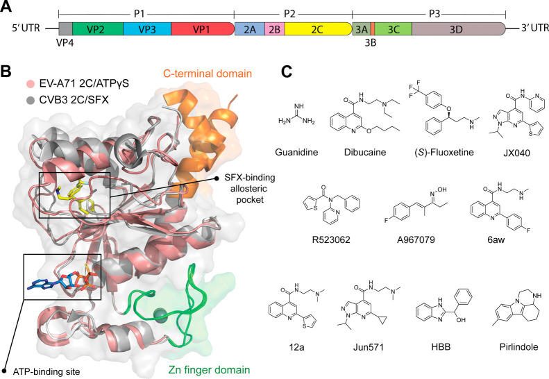
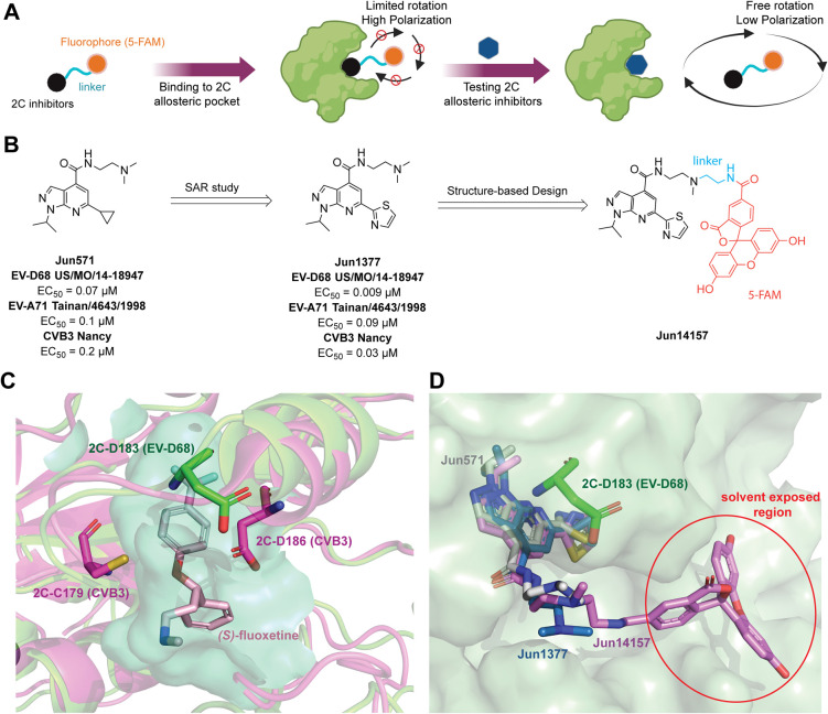
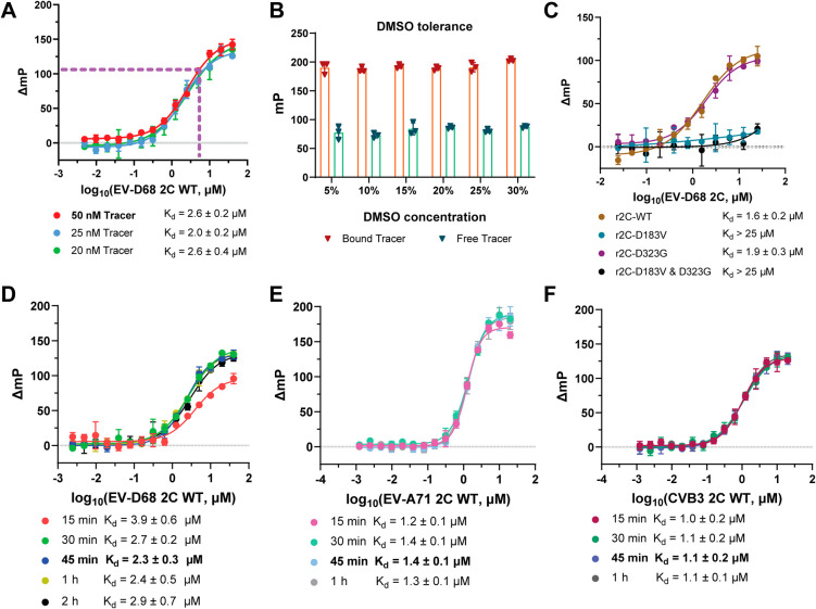
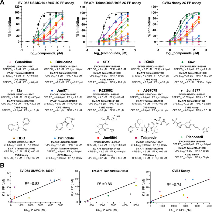
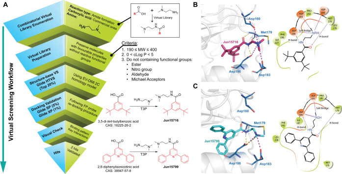
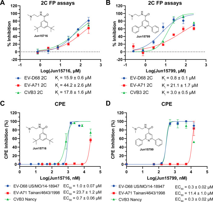
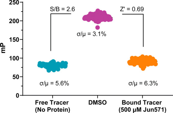
Enteroviruses (EVs), such as EV-D68, EV-A71, and CVB3, cause significant human disease; yet, no antivirals are currently approved. The highly conserved 2C protein, an essential AAA+ ATPase and helicase, is a prime antiviral target; however, it lacks suitable assays for inhibitor screening. Here, we report a fluorescence polarization (FP) assay using a rationally designed probe, Jun14157, which binds a conserved allosteric site in 2C with high affinity. This assay enables the quantitative assessment of binding to diverse 2C inhibitors with high signal-to-background ratios, DMSO tolerance, and a strong correlation between FP Ki and cellular EC50. Using this platform, we validated hits from virtual screening and identified two novel inhibitors, Jun15716 and Jun15799. This FP assay offers a robust and scalable tool for the mechanistic characterization and high-throughput screening of 2C-targeting antivirals.


Figures

1
1
Genome structure of enterovirus, structure of 2C, and reported 2C inhibitors. (A) The EV genome contains ORF encoding structural proteins VP1, VP2, VP3, and VP4 and nonstructural proteins 2A, 2B, and 2C and 3A, 3B, 3C, and 3D. (B) Alignment of the crystal structure of CVB3 2C in complex with SFX (PDB ID 6S3A, colored in gray) and EV-A71 2C in complex with ATPγS (PDB ID 5GRB, colored in wheat). The C-terminal part of 2C is highlighted in orange, the zinc finger domain is highlighted in green, ATPγS is shown in blue sticks, and SFX is shown in yellow sticks. (C) Chemical structures of reported enterovirus 2C inhibitors.
2
2
Design of the 2C FP probe. (A) FP assay principle. (B) SAR study and design of the 2C FP probe Jun14157. (C) Superimposed structures of EV-D68 2C (homology model) with CVB3 2C in complex with SFX (PDB ID 6T3W). EV-D68 2C is colored in green, CVB3 2C is colored in magenta, and the cavity of the allosteric pocket is colored in cyan. SFX is shown as wheat sticks. (D) The superposition of docking poses of Jun571, Jun1377, and Jun14157 occurred at the allosteric site of EV-D68 2C. 2C is colored in split pea, Jun571 is shown as gray sticks, Jun1377 is shown as marine sticks, and Jun14157 is shown as pink sticks.
1
1. Synthetic Route of 2C FP Probe Jun14157
3
3
Optimization of the FP assay using the Jun14157 probe. (A) Binding curves of the Jun14157 probe at 20, 50, and 100 nM with increasing concentrations of EV-D68 2C after 30 min of incubation. K d values represent means ± standard deviation (SD) from triplicate experiments. (B) Effect of dimethyl sulfoxide (DMSO) concentration on the mP values of bound and free probes. The assay was performed in triplicate. (C) Binding curves of 50 nM Jun14157 with increasing concentrations of WT and EV-D68 2C mutants D183V, D323G, and D183V/D323G. K d values are the mean ± SD from duplicate experiments. (D–F) FP titration curves of Jun14157 with EV-D68 2C-WT (D), EV-A71 2C (E), and CVB3 2C (F) with different incubation times. K d values are presented as the mean ± SD from triplicate experiments. Each data point represents the mean ± SD.
4
4
FP assay validation with reported 2C inhibitors and the correlation between the FP K i values and the antiviral EC50 values for selected 2C inhibitors. (A) Competitive binding experiments measuring the displacement of the 2C FP probe Jun14157 by guanidine, dibucaine, SFX, JX040, 6aw, 12a, Jun571, R523062, A967079, Jun1377, Jun6504, HBB, and pirlindole. The EV-D68 2A protease inhibitor (telaprevir) and capsid inhibitor (pleconaril) were used as negative controls. Plotted values from the FP assay are the mean ± SD (n = 2), and plotted values from the CPE assay are the mean ± SD (n = 3). (B) Correlation plot between the K i values from the FP assay and the EC50 values from the CPE assay.
5
5
Virtual screening and hit validation of novel 2C inhibitors. (A) Graphical workflow of the virtual screening for identifying novel 2C inhibitors against enteroviruses. (B) Docking pose and 2D-binding pattern of Jun15716. (C) Docking pose and 2D-binding pattern of Jun15799. The H-bonds were shown as yellow dashed lines in docking poses and purple arrow lines in 2D-binding patterns. The salt bridges are shown as magenta dashed lines in docking poses and red-to-blue gradient lines in 2D-binding patterns.
6
6
(A–D) Hit validation with the 2C FP and CPE assays. For the IC50 obtained from FP assays, the plotted values are the mean ± SD (n = 2). For EC50 obtained from CPE assays, the plotted values are the mean ± SD (n = 3).
7
7
Determination of the Z′ factor in the FP assay. Five hundred μM Jun571 and DMSO were treated as positive and negative controls, respectively. For each group, the data points were obtained in 96 replicates with a sample size (n = 96).

Similar articles

References

    1. Nikonov O. S., Chernykh E. S., Garber M. B., Nikonova E. Y.. Enteroviruses: Classification, Diseases They Cause, and Approaches to Development of Antiviral Drugs. Biochemistry. Biokhimiia. 2017;82(13):1615–1631. doi: 10.1134/S0006297917130041. - DOI - PMC - PubMed
    1. Hyypiä T., Hovi T., Knowles N. J., Stanway G.. Classification of enteroviruses based on molecular and biological properties. J. Gen. Virol. 1997;78(Pt 1):1–11. doi: 10.1099/0022-1317-78-1-1. - DOI - PubMed
    1. Holm-Hansen C. C., Midgley S. E., Fischer T. K.. Global emergence of enterovirus D68: a systematic review. Lancet Infect Dis. 2016;16(5):e64–e75. doi: 10.1016/S1473-3099(15)00543-5. - DOI - PubMed
    1. Fall A., Kenmoe S., Ebogo-Belobo J. T., Mbaga D. S., Bowo-Ngandji A., Foe-Essomba J. R., Tchatchouang S., Amougou Atsama M., Yengue J. F., Kenfack-Momo R.. et al. Global prevalence and case fatality rate of Enterovirus D68 infections, a systematic review and meta-analysis. PLoS Negl Trop Dis. 2022;16(2):e0010073. doi: 10.1371/journal.pntd.0010073. - DOI - PMC - PubMed
    1. Morens D. M., Folkers G. K., Fauci A. S., Casadevall A.. Acute Flaccid Myelitis: Something Old and Something New. Mbio. 2019;10(2):e00521-19. doi: 10.1128/mBio.00521-19. - DOI - PMC - PubMed

MeSH terms

LinkOut - more resources