

Expression of growth factors in buffalo ovarian tissue across different follicular developmental stages
- PMID: 40542852
- PMCID: PMC12374900
- DOI: 10.1007/s00404-025-08090-8
Expression of growth factors in buffalo ovarian tissue across different follicular developmental stages
Abstract
Background: In assisted reproduction, poor ovarian response to stimulation negatively affects oocyte yield and is influenced by genetic factors.
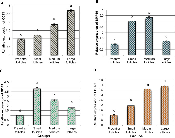
Objective: This study aimed to quantify the mRNA expression of key growth markers (BMP15, GDF9, OCT4, and FGFR2) in ovarian tissue according to the developmental stages of the follicle.
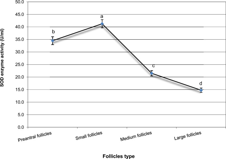
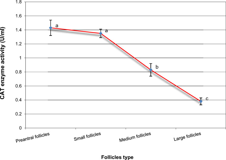
Methods: Samples were collected from ovarian tissue. Gene expression levels were analyzed using RT-qPCR. In addition, ELISA was used to measure the concentrations of catalase (CAT), glutathione peroxidase (GPx), and superoxide dismutase (SOD), along with reactive oxygen species (ROS) and malondialdehyde (MDA) as oxidative stress markers.
Results: OCT4 expression was similar in preantral and small follicles but significantly upregulated in medium and large follicles. GDF9 expression and SOD activity were highest in small follicles (P < 0.05). BMP15 levels were significantly elevated in small and medium follicles compared to preantral follicles but remained unchanged in large follicles (P < 0.05). FGFR2 expression increased progressively with follicle size (P < 0.05). GPx activity was directly proportional to follicle size, with the lowest levels in preantral follicles. Conversely, ROS, MDA, and CAT concentrations decreased as follicle size increased.
Conclusion: These findings provide insights into the molecular regulation of follicular development in buffalo, which could aid in improving reproductive efficiency in assisted reproduction programs.
Keywords: Buffalo; Follicle development; Ovarian tissue; Oxidative stress; mRNA transcription.
© 2025. The Author(s).
Conflict of interest statement
Declarations. Conflict of interest: The authors declare no competing interests. Ethical approval: Experiments were approved by “the Institutional Medical Research Ethical Committee” at the National Research Centre in Egypt (Number 05410723).
Figures

References
-
- Williams CJ, Erickson GF, Feingold KR, Anawalt B, Blackman MR, Boyce A, Chrousos G, Corpas E (2000) Morphology and physiology of the ovary. In: (eds) Endotext. South Dartmouth (MA). https://www.ncbi.nlm.nih.gov/books/NBK278951 - PubMed
MeSH terms
Substances
LinkOut - more resources
Full Text Sources
Miscellaneous
