

Predicting intraoperative meningioma consistency using features from standard MRI sequences: a preoperative evaluation
- PMID: 40542946
- PMCID: PMC12182493
- DOI: 10.1007/s00701-025-06582-9
Predicting intraoperative meningioma consistency using features from standard MRI sequences: a preoperative evaluation
Abstract
Background: Symptomatic meningiomas may require surgical resection to save or improve neurological function. The extent of tumor resection depends on multiple factors, including the tumor's consistency, its location, and the patient's overall condition. This prospective study aims to explore new criteria in combination with previously proposed tumor features on MRI to establish a rapid approach to tumor consistency characterization pre-operatively.
Methods: Forty-eight patients with meningiomas were prospectively included and underwent a dedicated MRI protocol prior to surgery. Qualitative and quantitative MRI characteristics of the tumor were correlated to a previously proposed surgical tumor consistency grading.
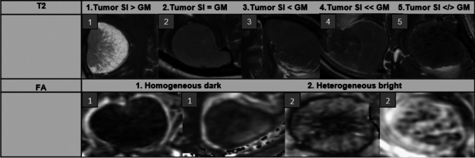
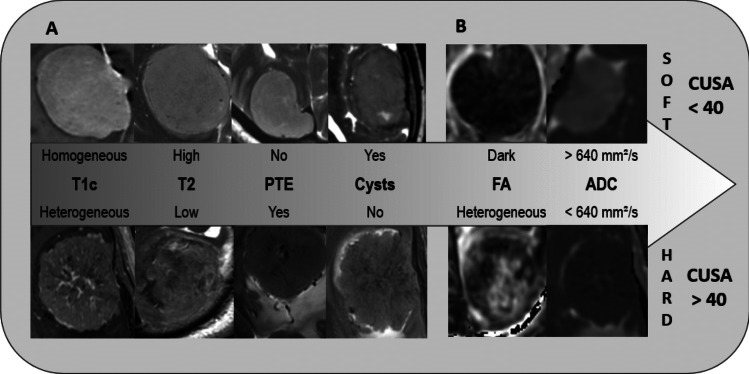
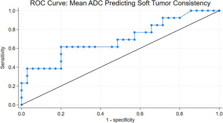
Results: Soft tumors were associated with homogeneous contrast enhancement, high T2 signal, absence of peritumoral edema (PTE), the presence of tumor cysts, and a uniformly dark appearance on fractional anisotropy (FA) maps. In contrast, firmer tumors were characterized by heterogeneous contrast enhancement, low T2 signal, the presence of PTE, absence of tumor cysts and a heterogeneous appearance on FA maps, requiring supranormal ultrasonic aspirator settings. Tumor signal quantification on T2 and Apparent Diffusion Coefficient maps (ADC) correlated moderately to tumor consistency. T1 sequences did not contribute in determining tumor consistency.
Conclusion: An array of simple qualitative meningioma characteristics on MRI can assist in swift discrimination of soft and hard tumors preoperatively. These have been displayed in a figure that can easily be implemented clinically for optimal surgical planning.
Keywords: Consistency; MRI; Meningioma; Tumor.
© 2025. The Author(s).
Conflict of interest statement
Declarations. Ethics approval: All procedures involving human participants in this study were conducted according to the ethical standards of the 1964 Declaration of Helsinki and its later amendments, or comparable ethical standards. The study was approved by the Regional Research Ethics Committee (REC reference number 20446 (2017/1875)) of Helse Sør-Øst and the Institutional Review Board (IRB) of Oslo University Hospital (PVO reference number 20/00819). Consent to participate: All patient’s informed written consent to participate in this study was collected. Consent for publication: Informed consent was obtained from all individual participants in the study. Competing interest: The authors declare no competing interests. The authors declare no competing interests.
Figures
Similar articles
-
Intracranial meningioma: A review of recent and emerging data on the utility of preoperative imaging for management.J Neuroimaging. 2024 Sep-Oct;34(5):527-547. doi: 10.1111/jon.13227. Epub 2024 Aug 7. J Neuroimaging. 2024. PMID: 39113129 Review.
-
Can MRI predict meningioma consistency?: a correlation with tumor pathology and systematic review.Neurosurg Rev. 2018 Jul;41(3):745-753. doi: 10.1007/s10143-016-0801-0. Epub 2016 Nov 21. Neurosurg Rev. 2018. PMID: 27873040 Free PMC article.
-
Preoperative Assessment of Meningioma Consistency Using a Combination of MR Elastography and DTI.AJNR Am J Neuroradiol. 2024 Nov 7;45(11):1755-1761. doi: 10.3174/ajnr.A8385. AJNR Am J Neuroradiol. 2024. PMID: 38906671
-
Angiographic Features of Meningiomas Predicting Extent of Preoperative Embolization.Neurosurgery. 2024 Nov 1;95(5):1010-1025. doi: 10.1227/neu.0000000000003054. Epub 2024 Aug 1. Neurosurgery. 2024. PMID: 39087784 Free PMC article.
-
Magnetic resonance perfusion for differentiating low-grade from high-grade gliomas at first presentation.Cochrane Database Syst Rev. 2018 Jan 22;1(1):CD011551. doi: 10.1002/14651858.CD011551.pub2. Cochrane Database Syst Rev. 2018. PMID: 29357120 Free PMC article.
References
-
- Brabec J, Szczepankiewicz F, Lennartsson F, Englund E, Pebdani H, Bengzon J, Knutsson L, Westin CF, Sundgren PC, Nilsson M (2022) Histogram analysis of tensor-valued diffusion MRI in meningiomas: relation to consistency, histological grade and type. NeuroImage Clinical 33:102912. 10.1016/j.nicl.2021.102912 - PMC - PubMed
-
- Chen TC, Zee CS, Miller CA, Weiss MH, Tang G, Chin L, Levy ML, Apuzzo ML (1992) Magnetic resonance imaging and pathological correlates of meningiomas. Neurosurgery 31:1015–1021. 10.1227/00006123-199212000-00005. (discussion 1021–1012) - PubMed
MeSH terms
LinkOut - more resources
Full Text Sources
Medical
