. 2025 Jul;301(7):110392.
doi: 10.1016/j.jbc.2025.110392. Epub 2025 Jun 19.

Acyl chains stabilize the acylated domain and determine the receptor-mediated interaction of the Bordetella adenylate cyclase toxin with cell membrane

Affiliations

Acyl chains stabilize the acylated domain and determine the receptor-mediated interaction of the Bordetella adenylate cyclase toxin with cell membrane

Carlos Espinosa-Vinals et al. J Biol Chem. 2025 Jul.

Abstract

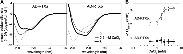
Acylated domains (ADs), like that of the Bordetella pertussis adenylate cyclase toxin (CyaA), are structures found in all pore-forming toxins from the family of Repeat-in-ToXin (RTX) proteins. These AD segments are fatty-acylated on ε-amino groups of conserved lysine residues, such as the K860 and K983 residues of CyaA. The ε-amide-linked acyl chains are essential for toxin activity and promote irreversible membrane insertion of the CyaA molecule, thus enabling the toxin to translocate its N-terminal adenyl cyclase enzyme domain into the host cell cytoplasm. In parallel, the membrane-inserted CyaA molecules can oligomerize into cation-selective pores in the plasma membrane. Here, we show that the attached acyl chains are not only crucial for membrane insertion of the toxin but also play an important role in CyaA folding. We demonstrate that assembly of the noncanonical β-roll structure in the C-terminal segment of the AD of CyaA is cooperatively directed by the Ca2+-driven folding of the adjacent RTX domain. In contrast, the N-terminal AD segment consists of an α-helical structure that folds independently of Ca2+ ion binding and may form one or two acyl binding site(s) accommodating the acyl chains protruding from the C-terminal AD segment. This acyl-mediated interaction between the N- and C-terminal segments promotes local structural rearrangements within the AD that significantly enhances the stability of the toxin molecule. These findings highlight the critical role of the acyl modification in membrane interaction capacity and structural stability of the CyaA toxin.

Keywords: Bordetella pertussis; RTX toxin; acylation; adenylate cyclase toxin; protein folding.


Conflict of interest statement

Conflict of interest The authors declare that they have no conflicts of interest with the contents of this article.

Figures

Figure 1
Figure 1
Schematic representation of the adenylate cyclase toxin and CyaA-derived constructs used in this study. CyaA consists of the N-terminal enzymatic adenylyl cyclase (AC) domain (green) linked to the C-terminal hemolysin moiety harboring the hydrophobic pore-forming domain (gray), the acylated domain (AD, in orange) with two acylation sites (K860 and K983, magenta), and the RTX domain constituted by five blocks of nonapeptide RTX repeats (I–V). The blue bars represent β-strands, which, within the RTX domain, are organized into a calcium-loaded parallel β-roll structure. The inset represents the high-resolution density map of the C-terminal RTX751 fragment of CyaA (residues 754–1488) obtained by cryo-EM (6). Yellow balls represent calcium ions. RTX, Repeat-in-ToXins.
Figure 2
Figure 2
Structural consecutiveness of the RTX repeats affects folding of the AD. A, far-UV CD spectra of the AD-RTXa (left panel) and AD-RTXb (right panel) constructs in the absence (thin line), and the presence of 0.5 mM CaCl2 (intermediate line) and 5 mM CaCl2 (thick line). B, Ca2+-induced folding of the AD-RTXa (black squares) and AD-RTXb (open circles) constructs. The proteins (100 μg/ml) were titrated with increasing concentrations of CaCl2, and changes in the mean residue ellipticity were followed at 218 nm (Θ218 nm) as a function of Ca2+ concentration. The data represent mean values ± SD from three independent experiments. AD, acylated domain; RTX, Repeat-in-ToXins.
Figure 3
Figure 3
Folding of the AD proceeds from the RTX β-roll structure. A, far-UV CD spectra of RTX1008, and the acylated and nonacylated variants of the RTX770 and RTX719 constructs upon folding of the proteins in the absence (thin line) and the presence of 1.5 mM CaCl2 (thick line). The dashed lines represent the far-UV CD spectra of the Ca2+-loaded proteins treated with 1.6 mM EGTA. B, the nanoDSF thermal unfolding first derivative curves calculated as the ratio of intrinsic fluorescence intensities recorded at 350 and 330 nm (F350 nm/F330 nm). The Ca2+-loaded proteins were supplemented with different concentrations of EGTA (0–1.6 mM) and subjected to a linear temperature ramp in the range of 20 to 95 °C. The data are representative of two independent experiments performed in duplicate. AD, acylated domain; nanoDSF, nanoscale differential scanning fluorimetry.
Figure 4
Figure 4
Acyl chains affect the overall stability of the RTX719 constructs. A, far-UV CD spectra of the RTX719 protein constructs upon folding in the presence of 1.5 mM CaCl2 (thick line) and unfolding in the presence of 1.6 mM EGTA (dashed line). B, the first derivative of the nanoDSF thermal unfolding curves of the RTX719 protein constructs calculated as the ratio of intrinsic fluorescence intensities recorded at 350 and 330 nm (F350/F330). The Ca2+-loaded proteins were supplemented with different concentrations of EGTA (0–1.6 mM) and subjected to a linear temperature ramp in the range of 20 to 95 °C. Data are representative of two independent experiments performed in duplicate. C, overlay of the size-exclusion chromatography elution profiles of the acylated RTX719 (upper panel) and the nonacylated proRTX719 (lower panel) proteins in the absence (thin line) or the presence of 1.5 mM CaCl2 (thick line). The dashed lines represent the chromatograms of the Ca2+-loaded proteins treated with 1.7 mM EGTA. nanoscale differential scanning fluorimetry.
Figure 5
Figure 5
Small-angle X-ray scattering of the Ca2+-loaded RTX719 protein constructs. A, experimental scattering curves for RTX719 (red), RTX719-K860R (yellow), RTX719-K983R (green), and proRTX719 (blue). B, the pair distance distribution function, and (C) the normalized Kratky plot of the experimental SAXS profiles of the RTX719 protein constructs. D, EOM analysis of the RTX719 protein constructs. The distribution of Rg values for the initial random pool (gray) and the selected ensembles for RTX719 (red), RTX719-K860R (yellow), RTX719-K983R (green), and proRTX719 (blue) proteins. E, representative conformers of the Ca2+-loaded RTX719 protein constructs, as revealed by EOM analysis. Corresponding Rg values are indicated for each model. EOM, Ensemble Optimization Method.
Figure 6
Figure 6
The pore-forming domain is dispensable for the receptor-dependent anchoring of CyaA into the target cell membrane. The CHO-CR3 cells were preincubated with the indicated concentrations of the CyaA variants for 15 min at 4 °C before supplemented with the Dy495-labeled CyaA-AC (A) or the Dy495-labeled RTX719 (B) proteins and incubated for an additional 15 min at 4 °C. The fluorescence intensity of the cell-bound Dy495-labeled proteins was then analyzed by flow cytometry. The data are expressed as the percentage of binding of the Dy495-labeled proteins, with 100% corresponding to the binding of the Dy495-labeled proteins in the absence of the competitor protein. The data represent the mean ± SD from three independent experiments. AC, adenylyl cyclase; CyaA, adenylate cyclase toxin.

References

    1. Linhartova I., Bumba L., Mašín J., Basler M., Osička R., Kamanová J., et al. RTX proteins: a highly diverse family secreted by a common mechanism. FEMS. Microbiol. Rev. 2010;34:1076–1112. - PMC - PubMed
    1. Baumann U. Structure-function relationships of the repeat domains of RTX toxins. Toxins. 2019;11 - PMC - PubMed
    1. Guermonprez P., Khelef N., Blouin E., Rieu P., Ricciardi-Castagnoli P., Guiso N., et al. The adenylate cyclase toxin of Bordetella pertussis binds to target cells via the alpha(M)beta(2) integrin (CD11b/CD18) J. Exp. Med. 2001;193:1035–1044. - PMC - PubMed
    1. El-Azami-El-Idrissi M., Bauche C., Loucka J., Osicka R., Sebo P., Ladant D., Leclerc C. Interaction of Bordetella pertussis adenylate cyclase with CD11b/CD18: role of toxin acylation and identification of the main integrin interaction domain. J. Biol. Chem. 2003;278:38514–38521. - PubMed
    1. Osicka R., Osickova A., Hasan S., Bumba L., Cerny J., Sebo P. Bordetella adenylate cyclase toxin is a unique ligand of the integrin complement receptor 3. Elife. 2015;4 - PMC - PubMed

LinkOut - more resources