. 2025 Jun 21;11(1):284.
doi: 10.1038/s41420-025-02572-z.

Gasdermin E in glioblastoma -pyroptosis resistance and tumor-promoting functions

Affiliations

Gasdermin E in glioblastoma -pyroptosis resistance and tumor-promoting functions

Ege Solel et al. Cell Death Discov. .

Abstract

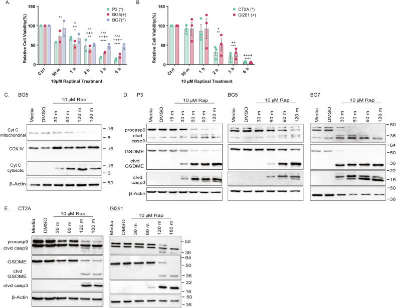
Treatment of glioblastoma (GB), the most common and most aggressive malignant brain tumor, has made little progress over the past two decades. Despite extensive research on apoptosis and autophagy, necrotic cell death mechanisms like pyroptosis, which have the potential to stimulate anti-tumor immune responses, remain largely underexplored in GB. Here, we investigated whether Gasdermin E (GSDME)-mediated pyroptosis can be induced in GB by employing the drug raptinal, an inducer of cytochrome c release. Using human patient-derived and mouse GB cell lines, we showed that raptinal promotes GSMDE cleavage. However, although a strong pyroptotic response was observed in mouse cell lines, it was weak in human cell lines. This resistance was partially reversed by the calcium chelator BAPTA-AM, indicating that membrane repair mechanisms may counteract the pyroptotic response. Gsdme knockout (KO) in mouse GB cells unexpectedly prolonged the survival of immunocompetent mice, demonstrating a tumor-promoting role of GSDME independent of its pyroptotic function. Analysis of the immune microenvironment revealed that Gsdme KO promoted infiltration of T cells, which was confirmed by spatial transcriptomic analysis of GB patient samples. In addition, Gsdme/GSMDE KO reduced the invasive capacity of mouse/human GB cells. In conclusion, active membrane repair mechanisms may impair the pyroptotic efficacy in GB. GSDME has a tumor-promoting role in GB by suppressing T cell infiltration and increasing tumor cell invasion.


Conflict of interest statement

Competing interests: The authors declare no competing interests. Ethics statement: The study was conducted in accordance with the Declaration of Helsinki and approved by the Regional Ethical Committee (REK 151825, 10.09.2020). Informed consent was obtained from all subjects involved in the study. The Regional Ethics Committee approved the animal study protocol. Animals were treated in accordance with the Norwegian Animal Act.

Figures

Fig. 1
Fig. 1. GSDME is expressed in GB at high levels.
A TCGA data extracted from proteinatlas.org [10] shows higher expression levels of GSDME in GB compared to other cancer types. RNA expression overview demonstrates RNA-seq data from TCGA, with expression levels presented as Transcripts Per Million (TPM) on the y-axis. B GSDME is highly expressed in GB cells, with levels comparable to those found in neurons and oligodendrocyte precursor cells (OPCs). Data derived Darmanis et al. [11]. C Analysis of histologically defined regions in GB from the Ivy GB Atlas [12] reveals the highest expression of GSDME in regions around necrosis with pseudopalisading cells. D Western blots of human GB cell lines and GSC lines show high GSDME expression in all samples. β-actin was used as loading control.
Fig. 2
Fig. 2. Raptinal induces cytochrome c release and GSDME cleavage in GB cell lines.
A WST-1 assay of human GSC lines treated with 10 μM raptinal. Time points after the start of treatment are indicated. Data represented as mean ± SEM (N = 2). ** (and corresponding signs) p < 0.01; *** (and corresponding signs) p < 0.001; *** (and corresponding signs) p < 0.0001. B WST-1 assay of mouse GB lines treated with 10 μM raptinal. Time points after the start of treatment are indicated. Data represented as mean ± SEM (N = 3). * (and corresponding signs) p < 0.05; ** (and corresponding signs) p < 0.01; *** (and corresponding signs) p < 0.001; *** (and corresponding signs) p < 0.0001. C Western blot of cytosolic and mitochondrial cytochrome C in BG5 cells treated with 10 μ μM raptinal. COXIV was used as a mitochondrial loading control, and β-actin as a cytosolic loading control. D Western blot for cleaved (clvd) Caspase 3, Caspase 9, clvd Caspase 9, GSDME, and clvd GSDME in GSC lines treated with 10 μM raptinal. Time points after the start of treatment are indicated. β-actin was used as loading control. E Western blot for cleaved (clvd) Capsase 3, Caspase 9, clvd Caspase 9, GSDME, and clvd GSDME in mouse GB lines treated with 10 μM raptinal. Time points after the start of treatment are indicated. β-actin was used as loading control.
Fig. 3
Fig. 3. Human GB cells show resistance to GSDME-mediated pyroptosis.
A Pyroptosis induction in human GSCs was quantified at the indicated time points post-raptinal treatment by microscopy, combining morphology and PI uptake. Data represented as mean ± SEM (N = 3). **p < 0.01; ***p < 0.001; ****p < 0.0001. The scale bar indicates 200 µm. B Pyroptosis induction in mouse GB lines was quantified at the indicated time points post-raptinal treatment by microscopy, combining morphology and PI uptake. Data represented as mean ± SEM (N = 3). *p < 0.05; **p < 0.01; ***p < 0.001; ****p < 0.0001. The scale bar indicates 200 µm. C Pyroptosis was measured by PI uptake in human GSC lines post-raptinal treatment at the indicated time points. BAPTA-AM was used as calcium chelator to block calcium-dependent plasma membrane repair mechanisms [16]. Data represented as mean ± SEM (N = 3). **p < 0.01; ***p < 0.001; ****p < 0.0001.
Fig. 4
Fig. 4. Knockout of Gsdme prolongs survival in mouse GB and promotes infiltration of T cells in the GB microenvironment.
A Schematic overview of the in vivo experiment (made with Biorender). B Kaplan Meier survival curve (n = 4–8 mice/group). **P < 0.01. C Immunohistochemistry of FFPE tumor sections for the leukocyte antigen CD45 and T cell antigens CD3, CD4, CD8, and Granzyme B. Quantifications and analyses of pooled ctrl and Gsdme KO groups are shown below the images. Data represented as mean ± SEM (n = 3–8 mice/group). *p < 0.05; **p < 0.01. The scale bar indicates 50 µm. D Immunohistochemistry of FFPE tumor sections for macrophage antigen F4/80. Quantifications and analyses of pooled ctrl and Gsdme KO groups are shown below the images. Data represented as mean ± SEM (n = 3–8 mice/group). The scale bar indicates 50 µm. E Dot plot showing average spatial correlation between GSDME, GB tumor cells, CD4, and CD8 T cells. CD3D and CD3E were used as CD4 T cell markers, CD8A was used as CD8 marker, while GFAP, SOX2, MKI67, and EGFR were used as GB tumor cell markers. F Surface plots showing spatial expression of GSDME, GB tumor cells, CD4, and annotated CD8 T cells in one representative patient (269UKF).
Fig. 5
Fig. 5. GSDME promotes invasion of GB cells.
A Immunohistochemistry of FFPE tumor sections (in vivo experiment from Fig. 4 A) for proliferation antigen Ki67. Quantification is shown next to the images. Data represented as mean ± SEM (n = 3–8 mice/group). B Wound healing assay with GL261 and GL261 Gsdme KO mouse GB cells. Wound closure was quantified at indicated time points. Data represented as mean ± SEM (N = 3). *p < 0.05; **p < 0.01. The scale bar indicates 400 µm. C Collagen invasion assay of GL261 and P3 and corresponding Gsdme/GSDME KO cells. The invasion was quantified at the indicated time points. Data represented as mean ± SEM (N = 3). *p < 0.05. The scale bar indicates 500 µm.

References

    1. Aldape K, Brindle KM, Chesler L, Chopra R, Gajjar A, Gilbert MR, et al. Challenges to curing primary brain tumours. Nat Rev Clin Oncol. 2019;16:509–20. - PMC - PubMed
    1. Ravi VM, Will P, Kueckelhaus J, Sun N, Joseph K, Salie H, et al. Spatially resolved multi-omics deciphers bidirectional tumor-host interdependence in glioblastoma. Cancer Cell. 2022;40:639–55.e13. - PubMed
    1. Hwang YK, Lee DH, Lee EC, Oh JS. Importance of autophagy regulation in glioblastoma with temozolomide resistance. Cells. 2024;13:1332. - PMC - PubMed
    1. Zhang Z, Zhang Y, Xia S, Kong Q, Li S, Liu X, et al. Gasdermin E suppresses tumour growth by activating anti-tumour immunity. Nature. 2020;579:415–20. - PMC - PubMed
    1. Zaffaroni N, Beretta GL. The therapeutic potential of pyroptosis in melanoma. Int J Mol Sci. 2023;24:1285. - PMC - PubMed

LinkOut - more resources