. 2025 Jun 21;11(1):285.
doi: 10.1038/s41420-025-02570-1.

Truncated LKB1 nonenzymatically enhances Fas-induced apoptosis by acting as a surrogate of Smac

Affiliations

Truncated LKB1 nonenzymatically enhances Fas-induced apoptosis by acting as a surrogate of Smac

Yutaro Yamada et al. Cell Death Discov. .

Abstract

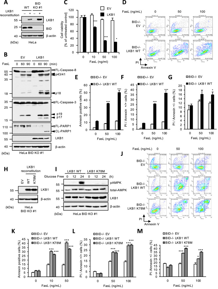
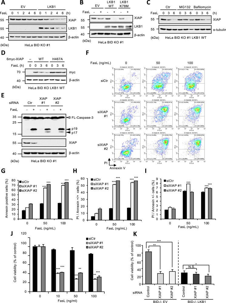
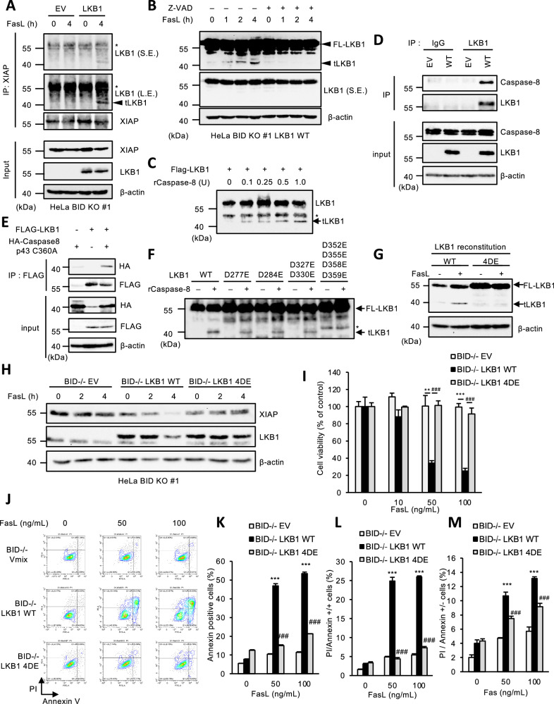
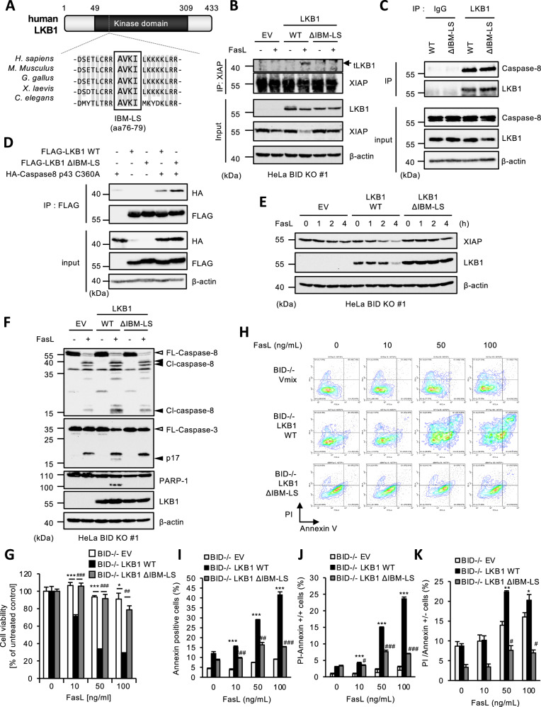
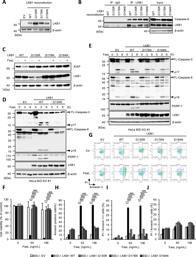
Although liver kinase B1 (LKB1) has been established as a tumor suppressor kinase, its mechanism of action is incompletely understood. Here we describe a novel nonenzymatic function of LKB1 in cell death induced by Fas/CD95. In BID knockout HeLa cells, inactivation of mitochondrial outer membrane permeabilization (MOMP) prevents Smac-induced inhibition of X-linked inhibitor of apoptosis (XIAP), causing resistance to Fas-induced apoptosis. However, reexpression of LKB1 in those cells naturally deficient for endogenous LKB1 restored apoptosis. Mechanistically, caspase-8 activated by Fas processed LKB1 to a truncated form, tLKB1. Both WT and kinase-inactive LKB1 antagonized XIAP to restore apoptosis, but somatic mutants of LKB1 found in Peutz-Jeghers syndrome (PJS) failed to do so. Thus, in addition to the known caspase-8 / tBid / Smac / XIAP pro-apoptotic axis, our results unveil a novel one, caspase-8 / tLKB1 / XIAP that potentially contributes to the antitumor functions of LKB1.


Conflict of interest statement

Competing interests: The authors declare no competing interests. Ethical approval and consent to participate: All methods were performed in accordance with the relevant guidelines and regulations. Recombinant DNA experiments were approved by ethics committee of Tohoku University (approval number: 2018PhR-019-02). All authors checked the study and agreed to participate in the manuscript. Consent for publication: All authors checked the study and agreed.

Figures

Fig. 1
Fig. 1. LKB1 nonenzymatically promotes Fas-induced apoptosis in BID KO HeLa cells.
A Immunoblot analysis of LKB1 in LKB1 reconstituted HeLa WT or BID KO cells. Cell lysates were subjected to immunoblotting with the indicated antibodies. β-actin was used as a loading control. B Empty vector (EV) or LKB1 reconstituted BID KO HeLa cells were treated with Fc-FasL (100 ng/mL) for indicated periods, and then cell lysates were subjected to immunoblotting with the indicated antibodies. C Empty vector (EV) or LKB1 reconstituted BID KO HeLa cells were treated with Fc-FasL (0,10,50,100 ng/mL) for 12 h. Cell viability was determined by PMS/MTS assay. Data shown are the mean ± SD (n = 3). Statistical significance was tested using an unpaired Student’s t test; *p < 0.05, ***p < 0.001, (vs. control cells). D Empty vector (EV) or LKB1 reconstituted BID KO HeLa cells were treated with Fc-FasL (0, 50,100 ng/mL) for 12 h. Apoptotic cells were labeled with annexin V-FITC and PI for 15 min, and analyzed by FACS. The data is presented as FITC-PE fluorescence density plots. E Quantification of the percentage of Annexin V-positive cells shown in Fig. 1D. Data shown are the mean ± SD (n = 3). Statistical significance was tested using an unpaired Student’s t test; ***p < 0.001, (vs. control cells). F Quantification of the percentage of PI and annexin V double-positive cells shown in Fig. 1D. Data shown are the mean ± SD (n = 3). Statistical significance was tested using an unpaired Student’s t test; ***p < 0.001, (vs. control cells). G Quantification of the percentage of PI-positive/Annexin V-negative cells shown in Fig. 1D. Data shown are the mean ± SD (n = 3). Statistical significance was tested using an unpaired Student’s t test; *p < 0.05, (vs. control cells). H Immunoblot analysis of LKB1 in LKB1 WT or K78M reconstituted HeLa BID KO cells. Cell lysates were subjected to immunoblotting with the indicated antibodies. I LKB1 WT or K78M reconstituted BID KO HeLa cells were cultured with glucose free medium for 0, 12 or 24 h, and then cell lysates were subjected to immunoblotting with the indicated antibodies. J Empty vector (EV) or LKB1 (WT or K78M) reconstituted BID KO HeLa cells were treated with Fc-FasL (0, 50,100 ng/mL) for 12 h. Apoptotic cells were labeled with annexin V-FITC and PI for 15 min, and analyzed by FACS. Data is presented as FITC-PE fluorescence density plots. K Quantification of the percentage of annexin V-positive cells shown in Fig. 1J. Data shown are the mean ± SD (n = 3). Statistical significance was tested using an unpaired Student’s t test; ***p < 0.001, (vs. control cells). L Quantification of the percentage of PI and annexin V double-positive cells shown in Fig. 1J. Data shown are the mean ± SD (n = 3). Statistical significance was tested using an unpaired Student’s t test; ***p < 0.001, (vs. control cells). M Quantification of the percentage of PI-positive/Annexin V-negative cells shown in Fig. 1J. Data shown are the mean ± SD (n = 3). Statistical significance was tested using an unpaired Student’s t test; ***p < 0.001, (vs. control cells). All data are representative of at least three biologically independent replicates.
Fig. 2
Fig. 2. LKB1 promotes Fas-induced apoptosis through the accelerated self-degradation of XIAP in BID KO HeLa cells.
A Empty vector (EV) or LKB1 reconstituted BID KO HeLa cells were treated with Fc-FasL (100 ng/mL) for 0, 1, 2, 4 or 6 h, and then cell lysates were subjected to immunoblotting with the indicated antibodies. B Empty vector (EV) or LKB1 (WT or K78M) reconstituted BID KO HeLa cells were treated with Fc-FasL (100 ng/mL) for 4 h, and then cell lysates were subjected to immunoblotting with the indicated antibodies. C LKB1 reconstituted BID KO HeLa cells were treated with Fc-FasL (100 ng/mL), and MG132 (10 μM) or bafilomycin (5 nM) for 0, 3 or 6 h, and then cell lysates were subjected to immunoblotting with the indicated antibodies. D LKB1 reconstituted BID KO HeLa cells were transfected with 6myc-XIAP (WT or H467A) for 24 h, and then treated with Fc-FasL (100 ng/mL) for 0, 3 or 6 h. Cell lysates were subjected to immunoblotting with the indicated antibodies. E BID KO HeLa cells were transfected with siRNA for negative control (siCtr) or XIAP (siXIAP #1 or siXIAP #2) for 24 h, and then treated with Fc-FasL (100 ng/mL) for 2 h. Cell lysates were subjected to immunoblotting with the indicated antibodies. F BID KO HeLa cells were transfected with siRNA for negative control (siCtr) or XIAP (siXIAP #1 or siXIAP #2) for 24 h, and then treated with Fc-FasL (0, 50, 100 ng/mL) for 12 h. Apoptotic cells were labeled with annexin V-FITC and PI for 15 min, and analyzed by FACS. Data is presented as FITC-PE fluorescence density plots. G Quantification of the percentage of annexin V-positive cells shown in Fig. 2F. Data shown are the mean ± SD (n = 3). Statistical significance was tested using an unpaired Student’s t test; ***p < 0.001, (vs. control cells). H Quantification of the percentage of PI and annexin V double-positive cells shown in Fig. 2F. Data shown are the mean ± SD (n = 3). Statistical significance was tested using an unpaired Student’s t test; ***p < 0.001, (vs. control cells). I Quantification of the percentage of PI-positive/Annexin V-negative cells shown in Fig. 2F. Data shown are the mean ± SD (n = 3). Statistical significance was tested using an unpaired Student’s t test; ***p < 0.001, (vs. control cells). J BID KO HeLa cells were transfected with siRNA for negative control (siCtr) or XIAP (siXIAP #1 or siXIAP #2) for 24 h, and then treated with Fc-FasL (0, 10, 50, 100 ng/mL) for 12 h. Cell viability was determined by PMS/MTS assay. Data shown are the mean ± SD (n = 3). Statistical significance was tested using an unpaired Student’s t test; **p < 0.01, ***p < 0.001, (vs. control cells). K Empty vector (EV) or LKB1 reconstituted BID KO HeLa cells were transfected with siRNA for negative control (Control) or XIAP (siXIAP #1 or siXIAP #2) for 24 h, and then treated with Fc-FasL (100 ng/mL) for 12 h. Cell viability was determined by PMS/MTS assay. Data shown are the mean ± SD (n = 3). Statistical significance was tested using an unpaired Student’s t test; *p < 0.05, ***p < 0.001, (vs. control cells). All data are representative of at least three biologically independent replicates.
Fig. 3
Fig. 3. Truncated LKB1 leads to the degradation of XIAP in BID KO HeLa cells.
A Empty vector (EV) or LKB1 reconstituted BID KO HeLa cells were treated with Fc-FasL (100 ng/mL) for 4 h, then immunoprecipitated with the anti-XIAP antibody, and subjected to immunoblotting with the indicated antibodies. The band indicated by an asterisk is a non-specific band that does not change upon LKB1 expression. The arrowhead indicates tLKB1. S.E.: short exposure. L.E.: long exposure. B LKB1 reconstituted BID KO HeLa cells were treated with Fc-FasL (100 ng/mL) and z-VAD (20 μM) for 0, 1, 2 or 4 h, and then cell lysates were subjected to immunoblotting with the indicated antibodies. C FLAG-LKB1, affinity-purified from 293 A cells overexpressing FLAG-LKB1, was reacted with recombinant active caspase-8 (0, 0.1, 0.25, 0.5, 1.0 units), and subjected to immunoblotting with the indicated antibodies. The band indicated with an asterisk is a non-specific band that is not affected by recombinant caspase-8. D Cell lysates from empty vector (EV) or LKB1-reconstituted BID KO HeLa cells were immunoprecipitated with protein G-Sepharose beads using the indicated antibodies, and subjected to immunoblotting with the indicated antibodies. E HEK293A cells were transfected with FLAG-LKB1 and/or HA-Caspase-8 p43 (C360A) plasmid for 24 h, and immunoprecipitated anti-FLAG-tagged agarose beads, and subjected to immunoblotting with the indicated antibodies. F FLAG-LKB1 (WT, D277E, D284E, D327/330E, D352/355/358/359E), affinity-purified from 293 A cells overexpressing FLAG-LKB1, were reacted with recombinant active caspase-8 (0.5 units), and subjected to immunoblotting with the indicated antibodies. The band indicated by an asterisk is a non-specific band that is not affected by recombinant caspase-8. G FLAG-LKB1 (WT or 4DE) reconstituted BID KO HeLa cells were treated with Fc-FasL (100 ng/mL) for 4 h, and then cell lysates were subjected to immunoblotting with the indicated antibodies. H Empty vector (EV) or LKB1 (WT or 4DE) reconstituted BID KO HeLa cells were treated with Fc-FasL (100 ng/mL) for 0, 2 or 4 h, and then cell lysates were subjected to immunoblotting with the indicated antibodies. I Empty vector (EV) or LKB1 (WT or 4DE) reconstituted BID KO HeLa cells were treated with Fc-FasL (0, 10, 50, 100 ng/mL) for 12 h. Cell viability was determined by PMS/MTS assay. Data shown are the mean ± SD (n = 3). Statistical significance was tested using an unpaired Student’s t test; ***p < 0.001, (vs. EV cells), ###p < 0.001 (vs. LKB1 WT cells). J Empty vector (EV) or LKB1 (WT/4DE) reconstituted BID KO HeLa cells were treated with Fc-FasL (0, 50, 100 ng/mL) for 12 h. Apoptotic cells were labeled with annexin V-FITC and PI for 15 min, and analyzed by FACS. Data is presented as FITC-PE fluorescence density plots. K Quantification of the percentage of Annexin V-positive cells shown in Fig. 3J. Data shown are the mean ± SD (n = 3). Statistical significance was tested using an unpaired Student’s t test; ***p < 0.001, (vs. EV cells), ###p < 0.001 (vs. LKB1 WT cells). L Quantification of the percentage of PI and annexin V double-positive cells shown in Fig. 3J. Data shown are the mean ± SD (n = 3). Statistical significance was tested using an unpaired Student’s t test; ***p < 0.001, (vs. EV cells), ###p < 0.001 (vs. LKB1 WT cells). M Quantification of the percentage of PI-positive/Annexin V-negative cells shown in Fig. 3J. Data shown are the mean ± SD (n = 3). Statistical significance was tested using an unpaired Student’s t test; ***p < 0.001, (vs. EV cells), ###p < 0.001 (vs. LKB1 WT cells). All data are representative of at least three biologically independent replicates.
Fig. 4
Fig. 4. A four amino acid sequence of LKB1 resembling an IAP binding motif is required for Fas-induced apoptosis in BID KO HeLa cells.
A Alignment of an IAP binding motif-like sequence (IBM-LS) of LKB1 for the indicated species. aa76-79 of human LKB1 correspond to the IBM-LS. B Empty vector (EV) or LKB1 (WT or ΔIBM-LS) reconstituted BID KO HeLa cells were treated with Fc-FasL (100 ng/mL) for 4 h, then immunoprecipitated with the anti-XIAP antibody, and subjected to immunoblotting with the indicated antibodies. C Cell lysates from LKB1 (WT/ΔIBM-LS) reconstituted BID KO HeLa cells were immunoprecipitated with protein G-Sepharose beads using the indicated antibodies, and subjected to immunoblotting with the indicated antibodies. D HEK293A cells were transfected with FLAG-LKB1 (WT/ΔIBM-LS) and/or HA-Caspase-8 p43 (C360A) plasmid for 24 h, and immunoprecipitated anti-FLAG-tagged agarose beads, and subjected to immunoblotting with the indicated antibodies. E Empty vector (EV) or LKB1 (WT or ΔIBM-LS) reconstituted BID KO HeLa cells were treated with Fc-FasL (100 ng/mL) for 0, 1, 2 or 4 h, and then cell lysates were subjected to immunoblotting with the indicated antibodies. F Empty vector (EV) or LKB1 (WT or ΔIBM-LD) reconstructed BID KO HeLa cells were treated with Fc-FasL (100 ng/mL) for 3 h, and then the cell lysates were subjected to immunoblotting with the indicated antibodies. G Empty vector (EV) or LKB1 (WT or ΔIBM-LS) reconstituted BID KO HeLa cells were treated with Fc-FasL (0, 10, 50, 100 ng/mL) for 12 h. Cell viability was determined by PMS/MTS assay. Data shown are the mean ± SD (n = 3). Statistical significance was tested using an unpaired Student’s t test; *p < 0.05, ***p < 0.001, (vs. EV cells), ##p < 0.01, ###p < 0.001 (vs. LKB1 WT cells) (H) Empty vector (EV) or LKB1 (WT or ΔIBM-LS) reconstituted BID KO HeLa cells were treated with Fc-FasL (0, 10, 50, 100 ng/mL) for 12 h. Apoptotic cells were labeled with annexin V-FITC and PI for 15 min, and analyzed by FACS. Data is presented as FITC-PE fluorescence density plots. I Quantification of the percentage of annexin V-positive cells shown in Fig. 4H. Data shown are the mean ± SD (n = 3). Statistical significance was tested using an unpaired Student’s t test; ***p < 0.001, (vs. EV cells), ###p < 0.001 (vs. LKB1 WT cells). J Quantification of the percentage of PI and annexin V double-positive cells shown in Fig. 4H. Data shown are the mean ± SD (n = 3). Statistical significance was tested using an unpaired Student’s t test; ***p < 0.001, (vs. EV cells), ###p < 0.001 (vs. LKB1 WT cells). K Quantification of the percentage of PI-positive/Annexin V-negative cells shown in Fig. 4H. Data shown are the mean ± SD (n = 3). Statistical significance was tested using an unpaired Student’s t test; ***p < 0.001, (vs. EV cells), ###p < 0.001 (vs. LKB1 WT cells). All data are representative of at least three biologically independent replicates.
Fig. 5
Fig. 5. Several LKB1 mutants found in PJS failed to promote Fas-induced apoptosis in BID KO HeLa cells.
A BID KO HeLa cells were reconstituted with LKB1 WT, G135R, D176N, or D194N. Cell lysates were subjected to immunoblotting with the indicated antibodies. B Cell lysates from LKB1 (WT/G135R/D176N/D194N) reconstituted BID KO HeLa cells were immunoprecipitated with protein G-Sepharose beads using the indicated antibodies, and subjected to immunoblotting with the indicated antibodies. C LKB1 (WT, G135R, D176N, or D194N) reconstituted BID KO HeLa cells were treated with Fc-FasL (100 ng/mL) for 3 h, and then cell lysates were subjected to immunoblotting with the indicated antibodies. D Empty vector (EV) or LKB1 (WT or G135R) reconstructed BID KO HeLa cells were treated with Fc-FasL (100 ng/mL) for 0, 3 or 6 h, and then cell lysates were subjected to immunoblotting with the indicated antibodies. E Empty vector (EV) or LKB1 (WT, D176N, or D194N) reconstituted BID KO HeLa cells were treated with Fc-FasL (100 ng/mL) for 0, 3 or 6 h, and then cell lysates were subjected to immunoblotting with the indicated antibodies. F Empty vector (EV) or LKB1 (WT, G135R, D176N, or D194N) reconstituted BID KO HeLa cells were treated with Fc-FasL (0, 50, 100 ng/mL) for 12 h. Cell viability was determined by PMS/MTS assay. Data shown are the mean ± SD (n = 3). Statistical significance was tested using an unpaired Student’s t test; ***p < 0.001, (vs. EV cells), ###p < 0.001 (vs. LKB1 WT cells) (G) Empty vector (EV) or LKB1 (WT, G135R, D176N, or D194N) reconstituted BID KO HeLa cells were treated with Fc-FasL (0, 50, 100 ng/mL) for 12 h. Apoptotic cells were labeled with annexin V-FITC and PI for 15 min, and analyzed by FACS. Data is presented as FITC-PE fluorescence density plots. H Quantification of the percentage of V-positive cells shown in Fig. 5G. Data shown are the mean ± SD (n = 3). Statistical significance was tested using an unpaired Student’s t test; ***p < 0.001, (vs. EV cells), ###p < 0.001 (vs. LKB1 WT cells). I Quantification of the percentage of PI and annexin V double-positive cells shown in Fig. 5G. Data shown are the mean ± SD (n = 3). Statistical significance was tested using an unpaired Student’s t test; ***p < 0.001, (vs. EV cells), ###p < 0.001 (vs. LKB1 WT cells). J Quantification of the percentage of PI-positive/Annexin V-negative cells shown in Fig. 5G. Data shown are the mean ± SD (n = 3). Statistical significance was tested using an unpaired Student’s t test; ***p < 0.001, (vs. EV cells), ###p < 0.001 (vs. LKB1 WT cells). All data are representative of at least three biologically independent replicates.
Fig. 6
Fig. 6. Schematic model to explain our study.
Upon Fas activation, caspase-8 induces MOMP by cleaving BID, and then promotes the cytoplasmic release of Smac, resulting in XIAP inactivation and apoptosis (Left part). XIAP can also be degraded in a parallel pathway independent on MOMP and Smac release. In this case, caspase-8 cleaves LKB1 within its C-terminal region to release tLKB1. tLKB1 binds to and induces proteasomal degradation of XIAP. Lowering XIAP levels releases the break imposed on caspase-3 activation and apoptosis induction (Right part).

Similar articles

References

    1. Kerr JF, Wyllie AH, Currie AR. Apoptosis: a basic biological phenomenon with wide-ranging implications in tissue kinetics. Br J Cancer. 1972;26:239–57. - PMC - PubMed
    1. Hengartner MO. The biochemistry of apoptosis. Nature. 2000;407:770–6. - PubMed
    1. Taylor RC, Cullen SP, Martin SJ. Apoptosis: controlled demolition at the cellular level. Nat Rev Mol Cell Bio. 2008;9:231–41. - PubMed
    1. Peter ME, Krammer PH. The CD95(APO-1/Fas) DISC and beyond. Cell Death Differ. 2003;10:26–35. - PubMed
    1. Nagata S. Fas ligand-induced apoptosis. Annu Rev Genet. 1999;33:29–55. - PubMed