. 2025 Jun 21;22(1):161.
doi: 10.1186/s12974-025-03483-7.

Pro-repair macrophages driven by CGRP rescue white matter integrity following intracerebral hemorrhage

Affiliations

Pro-repair macrophages driven by CGRP rescue white matter integrity following intracerebral hemorrhage

Huaping Huang et al. J Neuroinflammation. .

Abstract

Background: Intracerebral hemorrhage (ICH) triggers a dynamic immune response involving macrophages, However, the functional heterogeneity of these cells and the mechanisms through which they promote repair remain unclear. Although the neuropeptide CGRP has been shown to modulate macrophage phenotypes in other pathological contexts, its role in ICH recovery and white matter repair remains unexplored.

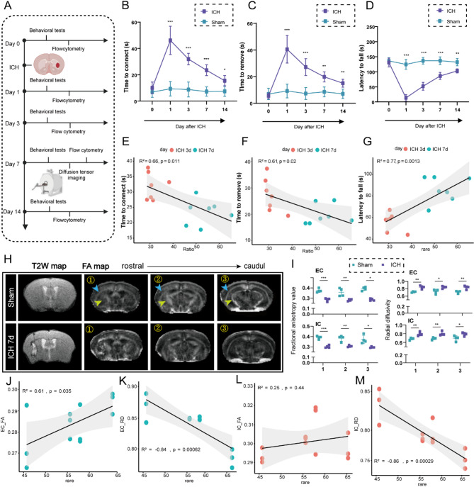
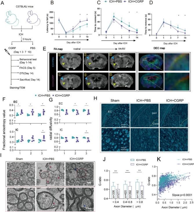
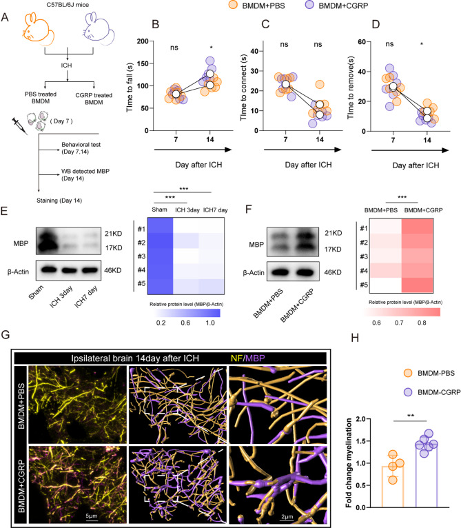
Methods: Single-cell RNA sequencing (scRNA-seq) of CD45 + cells from ICH mice (GSE167593 and GSE230414 datasets) identified macrophage subsets. Flow cytometry, diffusion tensor imaging (DTI), behavioral assays, and immunofluorescence validated macrophage dynamics and white matter (WM) integrity. Bone marrow and skull analyses traced macrophage origins. The role of calcitonin gene-related peptide (CGRP) was tested via intraperitoneal administration in ICH mice, with outcomes assessed through transcriptomics, ultrastructural imaging, and functional recovery.

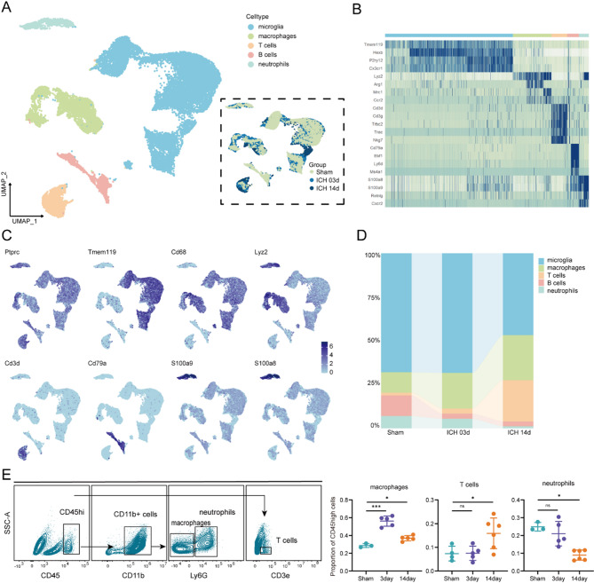
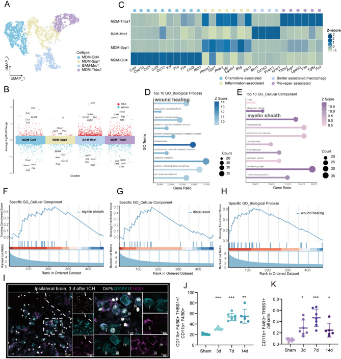
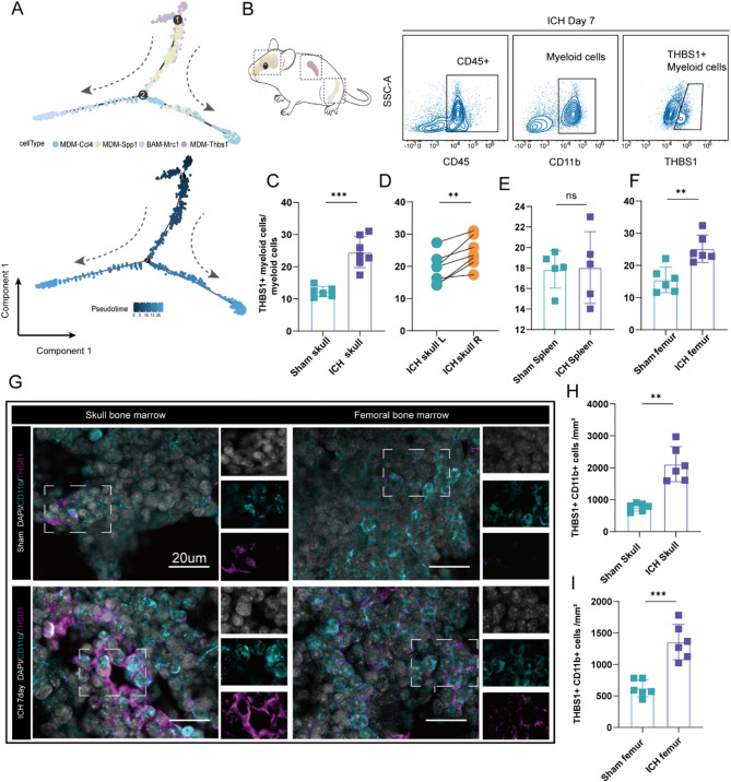
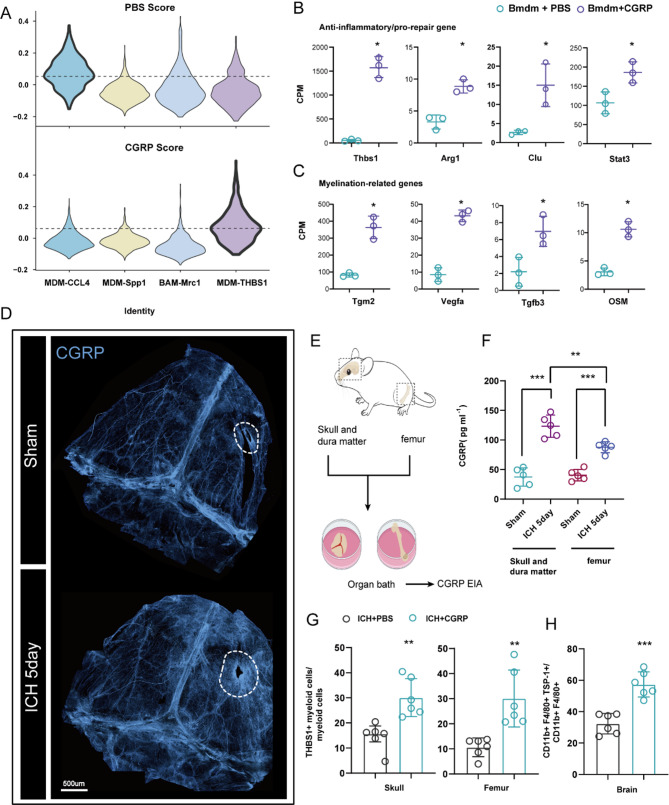
Results: scRNA-seq revealed sustained accumulation of THBS1 + macrophages post-ICH, correlating with WM repair and neurological recovery. These macrophages exhibited pro-repair and remyelination gene signatures (e.g., Arg1, Tgm2). Bone marrow-derived myeloid cells, particularly skull-resident populations, served as the primary source of THBS1 + macrophages. CGRP, elevated in meninges and bone marrow post-ICH, drove macrophage polarization toward THBS + phenotypes. CGRP administration expanded THBS1-positive macrophages in the bone marrow and brain, improving WM integrity (reduced radial diffusivity, higher fractional anisotropy) and sensorimotor function. Ultrastructural analysis confirmed enhanced myelin regeneration (lower g-ratio) in CGRP-treated mice.

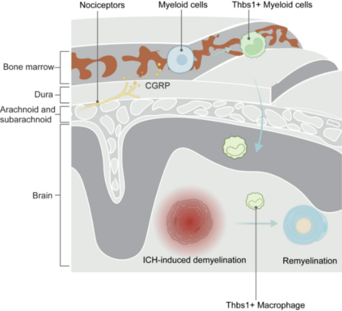
Conclusions: This study identifies a neuroimmune axis wherein CGRP promotes bone marrow-derived THBS1 + macrophages to facilitate WM repair and functional recovery after ICH. Targeting CGRP-macrophage signaling offers a therapeutic strategy to enhance recovery in hemorrhagic brain injury.

Supplementary Information: The online version contains supplementary material available at 10.1186/s12974-025-03483-7.

Keywords: Calcitonin gene-related peptide (CGRP); Intracerebral hemorrhage (ICH); Macrophage; Neuroimmune axis; THBS1; White matter repair.


Conflict of interest statement

Declarations. Ethics approval and consent to participate: All animal procedures were authorized by the Animal Ethics Committee of the Second Affiliated Hospital of Zhejiang University (Approval No. 2020 − 360) and conducted by the Guiding Principles for the Care and Use of Laboratory Animals, as endorsed by the National Science and Technology Committee of China. Consent for publication: All authors have read and approved the publication of this manuscript. Competing interests: The authors declare no competing interests.

Figures

Fig. 1
Fig. 1
Dynamic changes in immune cells following ICH. A UMAP visualization of immune cell heterogeneity in the brain following ICH at sham, 3 days, and 14 days post-injury. B Heatmap illustrates the top marker genes for each cluster. C Expression levels of selected known marker genes across unsorted cells, illustrated in UMAP plots, in Sham and ICH mice brains. D Composition of immune cell clusters in the brain following ICH. E Flow cytometry analysis of immune cell infiltration in the ICH brain at 3 days, 14 days post-ICH, and sham brains, *p < 0.05, ***p < 0.001; ns, not significant, one-way ANOVA and Bonferroni or Kruskal-Wallis and Dunn’s, n = 3–6. Data are mean ± SD
Fig. 2
Fig. 2
THBS1 + macrophages expressing pro-repair phenotype aggregate in the periphery of the hematoma following ICH. A UMAP representation of macrophage sub-clusters. B Manhattan plot of gene expression log2 fold-change values in macrophage sub-clusters. C Heatmap of selected genes representing standardized gene expression values (z-scores) in macrophage sub-clusters. Genes are classified by known functions, as indicated by colored circles below the heatmap for functional classification. D, E GO BP (D) and GO CC (E) enrichment analysis. Results were generated by gene set enrichment analyses of the significant DE genes for THBS1 + macrophage (log2FC > 1, adjusted < 0.05). F-H GSEA of THBS1 + macrophages reveals enrichment of myelin sheath, distal axon, and wound healing pathways. I Representative immunofluorescence images showing THBS1⁺ macrophages in the peri-hematomal region at 3 days after ICH, n = 3. J, K Flow cytometry quantification of CD11b + F4/80 + THBS1 + macrophages in ipsilateral brain tissue 3, 7, 14 days after ICH versus sham with mean ± SD, *p < 0.05, **p < 0.01, ***p < 0.001, Welch’s ANOVA and Dunnett’s or Kruskal-Wallis and Dunn’s, n = 6–9. Data are mean ± SD
Fig. 3
Fig. 3
THBS1 + macrophages correlate with improved neurological outcomes and enhanced white matter function. A Experimental design to investigate the function of THBS1 + macrophages. B-D Assessment of neurological function following ICH by adhesive removal (B, C) and rotarod (D) tests, *p < 0.05, **p < 0.01, and ***p < 0.001, two-way repeated-measures ANOVA and Bonferroni, n = 6–7. E-G Correlation analysis revealed a significant positive association between the THBS1 + macrophage ratio and adhesive removal test results and rotarod test performance following ICH (n = 7). H Representative axial views of T2W and DTI FA maps acquired 7 days post-ICH. I Quantification of FA and RD in the EC and IC at three levels (n = 4), *p < 0.05, **p < 0.01, ***p < 0.001; Student’s t-test, Welch’s t-test, or Mann-Whitney test. J-M Pearson correlation analysis revealed a significant positive association between the THBS1 + macrophage ratio and the quantification of FA and RD in the EC and IC following ICH (n = 4). Data are mean ± SD
Fig. 4
Fig. 4
Skull bone marrow remodeling following ICH is a major source of THBS1 + cells. A Pseudotime analysis of four macrophage subclusters utilizing Monocle. The trajectories of the clusters are represented by lines (above), while the corresponding changes in pseudotime are indicated by color gradients (below). B-F Flow cytometry analysis of THBS1 + myeloid cells in the skull (C, D), spleen(E), and femur(F) following ICH. **P < 0.01, ***P < 0.001, ns representing non-significant, Student t-test, Paired t-test, or Welch’s t-test, n = 5–7. G Representative images depicting THBS1 + myeloid cells in the skull and femur bone marrow at 7 days following ICH.H, I Quantification of THBS1 + CD11b + cells in the skull (H) and femur (I) bone marrow; **p < 0.01, ***p < 0.001, Welch’s t-test or Student’s t-test, n = 6. Data are mean ± SD
Fig. 5
Fig. 5
CGRP promotes the phenotypic transition of macrophages toward a THBS1 + pro-repair phenotype. A Violin plot depicting macrophage subset transcriptional enrichment for gene signatures identified in RNA-seq analyses from GSE255049. B, C The expression of anti-inflammatory/pro-repair (B) and remyelination-related(C) genes was increased in CGRP-stimulated macrophages versus PBS, *adjusted p < 0.05, moderated t-test and Benjamini-Hochberg false discovery rate, n = 3. D Whole-mount confocal imaging of CGRP + nociceptor innervation in the dura mater in sham (above) and ICH (below) mice. E llustration of skull and femur CGRP assays from sham or ICH mice. F CGRP levels released from the skull and femur were measured by ELISA, **p < 0.01, ***p < 0.001, one-way ANOVA and Bonferroni, n = 5. G Quantification of THBS1 + myeloid cells(CD45 + CD11b+)ratio in CGRP-treated and PBS-treated mice skull (left) and femur bone(right) marrow at 5 days post-ICH, **p < 0.01, Student’s t test or Welch’s t test, n = 6. H Quantification of THBS1 + macrophage ratio in CGRP-treated and PBS-treated mice ipsilateral brain at 5 days post-ICH, **p < 0.01, Student’s t test, n = 6. Data are mean ± SD
Fig. 6
Fig. 6
Enhancing THBS1 + macrophage populations post-treatment with CGRP promotes white matter repair following ICH. A Experimental design for evaluating the effects of CGRP treatment in a mouse model of ICH. B-D Adhesive removal tests (B, C) and rotarod performance (D) indicate that mice treated with CGRP exhibited better long-term recovery of sensory and motor functions compared to PBS following ICH (n = 8),*P < 0.05, two-way repeated-measures ANOVA and Bonferroni. E Representative axial views of DTI fractional FA maps and a single plane of the DEC map are presented for the same brains collected 14 days after ICH. F, G Quantitative analysis of FA and RD was conducted in the EC (F) and IC(G) at three distinct anatomical levels (n = 5), *p < 0.05, Student’s t-test, Welch’s t-test, or Mann-Whitney test. H MBP staining in brain Sect. 14 days post-ICH (n = 4–6). White dashed lines indicate the region of the ipsilateral EC. I Representative TEM images at 14 d post-ICH with red squares highlighting the areas that have been enlarged. Red arrows, defects in the myelin sheath. Blue arrowheads, remyelinated axons. J The g-ratios of myelinated axons were quantified to axon diameters. The counts of axons from three mice are indicated.***p < 0.001, Mann-Whitney test. K Scatterplot illustrating the individual g ratio values and the distribution of axonal sizes. Data are mean ± SD
Fig. 7
Fig. 7
CGRP-induced repair-associated macrophages promote remyelination after ICH. A Experimental Design for the Adoptive Transfer of PBS- or CGRP-Induced Macrophages into ICH Mice. B-D Rotarod performance (B) and adhesive removal tests (C, D) showed that mice receiving CGRP-induced macrophages exhibited significantly improved long-term sensory and motor recovery compared to those receiving PBS-induced macrophages following ICH, *p < 0.05, Student’s t test, n = 6. E Western blot and heatmap show the protein expression of MBP in the hematoma region, ***p < 0.001, one-way ANOVA and Bonferroni, n = 5. F Western blot and heatmap show the protein expression of MBP in the adoptive transfer of PBS- or CGRP-induced Macrophages into 14 days post-ICH Mice, ***p < 0.001, Student’s t test, n = 6. G Representative immunofluorescence images of peri-hematomal region stained for NF and MBP. H Fold change MBP + NF + axons of total NF + axons, **p < 0.01, Student’s t test, n = 4–6. Data are mean±SD
Fig. 8
Fig. 8
Neuro-immune axis mediated by CGRP enhances macrophage-driven white matter repair in ICH

Similar articles

References

    1. Keep RF, Hua Y, Xi G. Intracerebral haemorrhage: mechanisms of injury and therapeutic targets. Lancet Neurol. 2012;11:720–31. - PMC - PubMed
    1. van Asch CJ, Luitse MJ, Rinkel GJ, van der Tweel I, Algra A, Klijn CJ. Incidence, case fatality, and functional outcome of intracerebral haemorrhage over time, according to age, sex, and ethnic origin: a systematic review and meta-analysis. Lancet Neurol. 2010;9:167–76. - PubMed
    1. Magid-Bernstein J, Girard R, Polster S, Srinath A, Romanos S, Awad IA, et al. Cerebral hemorrhage: pathophysiology, treatment, and future directions. Circul Res. 2022;130:1204–29. - PMC - PubMed
    1. Puy L, Parry-Jones AR, Sandset EC, Dowlatshahi D, Ziai W, Cordonnier C. Intracerebral haemorrhage. Nat Rev Dis Primers. 2023;9:1–18. - PubMed
    1. Durocher M, Ander BP, Jickling G, Hamade F, Hull H, Knepp B, et al. Inflammatory, regulatory, and autophagy co-expression modules and hub genes underlie the peripheral immune response to human intracerebral hemorrhage. J Neuroinflamm. 2019;16:56. - PMC - PubMed

LinkOut - more resources