. 2025 Jun 21;19(1):58.
doi: 10.1186/s13036-025-00528-6.

3D-printed magnesium/strontium-co-doped calcium silicate scaffolds promote angiogenesis and bone regeneration through synergistic bioactive ion stimulation

Affiliations

3D-printed magnesium/strontium-co-doped calcium silicate scaffolds promote angiogenesis and bone regeneration through synergistic bioactive ion stimulation

Chia-Che Ho et al. J Biol Eng. .

Abstract

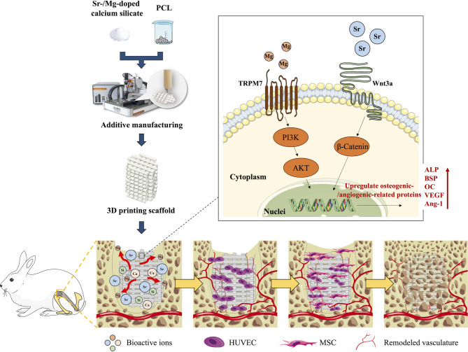
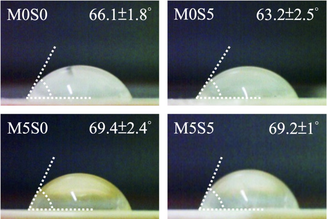
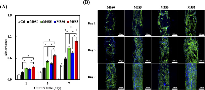
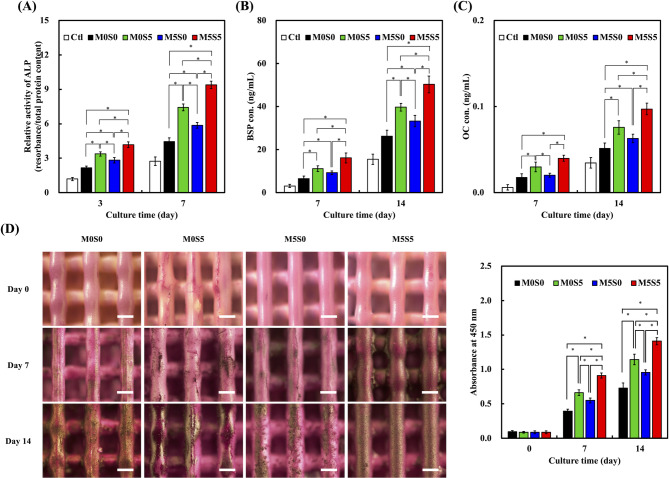
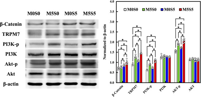
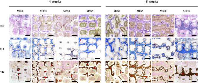
Bone defects resulting from trauma, infection, or surgical resection require biomaterials that support osteogenesis and vascularization for effective regeneration. In this study, we developed a 3D-printed magnesium- and strontium-co-doped calcium silicate (MSCS) scaffold using direct ink writing to optimize its bioactivity and structural integrity. X-ray diffraction confirmed the successful incorporation of Sr and Mg, leading to phase modifications that influenced ion release and degradation. Wettability and mechanical testing showed that Sr improved the stability, while Mg accelerated degradation, with M5S5 co-doping exhibiting a balanced degradation profile. In vitro, Wharton's jelly mesenchymal stromal cells cultured on M5S5 scaffolds displayed enhanced proliferation, cytoskeletal organization, and osteogenic differentiation, as evidenced by increased alkaline phosphatase activity and bone matrix protein expression. Angiogenesis assays using human umbilical vein endothelial cells revealed that Sr and Mg co-doping synergistically enhanced vascular endothelial growth factor and angiopoietin-1 secretion, thereby promoting endothelial tube formation. In vivo micro-computed tomography and histological analysis of a rabbit femoral defect model confirmed that M5S5 facilitated extensive new bone formation, exhibiting superior trabecular architecture and mineralization. These findings highlight MSCS scaffolds as promising biomaterials for bone tissue engineering applications.

Keywords: 3D printing; Bone regeneration; Calcium silicate; Magnesium; Strontium.


Conflict of interest statement

Declarations. Ethics approval and consent to participate: Not applicable. Consent for publication: Not applicable. Competing interests: The authors declare no competing interests.

Figures

Fig. 1
Fig. 1
Schematic diagram of the MSCS scaffold and the release of ions affecting Wharton Jelly mesenchymal stromal cell osteogenesis and angiogenesis
Fig. 2
Fig. 2
(A) XRD patterns, (B) enlarged XRD patterns highlighting selected 2θ regions, and (C) Fourier transform infrared (FTIR) spectra of MSCS scaffolds with different compositions (M0S0, M0S5, M5S0, and M5S5)
Fig. 3
Fig. 3
Water contact angle measurements of MSCS scaffolds with different compositions
Fig. 4
Fig. 4
(A) Optical images of the MSCS scaffolds with different compositions. (B) Scanning electron microscopy images and energy-dispersive spectrometry element mapping of calcium (Ca), silicon (Si), magnesium (Mg), and strontium (Sr) in the MSCS scaffolds. Scale bar: 1.4 mm
Fig. 5
Fig. 5
(A) Porosity, (B) stress-strain curves, (C) ultimate break point, and (D) Young’s modulus of the MSCS scaffolds with different compositions. Data are presented as the means ± standard error of the mean; n = 6 for each group
Fig. 6
Fig. 6
(A) Scanning electron microscopy images of MSCS scaffolds after immersion in SBF for 0, 3, 7, and 14 days. Scale bar: 2 μm. (B) Weight loss profiles of the MSCS scaffolds over a 6-month immersion period in SBF. Data are presented as the means ± standard error of the mean; n = 6 for each group
Fig. 7
Fig. 7
(A) Cell proliferation of WJMSCs on the MSCS scaffolds at days 1, 3, and 7. Data are presented as the means ± standard error the mean; n = 6 for each group. The symbol “*” denotes statistically significant differences (p < 0.05) between the groups. (B) Fluorescence images of WJMSCs on the MSCS scaffolds at days 1, 3, and 7. F-actin is stained green, while nuclei are stained blue. Scale bar: 200 μm
Fig. 8
Fig. 8
(A) Tube formation assay of HUVECs cultured with conditioned media from MSCS scaffolds of varying compositions. Scale bar: 500 μm. (B) Quantification of branch points and (C) total loops formed by HUVECs. (D) VEGF and (E) Ang-1 secretion levels on days 7 and 14. Data are presented as the means ± standard error of the mean; n = 6 for each group. The symbol “*” denotes statistically significant differences (p < 0.05) between the groups
Fig. 9
Fig. 9
(A) Alkaline phosphatase activity, (B) bone sialoprotein concentration, and (C) osteocalcin concentrations in Wharton Jelly mesenchymal stromal cells cultured on the MSCS scaffolds at days 3, 7, and 14. (D) Alizarin Red S staining images of mineralized matrix deposition at days 7 and 14 and the corresponding quantification of absorbance at 450 nm. Data are presented as the means ± standard error of the mean; n = 6 for each group. The symbol “*” denotes statistically significant differences (p < 0.05) between the groups
Fig. 10
Fig. 10
Western blot results of β-catenin, TRPM7, p-PI3K, and p-Akt of WJMSCs cultured on different scaffolds. * indicates a significant difference (p < 0.05) between the groups
Fig. 11
Fig. 11
(A) Micro-computed tomography images of rabbit femoral defects implanted with MSCS scaffolds at 4 and 8 weeks. (B) Bone volume fraction (BV/TV) and (C) trabecular thickness (Tb.Th) at 4 and 8 weeks post-implantation. Data are presented as the means ± standard error of the mean; n = 6 for each group. The symbol “*” denotes statistically significant differences (p < 0.05) between the groups
Fig. 12
Fig. 12
Hematoxylin and eosin, Masson’s trichrome, and Von Kossa staining of rabbit femoral defects implanted with MSCS scaffolds at 4 and 8 weeks post-implantation. Scale bar: 500 μm. Red stars indicate newly formed bone tissue; red arrows highlight newly formed blood vessels

Similar articles

References

    1. Zheng H, Zhou Y, Zheng Y, Liu G. Advances in hydrogels for the treatment of periodontitis. J Mater Chem B. 2023;11:7321–33. - PubMed
    1. Li N, Xie L, Wu Y, Wu Y, Liu Y, Gao Y, et al. Dexamethasone-loaded zeolitic imidazolate frameworks nanocomposite hydrogel with antibacterial and anti-inflammatory effects for periodontitis treatment. Mater Today Bio. 2022;16:100360. - PMC - PubMed
    1. Jia L, Zhang P, Ci Z, Hao X, Bai B, Zhang W, et al. Acellular cartilage matrix biomimetic scaffold with immediate enrichment of autologous bone marrow mononuclear cells to repair articular cartilage defects. Mater Today Bio. 2022;15:100310. - PMC - PubMed
    1. Liu Z, He X, Chen S, Yu H. Advances in the use of calcium silicate-based materials in bone tissue engineering. Ceram Int. 2023;49:19355–63.
    1. Chen Y, Dai F, Deng T, Wang L, Yang Y, He C, et al. An injectable MB/BG@LG sustained release lipid gel with antibacterial and osteogenic properties for efficient treatment of chronic periodontitis in rats. Mater Today Bio. 2023;21:100699. - PMC - PubMed

LinkOut - more resources