. 2025 Jun 21;56(1):123.
doi: 10.1186/s13567-025-01555-3.

CD44 mediates the internalization of foot-and-mouth disease virus through macropinocytosis

Affiliations

CD44 mediates the internalization of foot-and-mouth disease virus through macropinocytosis

Xuefei Wang et al. Vet Res. .

Abstract

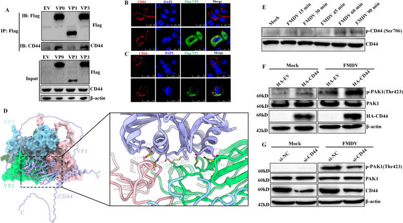
Foot-and-mouth disease virus (FMDV), a member of the Picornavirus family, poses a significant threat to global animal husbandry. While it is known that macropinocytosis plays a crucial role in the entry of FMDV into cells, the specific cellular proteins that regulate this process and the downstream signalling pathways associated with these proteins are not yet fully understood. In this study, we demonstrated that the membrane protein cluster of differentiation 44 (CD44) is essential for the internalization of FMDV in BHK-21 cells, while it is not involved in the attachment of the virus to the cell. In addition, we found that CD44 is internalized in response to FMDV entry. Interestingly, the use of a macropinocytosis inhibitor impaired both CD44 internalization and FMDV entry and infection, indicating a strong connection between these two processes. Further experiments revealed that CD44 facilitates FMDV internalization through macropinocytosis. Importantly, our study shows that CD44 interacts with FMDV capsid proteins VP2 and VP3 and becomes phosphorylated during the entry of the virus. The phosphorylation of CD44 subsequently promotes the phosphorylation of p21-activated kinase 1 (PAK1), which is a critical component in the macropinocytotic entry of FMDV. Overall, this study provides new insights into the role of CD44 in the invasion of pathogens into host cells and highlights potential strategies for improving FMDV vaccines.

Keywords: CD44; FMDV; internalization; macropinocytosis.


Conflict of interest statement

Declarations. Competing interests: The authors declare that they have no competing interests.

Figures

Figure 1
Figure 1
CD44 is required for FMDV internalization in BHK-21 cells. A, C PK-15 cells (A) or BHK-21 cells (C) transfected with plasmids encoding CD44 were infected with FMDV (10 MOI) at 4 ℃ for 1 h. After that, cells were washed and subjected to RT-qPCR. (B and D) PK-15 cells (B) or BHK-21 cells (D) pretreated with 10 μg/mL rabbit anti-CD44 antibody or rabbit IgG were infected for 1 h at 4 °C, and FMDV adsorption was determined by an RT-qPCR assay. E BHK-21 cells transfected with siRNA targetting CD44 were collected and assigned to western blot. F BHK-21 cells transfected with siRNA targeting CD44 were infected with FMDV (10 MOI) at 4 ℃ for 1 h. Cells were then washed and subjected to an RT-qPCR. (G and I) PK-15 cells G or BHK-21 cells (I) transfected with plasmids encoding CD44 were infected with FMDV (10 MOI) at 4 ℃ for 1 h. After that, cells were washed and shifted to 37 ℃ for 1 h. Cells were then treated with trypsin or proteinase K, and subjected to RT-qPCR. (H, J) PK-15 cells (H) or BHK-21 cells (J) pretreated with 10 μg/mL rabbit anti-CD44 antibody or rabbit IgG were infected with FMDV, and FMDV internalization was determined by an RT-qPCR assay. K BHK-21 cells transfected with siRNA targeting CD44 were infected with FMDV, and FMDV internalization was determined by an RT-qPCR assay. L BHK-21 cells transfected with plasmids encoding CD44 were infected with FMDV (10 MOI) for 1 h at 37 ℃, cells were then fixed, and localisation was determined using confocal microscopy. Data are means and SD of the results of three independent experiments. *P < 0.05; **P < 0.01; ns: not significant.
Figure 2
Figure 2
Inhibitor of macropinocytosis impairs CD44 internalization and type O FMDV entry. A BHK-21 cells transfected with plasmids encoding CD44 for 24 h were pre-treated with DMSO, EIPA (40 μM), CPZ (20 μM) or MβCD (1 mM) for 1 h, then infected with FMDV (10 MOI) at 37 ℃ in the presence of the inhibitors. At 1 h post-infection, cells were fixed and incubated with anti-HA antibodies and then with secondary antibodies conjugated with TRITC (red). Nuclei were counterstained with DAPI (blue), and localization was determined using confocal microscopy. B BHK-21 cells were pre-treated with DMSO or EIPA (40 μM) for 1 h, then infected with FMDV (10 MOI) at 37 ℃ in the presence of the inhibitor. At 1 h post-infection, the cell samples were collected and analysed by RT-qPCR assay to determine the viral mRNA. C, D BHK-21 cells were pre-treated with DMSO or EIPA (40 μM) for 1 h and then assigned to FMDV (1 MOI) for the indicated time points in the presence of the inhibitor. The cell lysates and supernatants were collected, and viral yields were determined by a TCID50 assay (C). The cells were collected and analysed by western blot for FMDV protein level (D). E BHK-21 cells were mock-infected or infected with FMDV (10 MOI) at 4 ℃ for 1 h. After that, cells were washed with cold PBS to remove unbound particles and the inoculum was replaced and incubated for 60 min at 37 °C. Cells were fixed and stained with 488-phalloidin, then subjected to confocal microscopy. Data are means and SD of the results of three independent experiments. *P < 0.05; ***P < 0.001.
Figure 3
Figure 3
CD44 promotes FMDV internalization via macropinocytosis. A BHK-21 cells were transfected with plasmids encoding HA-CD44. At 24 h post-transfection, cells were mock-infected and infected with FMDV (10 MOI) at 4 ℃ for 1 h. After that, cells were washed with cold PBS to remove unbound particles and the inoculum was replaced with medium containing dextran 10 K-Alexa Fluor 594 and incubated for 20 min at 37 °C, cells were then fixed and incubated with anti-FMDV and anti-HA antibodies and then with secondary antibodies conjugated with FITC (green) and Alexa Fluor 647 (pink). Nuclei were counterstained with DAPI (blue), and localization was determined using confocal microscopy. B BHK-21 cells were transfected with CD44 siRNA. At 36 h post-transfection, cells were mock-infected or infected with FMDV (10 MOI) at 4 ℃ for 1 h. After that, cells were washed with cold PBS to remove unbound particles and the inoculum was replaced with medium containing FITC-dextran and incubated for 60 min at 37 °C. Cells were then fixed with 4% paraformaldehyde. Dextran uptake was measured by confocal microscopy. C BHK-21 cells were transfected with CD44 siRNA. At 36 h post-transfection, cells were mock-infected or infected with FMDV (10 MOI) at 4 ℃ for 1 h. After that, cells were washed with cold PBS to remove unbound particles and the inoculum was replaced and incubated for 60 min at 37 °C. Cells were fixed and stained with 488-phalloidin, then subjected to confocal microscopy.
Figure 4
Figure 4
CD44 colocalises with rabankyrin-5 during FMDV internalization. A Cells mock-infected or infected with FMDV were lysed, and the cell lysates were immunoprecipitated with anti-CD44 antibody. The co-IP samples were then loaded to the SDS PAGE gel and subjected to silver staining. B BHK-21 cells transfected with EGFP-Rabankyrin-5 and HA-CD44 for 24 h were mock-infected or infected with FMDV (10 MOI) for 1 h at 37 °C. Cells were then fixed and incubated with anti-HA antibody and then with secondary antibodies conjugated with TRITC (red). Nuclei were counterstained with DAPI (blue), and localisation was determined using confocal microscopy. C The colocalisation of Rabankyrin-5 and CD44 was shown by Pearson’s coefficient. D BHK-21 cells transfected with EGFP-Rabankyrin-5 were mock-infected or infected with FMDV (10 MOI) for 1 h at 37 °C. The cells were then fixed and incubated with anti-FMDV serum and then with secondary antibodies conjugated with TRITC (red). Nuclei were counterstained with DAPI (blue), and localisation was determined using confocal microscopy. E BHK-21 cells transfected with EGFP-Rabankyrin-5 and HA-CD44 for 24 h were infected with FMDV (10 MOI) for 1 h at 37 °C. The cells were then fixed and incubated with anti-HA antibody and anti-FMDV serum, and then with secondary antibodies conjugated with TRITC (red) and Alexa Fluor 647 (pink). Nuclei were counterstained with DAPI (blue), and localisation was determined using confocal microscopy. Data are means and SD of the results of three independent experiments. ****P < 0.0001.
Figure 5
Figure 5
The process of CD44-mediated FMDV macropinocytotic entry. A BHK-21 cells were transfected with plasmids encoding FLAG-VP0, FLAG-VP1 and FLAG-VP3. At 24 hpt, cells were lysed on ice for 1 h and centrifuged at 15 000 × g for 20 min at 4 °C. Cell debris was discarded, and the supernatants were immunoprecipitated with anti-FLAG antibodies at 4 °C overnight. The immune complexes were incubated with protein G-agarose beads for 2 h, washed 5 times with lysis buffer, and eluted in SDS-PAGE buffer, then assigned to western blot analysis using anti-FLAG and anti-CD44 antibodies. B, C BHK-21 cells were transfected with plasmids encoding FLAG-VP0 (B) or FLAG-VP3 (C) and HA-CD44. At 24 hpt, cells were fixed and incubated with anti-FLAG and anti-HA antibodies and then with secondary antibodies conjugated with FITC (green) and TRITC (red), respectively. Nuclei were counterstained with DAPI (blue), and localization was determined using confocal microscopy. D Molecular docking model of CD44 with FMDV virion. FMDV capsid proteins are coloured with cyan (VP1), green (VP2), and pink (VP3), among which VP2 and VP3 interact with CD44, displayed with purple (left panel). The amino acid residues of the capsid protein interacting with CD44 are indicated (right panel). E BHK-21 cells were infected with FMDV at an MOI of 10 for the noted timepoints. The indicated protein expression levels were determined by western blot. F CD44 overexpressed exogenously in BHK-21 cells was mock-infected and infected with FMDV at an MOI of 10 for 60 min. The indicated protein expression levels were detected by western blot. G BHK-21 cells were transfected with CD44 siRNA. At 36 h post-transfection, cells were mock-infected and infected with FMDV at an MOI of 10 for 60 min. The indicated protein expression levels were detected by western blot.
Figure 6
Figure 6
Schematic model depicting CD44-mediated FMDV macropinocytotic entry. CD44 interacts with FMDV VP2 and VP3 and undergoes phosphorylation during FMDV entry. Subsequently, the activated CD44 initiates the activation of PAK1, a crucial element in FMDV macropinocytotic entry, thereby promoting macropinocytosis.

Similar articles

References

    1. Mercer J, Helenius A (2008) Vaccinia virus uses macropinocytosis and apoptotic mimicry to enter host cells. Science 320:531–535 - PubMed
    1. Kerr MC, Teasdale RD (2009) Defining macropinocytosis. Traffic 10:364–371 - PubMed
    1. Mercer J, Helenius A (2009) Virus entry by macropinocytosis. Nat Cell Biol 11:510–520 - PubMed
    1. Mercer J, Helenius A (2012) Gulping rather than sipping: macropinocytosis as a way of virus entry. Curr Opin Microbiol 15:490–499 - PubMed
    1. Han SC, Guo HC, Sun SQ, Jin Y, Wei YQ, Feng X, Yao XP, Cao SZ, Xiang Liu D, Liu XT (2016) Productive entry of foot-and-mouth disease virus via macropinocytosis independent of phosphatidylinositol 3-kinase. Sci Rep 6:19294 - PMC - PubMed

Substances

LinkOut - more resources