. 2025 Jun 21;22(1):202.
doi: 10.1186/s12985-025-02832-5.

Nuclear keratin 6A upregulates human papillomavirus oncogene expression through TEAD1 interaction

Affiliations

Nuclear keratin 6A upregulates human papillomavirus oncogene expression through TEAD1 interaction

Tomoya Miyamura et al. Virol J. .

Abstract

Background: Human papillomavirus (HPV) oncogenes, E6 and E7, play a critical role in cervical carcinogenesis, and their expression is regulated by cellular transcription factors bound to the long control region (LCR) in the HPV genome. To elucidate the mechanisms of HPV-induced carcinogenesis, it is important to identify host factors that control the LCR in cervical cancer cells. We previously reported that the LCR-bound transcription factor TEAD1 is critical for HPV oncogene expression. Here, we aimed to elucidate the role of stress-responsive keratin 6A (K6A), a potential cofactor for TEAD1, in HPV oncogene expression.

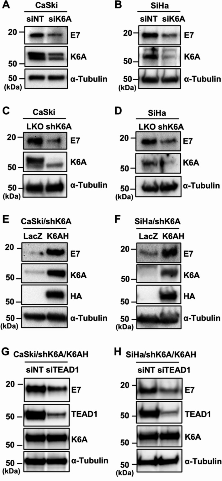
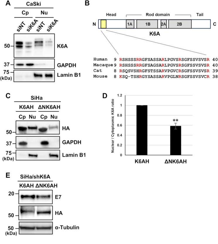
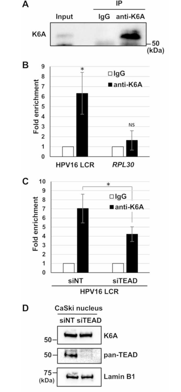
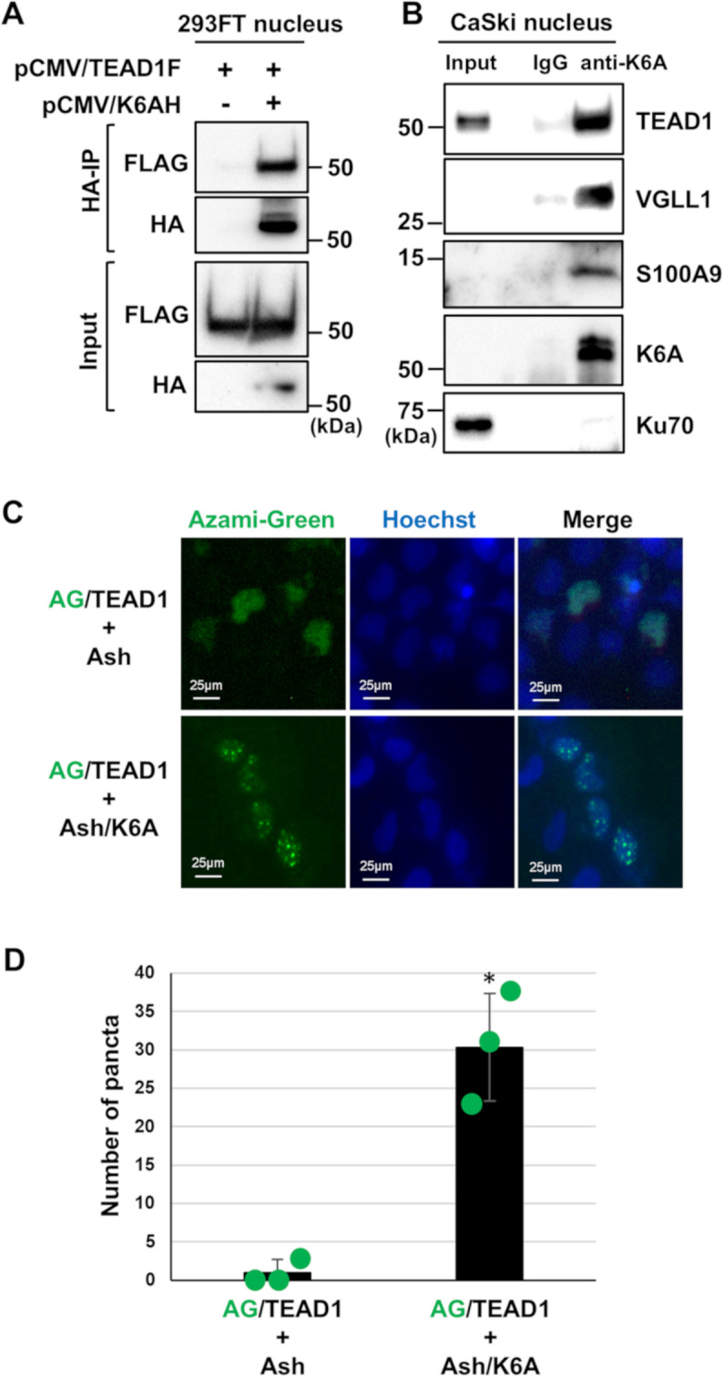
Methods: HPV16-positive cervical cancer cells were transfected with small interfering RNA (siRNA) or infected with a lentivirus expressing short hairpin RNA (shRNA) to downregulate the expression of K6A. HPV16 oncogene expression was examined by reverse transcription-quantitative polymerase chain reaction or Western blotting. Retroviral transduction was used to rescue the expression of K6A in K6A-depleted cells. Subcellular localization of K6A was analyzed by cellular fractionation followed by Western blotting. Chromatin immunoprecipitation (ChIP) assay was used to evaluate in vivo binding of K6A to the LCR. The physical interaction between K6A and TEAD1 was assessed by co-immunoprecipitation and fluorescence-based in vivo interaction assays.

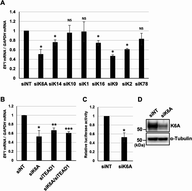
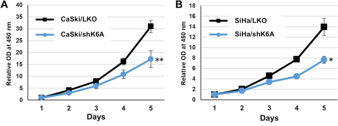
Results: Transfection of siRNA against K6A decreased levels of HPV16 E6*I mRNA and E7 protein in cervical cancer cells. Lentiviral delivery of shRNA against K6A also reduced E7 protein level and suppressed cell growth. Conversely, ectopic expression of shRNA-resistant K6A in the K6A-depleted cells restored E7 expression, and further siRNA knockdown of TEAD1 in the K6A-rescued cells attenuated E7 expression. K6A was detected in the nuclear fraction of cervical cancer cells with a functional bipartite nuclear localization signal in its N-terminus. ChIP assay showed that nuclear K6A bound to the HPV16 LCR in vivo, partially depending on the presence of the TEAD transcription factors. Finally, protein-protein interaction assays confirmed the association of K6A with TEAD1 in the nucleus.

Conclusions: Nuclear K6A associates with the LCR via TEAD1 and upregulates HPV16 oncogene expression, thereby contributing to cervical cancer development.

Supplementary Information: The online version contains supplementary material available at 10.1186/s12985-025-02832-5.

Keywords: Cervical cancer; Gene expression; Human papillomavirus; Keratin 6A; Viral oncogene.


Conflict of interest statement

Declarations. Ethics approval and consent to participate: Not applicable. Consent for publication: Not applicable. Competing interests: The authors declare no competing interests.

Figures

Fig. 1
Fig. 1
Involvement of keratins in HPV oncogene expression. (A, B) CaSki cells were transfected with the indicated small interfering RNAs (siRNAs). Two days after transfection, the mRNA levels of HPV16 E6*I were determined by reverse transcription-quantitative polymerase chain reaction (RT-qPCR), with normalization to the level of glyceraldehyde-3-phosphate dehydrogenase (GAPDH) mRNA. The data are averages from three independent experiments, with error bars representing standard deviations. P-values were determined by Student’s t-test. NS, not significant (P > 0.05); *, P < 0.05; **, P < 0.01; ***, P < 0.005. siNT, non-target control siRNA; siK6A, siRNA against KRT6A; siK14, siRNA against KRT14; siK10, siRNA against KRT10; siK1, siRNA against KRT1; siK16, siRNA against KRT16; siK9, siRNA against KRT9; siK2, siRNA against KRT2; siK78, siRNA against KRT78; and siTEAD1, siRNA against TEAD1. (C) HaCaT cells were transfected with siNT or siK6A. Six hours later, the cells were transfected with a reporter plasmid containing the HPV16 LCR-driven firefly luciferase gene (pGL3-P97), together with the Renilla luciferase plasmid. Two days after transfection, firefly luciferase activity was measured and normalized to the Renilla luciferase activity after background subtraction. The data are averages from three independent experiments, with error bars representing standard deviations. *, P < 0.05 (Student’s t-test). (D) HaCaT cells were transfected with siNT or siK6A, and the knockdown of K6A was verified by Western blotting with anti-K6A antibody. α-Tubulin was used as a loading control
Fig. 2
Fig. 2
Keratin 6A (K6A) induces E7 expression. (A, B) CaSki (A) and SiHa (B) cells were transfected with either non-target control small interfering RNA (siRNA) (siNT) or siRNA against KRT6A (siK6A). Two days after transfection, HPV16 E7 expression was detected by Western blotting with anti-HPV16 E7 antibody. (C, D) CaSki (C) and SiHa (D) cells stably expressing short hairpin RNA (shRNA) against KRT6A (CaSki/shK6A and SiHa/shK6A, respectively) or selection marker alone (CaSki/LKO and SiHa/LKO, respectively) were analyzed for expression of HPV16 E7 by Western blotting. The effects of the shRNA were verified by Western blotting for K6A. (E, F) CaSki/shK6A (E) and SiHa/shK6A (F) stably expressing hemagglutinin (HA)-tagged K6A (K6AH) (CaSki/shK6A/K6AH and SiHa/shK6A/K6AH, respectively) or β-galactosidase (LacZ) (CaSki/shK6A/LacZ and SiHa/shK6A/LacZ, respectively) were analyzed for HPV16 E7 expression by Western blotting. Ectopic expression of K6AH was verified by Western blotting with anti-K6A and anti-HA antibodies. (G, H) CaSki/shK6A/K6AH (G) and SiHa/shK6A/K6AH (H) cells were transfected with either siNT or siRNA against TEAD1 (siTEAD1). Two days after transfection, HPV16 E7 was detected by Western blotting. The effects of siTEAD1 on TEAD1 and K6A expression were verified by Western blotting with anti-TEAD1 and anti-K6A antibodies. α-Tubulin was used as a loading control
Fig. 3
Fig. 3
Keratin 6A (K6A) knockdown suppresses growth of cervical cancer cells. CaSki (A) and SiHa (B) cells stably expressing short hairpin RNA (shRNA) against KRT6A (CaSki/shK6A and SiHa/shK6A, respectively) or selection marker alone (CaSki/LKO and SiHa/LKO, respectively) were examined for cell viability using the Cell Counting Kit-8 (Dojindo) on the indicated days. The data are averages of relative optical density (450 nm) values obtained from triplicate experiments, with the error bars representing standard deviations. *, P < 0.05; **, P < 0.01 (Student’s t-test)
Fig. 4
Fig. 4
Keratin 6A (K6A) is present in the nucleus with a functional nuclear localization signal (NLS). (A) Cytoplasmic (Cp) and nuclear (Nu) fractions of CaSki cells transfected with non-target control small interfering RNA (siRNA) (siNT) or siRNA against KRT6A (siK6A) were analyzed by Western blotting for K6A. Glyceraldehyde-3-phosphate dehydrogenase (GAPDH) and lamin B1 were used as markers for the cytoplasmic and nuclear fractions, respectively. (B) A schematic diagram of K6A (upper). A bipartite NLS in the non-helical head domain is indicated by a yellow box. The helical rod domains are indicated by gray boxes. The amino acid sequence of the bipartite NLS of human K6A is aligned with those of other mammalian species (lower). The conserved lysine/arginine residues are highlighted in red. (C) Cytoplasmic (Cp) and nuclear (Nu) fractions of SiHa cells stably expressing hemagglutinin (HA)-tagged K6A (K6AH) or NLS-deleted K6AH (∆NK6AH) were analyzed by Western blotting with anti-HA antibody. GAPDH and lamin B1 were used as markers for the cytoplasmic and nuclear fractions, respectively. (D) HA-tagged protein levels in panel C in the nuclear fractions were quantified using Image Lab software (Bio-Rad) and normalized to those in the cytoplasmic fractions. The data are averages from three independent experiments, with error bars representing standard deviations. P-values were determined by Student’s t-test. **, P < 0.01. (E) SiHa/shK6A cells stably expressing shRNA-resistant K6AH (SiHa/shK6A/K6AH) or ∆NK6AH (SiHa/shK6A/∆NK6AH) were analyzed for HPV16 E7 expression by Western blotting. Ectopic expression of K6AH and ∆NK6AH was verified by Western blotting with anti-HA antibody. α-Tubulin was used as a loading control
Fig. 5
Fig. 5
Nuclear keratin 6A (K6A) binds to the HPV16 LCR via TEADs. (A) The nuclear extract isolated from CaSki cells was incubated with normal rabbit IgG or anti-K6A antibody. The input fraction (Input) and the precipitated fractions (IP) were analyzed by Western blotting with anti-K6A monoclonal antibody. (B) Cross-linked chromatin prepared from CaSki cells was assessed by chromatin immunoprecipitation (ChIP) assay using anti-K6A antibody or normal rabbit IgG, and the recovered DNA was quantified using real-time PCR with primers for the HPV16 LCR. The ribosomal protein L30 (RPL30) gene was used as a negative control (right). The level of K6A binding to the HPV16 LCR is shown as fold enrichment of the LCR DNA with anti-K6A antibody relative to that with normal IgG (left). (C) Cross-linked chromatin prepared from CaSki cells transfected with non-target control small interfering RNA (siRNA) (siNT) or a mixture of siRNA against TEAD1, TEAD3, and TEAD4 (siTEAD) was used to analyze the binding of K6A to the LCR by ChIP assay as described above. ChIP data are averages from four experiments performed using independent chromatin preparations, with the error bars representing standard deviations. NS, not significant; *, P < 0.05 (Student’s t-test). (D) The nuclear fraction of CaSki cells transfected with siNT or siTEAD was analyzed by Western blotting with anti-K6A and anti-pan-TEAD antibodies, the latter of which recognizes all TEAD family proteins. Lamin B1 was used as a loading control
Fig. 6
Fig. 6
Nuclear keratin 6A (K6A) interacts with TEAD1 and its transcriptional coactivators. (A) The nuclear extract prepared from 293FT cells transfected with the indicated expression plasmids was incubated with anti-hemagglutinin (HA) antibody. The input fractions (Input) and the precipitated fractions (HA-IP) were analyzed by Western blotting with anti-HA and anti-FLAG antibodies. (B) The nuclear extract prepared from CaSki cells was incubated with normal rabbit IgG or anti-K6A antibody. The input fraction (Input) and the precipitated fractions were analyzed by Western blotting with anti-TEAD1, anti-VGLL1, anti-S100A9, anti-K6A, and anti-Ku70 antibodies. (C) CaSki cells were co-transfected with the expression plasmids for a fusion protein of Azami-Green (AG) and TEAD1 (AG/TEAD1) and for Assembly Helper Tag (Ash) (upper panels) or with the expression plasmids for AG/TEAD1 and Ash-tagged K6A (Ash/K6A) (lower panels). Twenty-four hours after transfection, the nuclei were stained with Hoechst 33,342 and examined by fluorescence microscopy. Representative images are shown. (D) In experiment (C), the number of puncta in 20 randomly-selected fields of view was counted using the Analyze Particles tool in the ImageJ software. The data are averages from three independent experiments, with the error bars representing standard deviations. *, P < 0.05 (Student’s t-test)

Similar articles

References

    1. Doorbar J, Egawa N, Griffin H, Kranjec C, Murakami I. Human papillomavirus molecular biology and disease association. Rev Med Virol. 2015;25(Suppl):2–23. - PMC - PubMed
    1. International Agency for Research on Cancer. Cervical Cancer. https://www.iarc.who.int/cancer-type/cervical-cancer/. Accessed 28 February 2025.
    1. Perkins RB, Wentzensen N, Guido RS, Schiffman M. Cervical cancer screening: A review. JAMA. 2023;330:547–58. - PubMed
    1. Della Fera AN, Warburton A, Coursey TL, Khurana S, McBride AA. Persistent human papillomavirus infection. Viruses. 2021;13:321. - PMC - PubMed
    1. Boccardo E, Lepique AP, Villa LL. The role of inflammation in HPV carcinogenesis. Carcinogenesis. 2010;31:1905–12. - PubMed

LinkOut - more resources