. 2025 Jun 21;30(1):504.
doi: 10.1186/s40001-025-02787-x.

GDF-15 upregulates the SLC7A11/GPX4 signaling axis and promotes mitoxantrone resistance in AML cells

Affiliations

GDF-15 upregulates the SLC7A11/GPX4 signaling axis and promotes mitoxantrone resistance in AML cells

Qian-Wei Lu et al. Eur J Med Res. .

Abstract

Chemotherapy resistance poses a significant challenge in the initial treatment of acute myeloid leukemia (AML). Growth differentiation factor 15 (GDF-15) has been shown to play a critical role in cancer progression; however, the potential mechanisms by which GDF-15 contributes to AML progression and chemotherapy resistance remain unclear. We found that M2 macrophages secrete high levels of GDF-15, promoting resistance of AML cells to mitoxantrone (MTX). Furthermore, we demonstrated that MTX induces downregulation of the SLC7A11/GPX4 signaling axis in AML cells, mediating ferroptosis. GDF-15 enhances the expression of the SLC7A11/GPX4 axis, thereby inhibiting ferroptosis in AML cells and contributing to drug resistance. In addition, GDF-15 mitigates the decline in mitochondrial membrane potential and mitochondrial quality induced by MTX. In vivo experiments indicate that blocking GDF-15 effectively enhances the sensitivity of AML cells to mitoxantrone by reducing the expression of the SLC7A11/GPX4 axis.

Keywords: Acute myeloid leukemia; Chemoresistance; Ferroptosis; GDF-15.


Conflict of interest statement

Declarations. Ethics approval and consent to participate: This animal study was reviewed and approved by the Experimental Animal Welfare and Ethics Committee of the Third Military Medical University on March 5, 2021 (SYXK (Ethics) 20,170,002). Primary AML cells were obtained from the bone marrow blood samples of leukemia patients with informed consent. The study protocol was approved by the Ethics Committee of Shanghai Xinchao Biotechnology Co., Ltd. and complies with the Declaration of Helsinki (Permit No.: SHXC2021YF02). Institutional review board: Not applicable. Informed consent: Not applicable. Competing interest: The authors declare no competing interests.

Figures

Fig. 1
Fig. 1
GDF-15 is the key factor of M2 macrophage mediated MTX resistance in AML cells. A Primary AML cells were treated with mitoxantrone (0.2, 0.4, 0.8, 1.6 and 3.2 μM) and ferroptosis inducer RSL3 (1 μM) for 48 h, and then CCK-8 reagent was used to determine cell viability B M1 and M2 macrophages were differentiated from THP-1 cells, and the supernatants after 24 h of incubation were used to pretreat AML primary cells for 2 h, and then MTX (1.6 μM) was added, and CCK-8 was used to determine cell viability. C The supernatants of M1 and M2 macrophages were incubated for 24 h for solid phase antibody chip determination. D The expression level of GDF-15 in AML patients and the prognosis of different GDF-15 expression levels were analyzed using the NCBI patient database. E GDF-15 was knocked down in THP-1 cells. M2 macrophages were differentiated from ShGDF-15 THP-1 cells and wild-type THP-1 cells, respectively. The supernatants after 24 h of incubation were used to pretreat AML primary cells for 2 h, and then MTX (0.8, 1.6, 3.2 μM) was added. Cell viability was determined using CCK-8. F AML cells were pretreated with ferroptosis inhibitors (Fer-1, DFO) for 30 min, followed by MTX (1.6 μM) treatment, and then incubated for 48 h before performing the CCK-8 assay. Data are expressed as mean ± standard deviation (SD) from three independent experiments with n = 3 per group
Fig. 2
Fig. 2
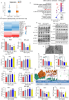
MTX mediates downregulation of the SLC7A11/GPX4 pathway in AML cells and induces ferroptosis. A M13 cells were treated with mitoxantrone (1.6 μM) for 24 h, followed by transcriptomic analysis and statistical analysis of differential mRNAs. B KEGG pathway enrichment was performed on the transcriptomic analysis results. C Heat map analysis of ferroptosis-related pathways was performed on the transcriptomic analysis results. D M13 cells were treated with mitoxantrone (0, 0.8, 1.6, 3.2 μM) and ferroptosis inducer RSL3 (1 μM) for 24 h, and then the protein levels of TFR1, FTL, SLC7A11, GPX4, ACSL4, and PRDX3 were measured. E M13 cells were treated with mitoxantrone (0, 1.6 μM) for 24 h, and then the morphological changes of mitochondria in M13 cells were observed by transmission electron microscopy. F M13 and MV-4–11 cells were treated with mitoxantrone (0, 0.8, 1.6, 3.2 μM) and RSL3 for 24 h, and then GSH levels were measured using a GSH kit. G Molecular docking of MTX and SLC7A11 was performed using AUTODOCK and visualized using P-MOL software. Data are expressed as mean ± standard deviation (SD) from three independent experiments with n = 3 per group
Fig. 3
Fig. 3
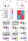
GDF-15 enhances the SLC7A11/GPX4 signaling axis in AML cells and inhibits MTX-mediated ferroptosis. A M13 cells were treated with mitoxantrone (1.6 μM) and GDF-15 (100 ng/mL) for 24 h, and then transcriptomic analysis was performed, and the differential mRNA was statistically analyzed. B The results of transcriptomic analysis were analyzed by volcano plot. C The results of transcriptomic analysis were enriched by KEGG pathways. D The results of transcriptomic analysis were analyzed by heat map analysis of ferroptosis-related pathways. EF M13 and MV-4–11 cells were treated with mitoxantrone (0, 0.8, 1.6 μM) and GDF-15 (100 ng/mL) for 24 h, and then the protein levels of SLC7A11, GPX4, ACSL4, and PRDX3 were measured. Data are expressed as mean ± standard deviation (SD) from three independent experiments with n = 3 per group
Fig. 4
Fig. 4
GDF-15 alleviates mitoxantrone-mediated mitochondrial damage in AML cells. A M13 and MV-4–11 cells were treated with mitoxantrone (1.6 μM) and RSL3 (1 μM) for 24 h, and then the mitochondrial membrane potential was measured using the JC-1 kit. B M13 and MV-4–11 cells were treated with mitoxantrone (1.6 μM) and RSL3 (1 μM) for 24 h, and then the mitochondrial mass was measured using the NonylAcridineOrange probe. C M13 and MV-4–11 cells were treated with mitoxantrone (1.6 μM) and GDF-15 (100 ng/mL) for 24 h, and then the mitochondrial membrane potential was measured using the JC-1 kit. D M13 and MV-4–11 cells were treated with mitoxantrone (0.8, 1.6 μM) and GDF-15 (100 ng/mL) for 24 h, and then the mitochondrial mass was measured using the NonylAcridineOrange probe. Data are expressed as mean ± standard deviation (SD) from three independent experiments with n = 3 per group
Fig. 5
Fig. 5
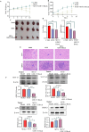
GDF-15 blocking can restore the sensitivity of AML to MTX. A After the xenograft tumor model was established, the weight changes of mice in each group were measured every other day. B The changes in tumor volume of mice in each group were measured every other day. C After continuous administration for 8 days, samples were taken and tumor size was photographed. D After the animals were taken, the liver and spleen weights were weighed and the liver and spleen organ indexes were calculated. E HE staining of liver and spleen tissues after administration. F Protein extraction was performed on tumor tissues in each group, and then Western Blot was used to determine the protein levels of GDF-15, SLC7A11, GPX4, and PRDX3. Data are expressed as mean ± standard deviation (SD) from three independent experiments with n = 5 per group
Fig. 6
Fig. 6
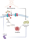
GDF-15 upregulates the SLC7A11/GPX4 signaling axis and enhances the resistance of AML cells to mitoxantrone. Mitoxantrone binds to SLC7A11, causing conformational changes and functional inactivation, leading to reduced cysteine uptake, blocked GSH synthesis, and GPX4 protein depletion, thereby inhibiting the lipid peroxidation clearance ability of AML cells. In addition, mitoxantrone also mediates mitochondrial damage in AML cells, leading to cell ferroptosis. GDF-15 weakens the ferroptosis-inducing effect of MTX in two ways: (1) enhancing SLC7A11 protein activity, and (2) enhancing mitochondrial metabolic balance, weakening the mitochondrial damage effect of mitoxantrone

Similar articles

Cited by

References

    1. Ladikou E, Sivaloganathan H, Pepper A, Chevassut T. Acute myeloid leukaemia in its niche: the bone marrow microenvironment in acute myeloid leukaemia. Curr Oncol Rep. 2020;22:1–9. - PMC - PubMed
    1. Tikhomirov AS, Shtil AA, Shchekotikhin AE. Advances in the discovery of anthraquinone-based anticancer agents. Recent Pat Anti-Cancer Drug Discovery. 2018;13(2):159–83. - PubMed
    1. Chen Z, Shi T, Zhang L, Zhu P, Deng M, Huang C, Hu T, Jiang L, Li J. Mammalian drug efflux transporters of the ATP binding cassette (ABC) family in multidrug resistance: a review of the past decade. Cancer Lett. 2016;370(1):153–64. - PubMed
    1. Shahar N, Larisch S. Inhibiting the inhibitors: targeting anti-apoptotic proteins in cancer and therapy resistance. Drug Resist Updates. 2020;52: 100712. - PubMed
    1. Lee M, Kim H-G. Anti-cancer strategy based on changes in the role of autophagy depending on the survival environment and tumorigenesis stages. Molecules. 2024;29(21):5134. - PMC - PubMed

MeSH terms

Substances

LinkOut - more resources