. 2025 Aug;66(8):100848.
doi: 10.1016/j.jlr.2025.100848. Epub 2025 Jun 20.

Optogenetic perturbation of lipid droplet localization affects lipid metabolism and development in Drosophila

Affiliations

Optogenetic perturbation of lipid droplet localization affects lipid metabolism and development in Drosophila

Xin Deng et al. J Lipid Res. 2025 Aug.

Abstract

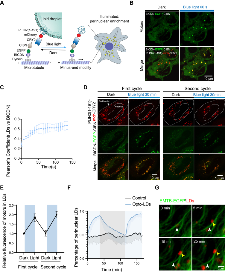
Lipid droplets (LDs) are dynamic organelles crucial for lipid storage and homeostasis. Despite extensive documentation of their importance, the causal relationship between LD localization and function in health and disease remains inadequately understood. Here, we developed optogenetics-based tools, termed "Opto-LDs," which facilitate the interaction between LDs and motor proteins in a light-dependent manner, enabling precise control of LD localization within cells. Utilizing these optogenetic modules, we demonstrated that light-induced relocation of LDs to the periphery of hepatocytes results in elevated very-low-density lipoprotein (VLDL) secretion, recapturing the beneficial effect of insulin in vitro. Furthermore, our studies in transgenic Drosophila revealed that proper LD localization is critical for embryonic development, with mistargeting of LDs significantly affecting egg hatching success. In summary, our work underscores the great importance of LD localization in lipid metabolism and development, and our developed tools offer valuable insights into the functions of LDs in health and disease.

Keywords: Drosophila; development; lipid droplet; metabolism; oogenesis; optogenetics.


Conflict of interest statement

Conflict of interest The authors declare that they have no conflicts of interest with the contents of this article.

Figures

Fig. 1
Fig. 1
Light-induced distribution of LDs to cell periphery. A: schematic representation of the predicted domain architecture of human PLIN2, with amino acid numbers indicated, and the diagrams of the generated PLIN2(1–191)-mCherry-CRY2 and KIF5A-EGFP-CIBN constructs. Note: The PLIN2(1–191) sequence contains a highly conserved PAT domain and an 11-mer repeat region, predicted to form amphipathic helices, which facilitate the targeting of co-expressed proteins to the LD surface. B. Schematic depiction of optogenetics-induced redistribution of LDs to the cell periphery. LDs (yellow) are tagged with PLIN2(1–191) fused to CRY2. CIBN is co-expressed with a truncated kinesin, KIF5A. Blue light exposure induces the interaction of CIBN and CRY2, recruiting kinesin motors to the LDs and driving their transport toward the microtubule plus end, thereby promoting peripheral localization. C-H: Live-cell imaging (C–D) and quantification (E–H) of COS-7 cells expressing PLIN2(1–191)-mCherry-CRY2 and KIF5A-EGFP-CIBN before and after 50 min of blue light illumination (5 mW 488 nm laser; 60 s interval). Cells underwent 2 cycles of blue light illumination (50 min each), followed by 60 min of recovery in the dark (see also Video S1). LDs were moving toward the cell periphery after blue light stimulation, as indicated by white arrows. D: Time-lapse images of LDs movement. The top panel showed COS-7 cells transfected with PLIN2(1–191)-mCherry and KIF5A-EGFP-CIBN (Control) and the bottom panel showed the Opto-LDs transfected cells under light simulation. E: Colocalization analysis of PLIN2(1–191)-mCherry-CRY2 (LDs) and KIF5A-EGFP-CIBN (KIF5A) over time (n = 10). F: The relative fluorescence of KIF5A-EGFP-CIBN (motors) upon LDs in both dark and light conditions under two cycles of stimulus. G: The cell periphery was defined as 70% distance from the cellular outline and the remaining fraction was defined as the perinuclear region. H: Graphs illustrating the percentage of cell periphery LDs in both Control and Opto-LDs transfected cells under either dark or light stimulated conditions. Data are represented as mean ± SEM from 10 cells for Opto-LDs and 8 cells for the Control group analyzed across 3 independent experiments. Scale bars, 20 μm.
Fig. 2
Fig. 2
Light-induced perinuclear localization of LDs. A: Schematic depiction of the optogenetics-induced perinuclear movement of LDs. LDs (yellow) are tagged with PLIN2(1–191) fused to CRY2. CIBN is linked to BICDN, a dynein/dynactin adaptor protein. Blue light illumination promotes heterodimerization between CIBN and CRY2, recruiting dynein to LDs and directing their movement toward the microtubule minus end, promoting perinuclear transport. B-C: Live-cell imaging (B) and colocalization analysis (C) of PLIN2(1–191)-mCherry-CRY2 (LDs) and BICDN-EGFP-CIBN (BICDN) over time. (B) The top panel showed the motors before and after 60 s of blue light stimulation. The bottom panel showed the merged images of motors and LDs in both dark and light simulated conditions. D-G: Live-cell imaging (D) and quantification (E–F) of COS-7 cells expressing PLIN2(1–191)-mCherry-CRY2 and BICDN-EGFP-CIBN, before and after blue light illumination (5 mW 488 nm laser; 60 s interval). Cells underwent 2 cycles of blue light illumination (30 min each), followed by 60 min of recovery in the dark (see also Video S2). E: The relative fluorescence of BICDN-EGFP-CIBN (motors) in LDs in both dark and light conditions under two cycles of stimulus. F: Graphs illustrating the percentage of cell perinuclear LDs in both Control and Opto-LDs transfected cells in either dark or light stimulated conditions. Data are represented as mean ± SEM from 13 cells for Opto-LDs and 8 cells for Control group analyzed across 3 independent experiments. G: LD movement towards the cell perinuclear region, indicated by yellow arrows, and the starting position marked by white arrows, occurs via light-induced recruitment of dynein along microtubules. Cells were co-transfected with BICDN-CIBN and PLIN2(1–191)–mCherry–CRY2, along with EMTB-EGFP, utilized to visualize microtubules.
Fig. 3
Fig. 3
Localization of LDs affects lipid metabolism in hepatocytes. A: Live-cell confocal imaging and quantification of Opto-LDs in LO2 hepatocytes transfected with PLIN2(1–191)-mCherry-CRY2 and either KIF5A-EGFP-CIBN (for peripheral enrichment) or BICDN-EGFP-CIBN (for perinuclear enrichment) before and after blue light stimulation for 30 min. B-C: Quantification of both intracellular (B) and secreted (C) triglyceride (TG) levels in LO2 cells transfected with PLIN2(1–191)-mCherry-CRY2 and KIF5A-EGFP-CIBN (Opto-LDs) or with PLIN2(1–191)-mCherry and KIF5A-EGFP-CIBN (Control group). Cells were subjected to pulsed blue light LED array illumination (1 min on, 1 min off, 2 mW/cm2) for 16 h. D–E: Quantification of both intracellular (D) and secreted (E) triglyceride (TG) levels in LO2 cells transfected with PLIN2(1–191)-mCherry-CRY2 and BICDN-EGFP-CIBN (Opto-LDs) or with PLIN2(1–191)-mCherry and BICDN-EGFP-CIBN (Control group). Cells were subjected to pulsed blue light LED array illumination (1 min on, 1 min off, 2 mW/cm2) for 16 h. F: Schematic representation of light-induced LD transport to the periphery in hepatocytes. Data are represented as mean ± SEM. ∗P < 0.05; ∗∗P < 0.01; ∗∗∗P < 0.001 by t test. Scale bars, 10 μm.
Fig. 4
Fig. 4
Sustained light-induced relocation of LDs in Drosophila oocyte. A: Overview of Drosophila oogenesis and LDs motility. Left: A schematic illustration of Drosophila oogenesis in one ovariole. Right: a representative mid-stage egg chamber. Oogenesis consists of 14 stages of follicle development. Each follicle consists of a layer of somatic epithelial cells, termed follicle cells, surrounding sixteen germline-derived cells; fifteen of these are support cells, termed nurse cells, and the other is the oocyte. Nurse cells supply the growing oocyte with nutrients, organelles, and signaling molecules. During Drosophila mid-oogenesis, many LDs are generated in the nurse cells. Bottom left: cytoplasmic streaming transports LDs from nurse cells through ring canals into the oocyte. Bottom right: In the oocyte cytoplasm, most LDs move passively by cytoplasmic streaming; a subset is actively transported by motors along microtubules. B: Schematic representation of the experimental treatment in Drosophila, where UASp-Lsd-1-mCherryP2A-KIF5A-CIBN (Control) or UASp-Lsd-1-mCherry-CRY2P2A-KIF5A-CIBN (Opto-LDs) transgenic flies were crossed with nanos-Gal4 flies and kept in a sustained 24-h blue light (488 nm, 2.2 mW/cm2) or a 24-h dark photoperiod. The resulting virgin females carrying both Gal4 and UAS transgenes were then mated with w1118 male flies and subjected to either a sustained 24-h blue light (488 nm, 2.2 mW/cm2) or a 24-h dark photoperiod. C: Confocal imaging of ovarioles from adult females expressing nanos-Gal4> Lsd-1-mCherryP2A-KIF5A-CIBN (Control) or nanos-Gal4> Lsd-1-mCherry-CRY2P2A-KIF5A-CIBN (Opto-LDs) flies in the dark and blue light stimulated conditions. Ovaries were stained with phalloidin-Alexa Fluor 488 and DAPI, with genotypes indicated above the images. mCherry signal indicating Lsd-1 labeled LDs. Scale bars, 100 μm, 50 μm or 20 μm as labeled. D: Quantification of the number of laid eggs in 1.5 h from crosses of 60 females and 20 males with indicated genotypes in the dark or after light stimulation. E: The hatching rate of eggs from Control and Opto-LDs expressing female flies crossed with w1118 male flies, subjected to sustained 24 h blue light or 24 h dark photoperiod. 30 eggs were examined in independent experiments (n = 7). Data are presented as mean ± SEM.

Similar articles

References

    1. Jin Y., Ren Z., Tan Y., Zhao P., Wu J. Motility plays an important role in the lifetime of mammalian lipid droplets. Int. J. Mol. Sci. 2021;22:3802. - PMC - PubMed
    1. Thiam A.R., Farese R.V., Jr., Walther T.C. The biophysics and cell biology of lipid droplets. Nat. Rev. Mol. Cel. Biol. 2013;14:775–786. - PMC - PubMed
    1. Prado L.G., Camara N.O.S., Barbosa A.S. Cell lipid biology in infections: an overview. Front. Cell. Infect. Microbiol. 2023;13 - PMC - PubMed
    1. Jarc E., Petan T. Lipid droplets and the management of cellular stress. Yale J. Biol. Med. 2019;92:435–452. - PMC - PubMed
    1. Kilwein M.D., Welte M.A. Lipid droplet motility and organelle contacts. Contact (Thousand Oaks) 2019;2 doi: 10.1177/2515256419895688. - DOI - PMC - PubMed

MeSH terms

LinkOut - more resources