. 2025 Jun 22;23(1):694.
doi: 10.1186/s12967-025-06666-1.

Notch3 enhances the synergistic effect of all-trans retinoic acid and calcipotriol in pancreatic stellate cell activation

Affiliations

Notch3 enhances the synergistic effect of all-trans retinoic acid and calcipotriol in pancreatic stellate cell activation

Zheng Lian et al. J Transl Med. .

Abstract

Background: Chronic pancreatitis (CP) is characterized by progressive fibrosis and the activation of pancreatic stellate cells (PSCs). As major producers of collagen-I and fibronectin, PSCs play important roles in pancreatic fibrosis, but few studies have explored methods to target activated PSCs. Notch3, a receptor in the Notch signaling pathway, is highly expressed in activated PSCs, but its specific effect on PSC activation needs to be confirmed. All-trans retinoic acid (ATRA) and the vitamin D analog calcipotriol were able to influence the activation of PSCs, but the relationship between ATRA, calcipotriol and Notch3 has not yet been clarified, and the effects of ATRA and calcipotriol on PSC activation need to be further enhanced.

Methods: The impact of Notch3 on pancreatic stellate cell (PSC) activation was evaluated by knocking down Notch3 in PSCs. PSCs were incubated with ATRA and calcipotriol individually or in combination to explore their effects on PSC activation. Notch3-knockdown PSCs were treated with ATRA or calcipotriol under various conditions in vitro to assess their effects on activation. Nuclear receptor inhibition was used to dissect the interplay between Notch3 signaling and ATRA/calcipotriol pathways. The roles of Notch3, ATRA, and calcipotriol were investigated in vivo using a chronic pancreatitis model. Different combinations of these interventions were tested in the chronic pancreatitis model to evaluate their in vivo efficacy.

Results: In this study, we confirmed the important role of Notch3 in PSC activation and found that ATRA and calcipotriol could regulate Notch3 expression. Furthermore, that ATRA and calcipotriol can synergistically prevent and reverse PSC activation, whereas knockdown of Notch3 can enhanced this synergistic effect. In CP model, we verified the effect of targeting Notch3 in combination with ATRA and calcipotriol. At last, we found that ATRA and calcipotriol do not regulate Notch3 through the nuclear receptors RARβ and VDR, but ATRA and calcipotriol depend on Notch3 to regulate PSC activation.

Conclusions: Notch3 is a target for inhibiting PSC activation, ATRA and calcipotriol regulate Notch3 expression in PSCs, and targeting Notch3 in combination with ATRA and calcipotriol against PSC activation holds promise as a novel therapeutic approach for treating pancreatic fibrosis in CP.

Keywords: All-trans retinoic acid; Calcipotriol; Chronic pancreatitis; Fibrosis; Notch3; Pancreatic stellate cells.


Conflict of interest statement

Declarations. Ethics approval and consent to participate: All animal experiments are conduct following ethical policies and procedures approved by the Capital Medical University ethics committee (Ethical inspection No. AEEI-2015-143). Consent for publication: Not applicable. Competing interests: The authors report no competing interests.

Figures
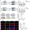
Fig. 1
Fig. 1
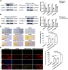
Notch3 affects PSC activation. A The expression of Notch3 was compared before and after PSC activation, as were the expression of the fibrosis-related genes fibronectin, collagen-I, and α-SMA (n = 3, *p < 0.05, **p < 0.01, ***p < 0.001). B Knockdown of Notch3 in activated PSCs and detection of fibrosis-related gene expression (n = 3, *p < 0.05, **p < 0.01, ***p < 0.001). C Changes in lipid droplets in activated PSCs, as well as changes in lipid droplets in activated PSCs after the knockdown of Notch3, were detected via Oil Red O staining, red arrows point to lipid droplets. Representative images were selected from three independent experiments. Scale bar, 400 µm (n = 3, *p < 0.05, **p < 0.01, ***p < 0.001). D BODIPY was used to determine whether activated PSCs were positive for lipid droplets, and morphological changes were evaluated via F-ACTIN staining. Knockdown of Notch3 in activated PSCs to detect lipid droplets and morphological changes. Representative images were selected from three independent experiments, where green represents lipid droplets, red represents F-ACTIN, and blue represents DAPI. Scale bar, 400 µm. Zoom with 60 × magnification (n = 3, *p < 0.05, **p < 0.01, ***p < 0.001)
Fig. 2
Fig. 2
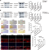
ATRA and calcipotriol synergistically prevent PSC activation and decrease Notch3 expression. A Changes in the expression of Notch3 and fibrosis-related genes after quiescent PSCs were incubated with ATRA (n = 3, *p < 0.05, **p < 0.01, ***p < 0.001). B Changes in Notch3 and fibrosis-related gene expression caused by incubation of quiescent PSCs with calcipotriol (n = 3, *p < 0.05, **p < 0.01, ***p < 0.001). C Preservation of lipid droplets in quiescent PSCs by ATRA and calcipotriol, as detected by Oil Red O staining, red arrows point to lipid droplets. Representative images were selected from three independent experiments. Scale bar, 400 µm (n = 3, *p < 0.05, **p < 0.01, ***p < 0.001). D BODIPY was used to detect the effects of ATRA or calcipotriol on lipid droplets in quiescent PSCs and their synergistic effects, while changes in cell morphology were detected by F-ACTIN staining. Representative images were selected from three independent experiments, where green represents lipid droplets, red represents F-ACTIN, and blue represents DAPI. Scale bar, 400 µm. Zoom with 60× magnification (n = 3, *p < 0.05, **p < 0.01, ***p < 0.001)
Fig. 3
Fig. 3
ATRA and calcipotriol synergistically reverse the activation of PSCs. A Effect of ATRA on the expression of Notch3 and fibrosis-related genes in activated PSCs (n = 3, *p < 0.05, **p < 0.01, ***p < 0.001). B Effect of calcipotriol on the expression of Notch3 and fibrosis-related genes in activated PSCs (n = 3, *p < 0.05, **p < 0.01, ***p < 0.001). C Re-accumulation of lipid droplets in activated PSCs by ATRA and calcipotriol, as measured by Oil Red O staining, red arrows point to lipid droplets. Representative images were selected from three independent experiments. Scale bar, 400 µm (n = 3, *p < 0.05, **p < 0.01, ***p < 0.001). D BODIPY was used to detect the effects of ATRA and calcipotriol on lipid droplet re-accumulation in activated PSCs, and F-ACTIN staining was used to detect changes in cellular morphology. Representative images were selected from three independent experiments, where green represents lipid droplets, red represents F-ACTIN, and blue represents DAPI. Scale bar, 400 µm. Zoom with 60× magnification (n = 3, *p < 0.05, **p < 0.01, ***p < 0.001)
Fig. 4
Fig. 4
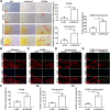
Knockdown of Notch3 enhances the synergistic reversal of activated PSCs by ATRA and calcipotriol. A Oil red O staining was used to assess the effects of Notch3 knockdown on the effects of ATRA or calcipotriol, as well as the synergistic effects of ATRA and calcipotriol, red arrows point to lipid droplets. Representative images were selected from three independent experiments. Scale bar, 400 µm (n = 3, *p < 0.05, **p < 0.01, ***p < 0.001). BODIPY was used to detect the number of activated PSCs that accumulated lipid droplets, and F-ACTIN staining was used to detect the degree of cellular morphological changes. B Knockdown of Notch3 via ethanol incubation. C, F Knockdown of Notch3 via incubation with ATRA. D, G Knockdown of Notch3 with Calcipotriol incubation. E, H Effect of Notch3 knockdown on the synergistic effects of ATRA and calcipotriol. Representative images were selected from three independent experiments, where green represents lipid droplets, red represents F-ACTIN, and blue represents DAPI. Scale bar, 400 µm. Zoom with 60× magnification (n = 3, *p < 0.05, **p < 0.01, ***p < 0.001)
Fig. 5
Fig. 5
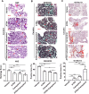
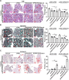
Targeting Notch3 in chronic pancreatitis reduces pancreatic injury and fibrosis. A Experimental flowchart of the CP mouse model and pAAV intraperitoneal injection as well as ATRA and calcipotriol treatment. B Ex vivo tissue fluorescence images showing mScarlet expression after intraperitoneal injection of pAAV. The scale bar represents the radiance (p/s/cm2/sr). C Representative images of H&E, MASSON, and Oil Red O staining of pancreatic histology in four groups of mice, where H&E staining was used to assess pancreatic injury, MASSON staining was used to detect pancreatic fibrosis, and Oil Red O staining was used to detect lipid droplets. Red arrows show areas of injury and fibrosis. Scale bar, 400 µm (n = 4, *p < 0.05, **p < 0.01, ***p < 0.001)
Fig. 6
Fig. 6
ATRA and calcipotriol synergistically reduce pancreatic injury and fibrosis in CP and reaccumulate lipid droplets. After the CP model establishment, ATRA or calcipotriol was intraperitoneally injected individually or in combination to detect the effects of ATRA and calcipotriol on the CP pancreas. A H&E staining was used to measure pancreatic injury. B MASSON staining was used to measure pancreatic fibrosis. C Oil Red O staining was used to detect lipid droplet accumulation. Representative images of histologic H&E, MASSON, and Oil Red O staining of the pancreas in the four groups of mice. Red arrows show injury, fibrosis and positive lipid droplet areas. Scale bar, 400 µm (n = 4, *p < 0.05, **p < 0.01, ***p < 0.001)
Fig. 7
Fig. 7
Targeting Notch3 enhances the synergistic effect of ATRA and calcipotriol on CP. pAAV-shNotch3 was combined with ATRA and calcipotriol in CP to determine the synergistic effects of pAAV-shNotch3 on ATRA and calcipotriol. A H&E staining was used to measure pancreatic injury. B MASSON staining was used to measure pancreatic fibrosis. C Oil Red O staining was used to detect lipid droplet accumulation. Representative images of pancreatic histology with H&E, MASSON, and Oil Red O staining in eight groups of mice. Red arrows show injury, fibrosis and positive lipid droplet areas. Scale bar, 400 µm (n = 4, *p < 0.05, **p < 0.01, ***p < 0.001)
Fig. 8
Fig. 8
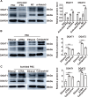
Notch3, ATRA, and calcipotriol influence the expression of diacylglycerol acyltransferases DGAT1 and DGAT2. A Expression of DGAT1 and DGAT2 in quiescent and activated PSCs. Effects of Notch3 knockdown on DGAT1 and DGAT2 (n = 3, *p < 0.05, **p < 0.01, ***p < 0.001). B Results of incubation of quiescent PSCs with ATRA or calcipotriol (n = 3, *p < 0.05, **p < 0.01, ***p < 0.001). C Incubation of activated PSCs with ATRA or calcipotriol (n = 3, *p < 0.05, **p < 0.01, ***p < 0.001)
Fig. 9
Fig. 9
ATRA and calcipotriol are independent of RARβ and VDR to regulate Notch3. Western blot analysis of the expression of Notch3 and the nuclear receptors RARβ and VDR after LE135 binding to ATRA or calcipotriol. A LE135 binding to ATRA for incubation of quiescent or activated PSCs (n = 3, *p < 0.05, **p < 0.01, ***p < 0.001). B Results of LE135 binding to calcipotriol for the incubation of quiescent or activated PSCs (n = 3, *p < 0.05, **p < 0.01, ***p < 0.001)
Fig. 10
Fig. 10
Summary. Knockdown of Notch3 enhances the synergistic effect of ATRA and calcipotriol on pancreatic stellate cell activation

Similar articles

References

    1. Xiao AY, Tan ML, Wu LM, Asrani VM, Windsor JA, Yadav D, Petrov MS. Global incidence and mortality of pancreatic diseases: a systematic review, meta-analysis, and meta-regression of population-based cohort studies. Lancet Gastroenterol Hepatol. 2016;1(1):45–55. - PubMed
    1. Vege SS, Chari ST. Chronic pancreatitis. N Engl J Med. 2022;386(9):869–78. - PubMed
    1. Bynigeri RR, Jakkampudi A, Jangala R, Subramanyam C, Sasikala M, Rao GV, Reddy DN, Talukdar R. Pancreatic stellate cell: Pandora’s box for pancreatic disease biology. World J Gastroenterol. 2017;23(3):382–405. - PMC - PubMed
    1. Jin G, Hong W, Guo Y, Bai Y, Chen B. Molecular mechanism of pancreatic stellate cells activation in chronic pancreatitis and pancreatic cancer. J Cancer. 2020;11(6):1505–15. - PMC - PubMed
    1. Beyer G, Habtezion A, Werner J, Lerch MM, Mayerle J. Chronic pancreatitis. Lancet (London, England). 2020;396(10249):499–512. - PubMed

LinkOut - more resources