. 2025 Jun;15(6):e70373.
doi: 10.1002/ctm2.70373.

Programmed death-ligand 1 expression and prognostic significance in bevacizumab treated ovarian cancer patients: Results from the phase IV MITO16A/MaNGO OV-2 translational study

Affiliations

Programmed death-ligand 1 expression and prognostic significance in bevacizumab treated ovarian cancer patients: Results from the phase IV MITO16A/MaNGO OV-2 translational study

Francesca Basso-Valentina et al. Clin Transl Med. 2025 Jun.
No abstract available


Conflict of interest statement

The authors declare no conflict of interest.

Figures
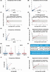
FIGURE 1
FIGURE 1
Correlation and agreements between different anti‐PD‐L1 antibodies and staining techniques. A/B Correlation analyses evaluating the scoring of Pathologist 1 (PAT1) and 2 (PAT2) in evaluating the staining of PD‐L1 using the 22C3 (A) or the E1L3N (B) antibodies in immunohistochemistry. C/D Graphs reporting the Bland‐Altman analysis evaluating the Limits of Agreement (LOAs) between PAT1 and PAT2 in evaluating PD‐L1 expression using the 22C3 (C) or the E1L3N (D) antibodies. (E) Correlation analyses evaluating the scoring of PD‐L1 using the 22C3 or the E1L3N antibodies in immunohistochemistry. (F) Graphs reporting the Bland‐Altman analysis evaluating the LOAs between the 22C3 and the E1L3N antibodies used in immunohistochemistry to evaluate PD‐L1 expression. G/H Graphs (G) and Table (H) reporting the number of PD‐L1 positive cells in EOC samples stained with the 22C3 and the E1L3N antibodies and evaluated by PAT1 and PAT2, as indicated. IQR = Inter Quartile Range; ICC = Intraclass Correlation Coefficient. (H) Table reporting the number of positive cells. (I) Correlation analyses evaluating the scoring of PD‐L1 using the E1L3N antibody in immunohistochemistry (IHC) or in multiplex immunofluorescence (MIF). (L) Graph reporting the Bland‐Altman analysis evaluating the LOAs between immunohistochemistry (IHC) or in multiplex immunofluorescence (MIF) in assessing PD‐L1 expression.
FIGURE 2
FIGURE 2
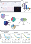
Multiplex staining evaluating tumour and stroma immune infiltration in samples from the MITO16A case material. (A) Typical image of the multiplex analyses using the OPAL Multiplex Assay coupled with multispectral image acquisition. Centralized tumour samples were processed and single FFPE tissue slides were stained for the indicated antibodies. Stained sections were acquired with a Nikon microscope coupled with a multispectral camera and acquired images were analyzed with the MANTRA software to define the number of immune cells infiltrating the tumours (PanCK‐positive areas) and the surrounding stroma. (B) Graph reporting the distribution of PD‐L1 positive cells in the analyzed samples. (C) Pie charts reporting the subtypes of PD‐L1 positive cells in the analyzed samples. (D) Kaplan Meier curves evaluating patients’ progression‐free survival based on PD‐L1 expression in the tumour (left panels), in the stroma (middle panels) or in the whole section (right panels), using the identified best cutoff.

References

    1. Bagchi S, Yuan R, Engleman EG. Immune checkpoint inhibitors for the treatment of cancer: clinical impact and mechanisms of response and resistance. Ann Rev Pathol: Mech Dis. 2021;16:223‐249. doi: 10.1146/annurev-pathol-042020-042741 - DOI - PubMed
    1. Peng Z, Li M, Li H, Gao Q. PD‐1/PD‐L1 immune checkpoint blockade in ovarian cancer: dilemmas and opportunities. Drug Discov Today. 2023;28:103666. doi: 10.1016/j.drudis.2023.103666 - DOI - PubMed
    1. Ghisoni E, Imbimbo M, Zimmermann S, Valabrega G. Ovarian cancer immunotherapy: turning up the Heat. Int J Mol Sci. 2019;20:2927. doi: 10.3390/ijms20122927 - DOI - PMC - PubMed
    1. Moore KN, Bookman M, Sehouli J, et al. Atezolizumab, bevacizumab, and chemotherapy for newly diagnosed stage III or IV ovarian cancer: placebo‐controlled randomized phase III trial (IMagyn050/GOG 3015/ENGOT‐OV39). J Clin Oncol. 2021;39:1842‐1855. doi: 10.1200/JCO.21.00306 - DOI - PMC - PubMed
    1. Freyer G, Floquet A, Tredan O, et al. Bevacizumab, olaparib, and durvalumab in patients with relapsed ovarian cancer: a phase II clinical trial from the GINECO group. Nat Commun. 2024;15:1985. doi: 10.1038/s41467-024-45974-w - DOI - PMC - PubMed

LinkOut - more resources