. 2025 Jun;14(6):e70098.
doi: 10.1002/jev2.70098.

Metabolically Engineered Extracellular Vesicles Released From a Composite Hydrogel Delivery System Regulate the Microenvironment for Periprosthetic Osteolysis Treatment

Affiliations

Metabolically Engineered Extracellular Vesicles Released From a Composite Hydrogel Delivery System Regulate the Microenvironment for Periprosthetic Osteolysis Treatment

Chenchen Wang et al. J Extracell Vesicles. 2025 Jun.

Abstract

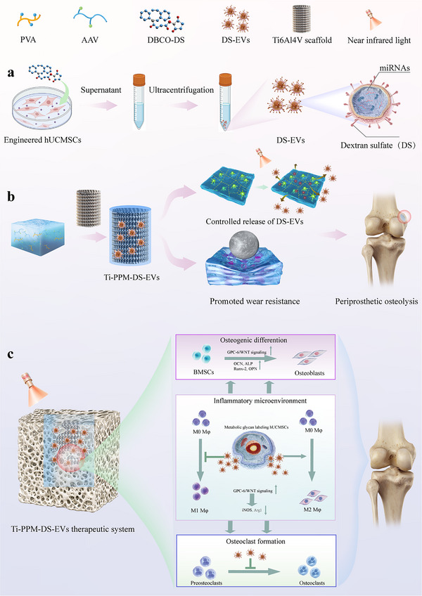
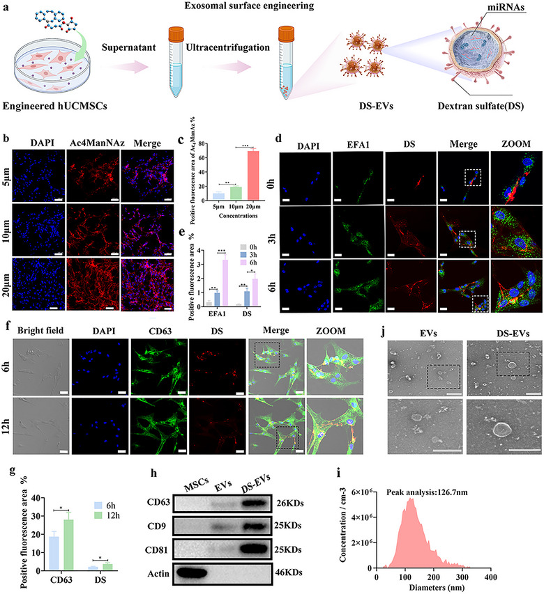
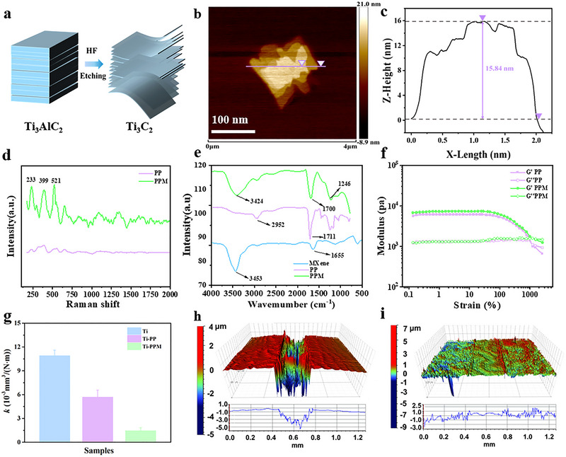
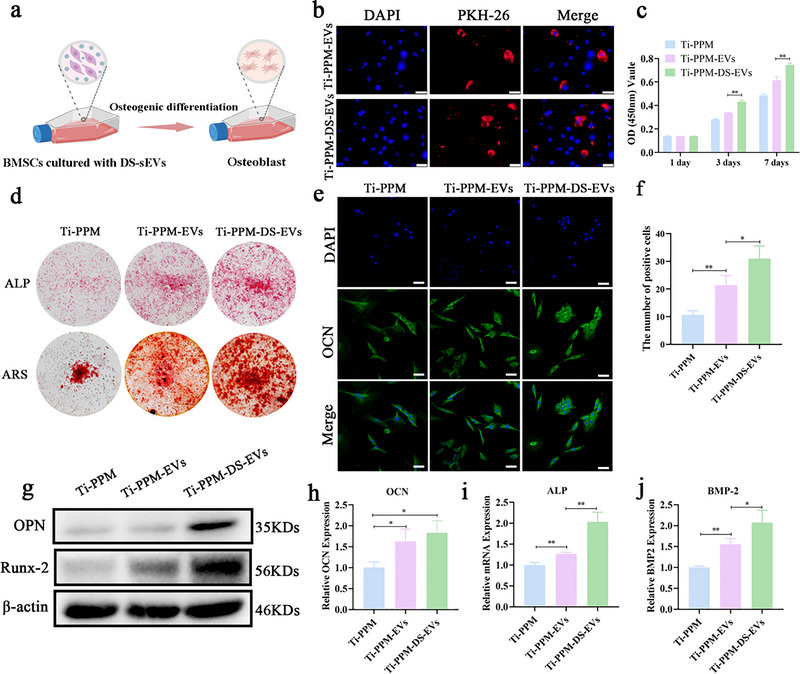
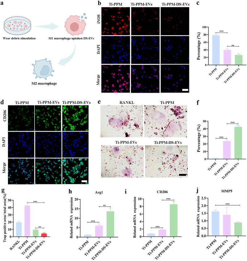
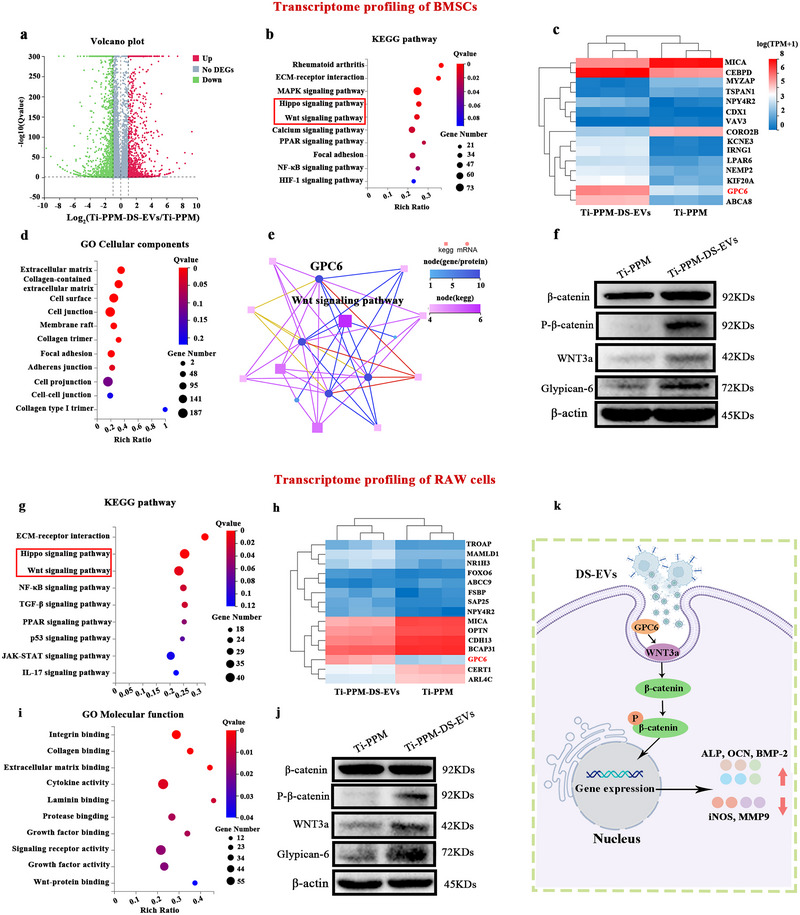
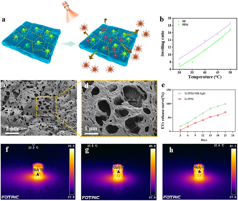
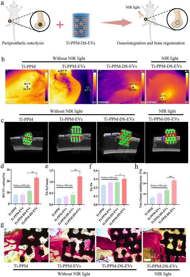
Despite remarkable progress in total joint arthroplasty, aseptic loosening of titanium (Ti) alloy persists as a critical clinical challenge due to the poor wear resistance and biological inertness of such implants. Targeting of inflammatory osteolysis and remodelling of the osseointegration environment represent promising therapeutic approaches to address this issue. In this study, we developed a novel engineered extracellular vesicles (EVs) with a tag of dextran sulfate (DS-EVs) via metabolic glycan labelling (MGL)-mediated click chemistry. This targeted delivery of EVs, derived from metabolically engineered stem cells, establishes a new cell-free therapeutic system for periprosthetic osteolysis. DS-EVs demonstrated specific macrophage tropism, effectively reprogramming macrophage polarisation from pro-inflammatory M1 to regenerative M2 phenotypes. This phenotypic shift attenuated osteoclastogenesis while enhancing osseointegration through GPC6/Wnt pathway activation in vitro. Furthermore, we designed a multifunctional 3D titanium alloy scaffold with MXene-PVA composite hydrogel coatings (Ti-PPM scaffold). The multifunctional Ti-PPM composite scaffold, incorporating DS-EVs, provides a robust delivery system for periprosthetic osteolysis. This integrated system exhibits dual advantages of enhanced wear resistance and optimised interfacial adhesion, while enabling controlled EV release to maximize DS-EVs' osseointegration potential in vivo. Collectively, our findings establish DS-EVs as a transformative therapeutic modality for periprosthetic osteolysis through dual modulation of the osseointegration microenvironment and macrophage phenotypic heterogeneity.

Keywords: extracellular vesicles; hydrogel; macrophage; periprosthetic osteolysis; titanium alloys.


Conflict of interest statement

The authors declare no conflict of interest.

Figures

SCHEME 1
SCHEME 1
Schematic illustration of MXene‐PVA composite hydrogel delivery system with controllable release of metabolic glycan labelling extracellular vesicles enhanced titanium alloy osseointegration.
FIGURE 1
FIGURE 1
Metabolic glycan labelling (MGL) of hUCMSCs generates chemically tagged DS‐EVs. (a) Schematic of exosomal surface engineering. (b) Representative confocal images of DS‐modified hUCMSCs at different concentrations of Ac4ManNAz by efficient click chemistry. hUCMSCs were incubated with Ac4ManAz for 48 h to obtain the azide group. Then, hUCMSCs labelled with azide group were treated with Cy5.5‐labelled dibenzocyclooctyne‐conjugated dextran sulfate (DBCO‐DS) (10 µM) for 2 h. Scale bar: 100 µm. (c) Quantification of the positive fluorescence area of Ac4ManAz (n = 3, **p < 0.01, ***p < 0.001). (d) Representative confocal images of the early EVs marker EFA1 and Cy5.5‐marked DS in the metabolic glycan labelling hUCMSCs at different time points. Scale bar: 20 µm. (e) Positive fluorescence area of EFA1 and Cy5.5‐marked DS (n = 3, *p < 0.05, **p < 0.01, ***p < 0.001). (f) Confocal microscopy images reveal the intracellular colocalisation of CD63 and Cy5.5‐marked DS in the hUCMSCs. Scale bar: 20 µm. (g) Positive fluorescence area of CD63 and Cy5.5‐marked DS (n = 3, *p < 0.05, **p < 0.01, ***p < 0.001). (h) Western blot was used to characterise the surface markers of EVs and DS‐EVs. (i) Particle size of DS‐EVs measured via nanoparticle tracking analysis (NTA). (j) TEM images show the morphology of EVs and DS‐EVs. Scale bar: 500 nm.
FIGURE 2
FIGURE 2
Construction of anti‐wear coating on the 3D porous Ti6Al4V scaffolds to reduce wear debris. (a) Fabrication of Ti2C3 MXene. (b and c) AFM images of the Ti2C3 MXene. (d) Raman spectrum of PP and PPM hydrogel. (e) FTIR spectrum of MXene, PP and PPM hydrogel. (f) Strain amplitude sweep test (γ = 0.1%−1500%) at a fixed angular frequency (1 Hz) at 25°C. (g) Wear rates of Ti, Ti‐PP and Ti‐PPM samples. (h and i) 3D topography of wear scar surfaces of Ti and Ti‐PPM samples.
FIGURE 3
FIGURE 3
DS‐EVs influence macrophage polarisation and suppress osteoclastogenesis in vitro. (a) Mode pattern of macrophage polarisation from M1 to M2 phenotype induced by DS‐EVs stimulation. (b) Immunofluorescence staining of iNOS in RAW 264.7 cells. Ti‐PPM‐EVs and Ti‐PPM‐DS‐EVs groups respectively represent RAW 264.7 cells were treated with EVs or DS‐EVs in the presence of Ti‐PPM wear debris (2 mg/mL) and lipopolysaccharide (LPS, 500 ng/mL). Scale bar: 100 µm. (c) Quantitative analysis of iNOS‐positive cells. Immunofluorescence positive cells were analysed using Image J software (n = 3, **p < 0.01, ***P < 0.001). (d) Immunofluorescence staining of CD206 in RAW 264.7 cells. Ti‐PPM‐EVs and Ti‐PPM‐DS‐EVs groups respectively represent RAW 264.7 cells were treated with EVs or DS‐EVs in the presence of Ti‐PPM wear debris and LPS. Scale bar: 100 µm. (e) Representative photograph of tartrate‐resistant acid phosphatase (TRAP) staining. Ti‐PPM‐EVs and Ti‐PPM‐DS‐EVs groups respectively represent RAW 264.7 cells were treated with EVs or DS‐EVs in the presence of Ti‐PPM wear debris, LPS and RANKL. (f) Quantitative analysis of CD206‐positive cells. Immunofluorescence positive cells were analysed by Image J software (n = 3, ***p < 0.001). (g) Quantitative analysis of TRAP‐positive areas (n = 3, **p < 0.01, ***p < 0.001). (h‐j) qPCR analysis of Arg1, CD206 and MMP‐9 gene expression respectively (n = 3, ***p < 0.001). Data was presented as mean ± SD of three number of replicates. t‐test was applied to each group in order to compare mean beta values.
FIGURE 4
FIGURE 4
DS‐EVs are conducive to promote osteogenic differentiation of BMSCs in vitro. (a) Scheme of the osteogenic differentiation of BMSCs stimulated by DS‐EVs. (b) Representative fluorescence micrograph of PKH‐26 (red)‐labelled DS‐EVs internalised by BMSCs. Scale bar: 20 µm. (c) CCK‐8 assay of BMSCs cultured with DS‐EVs. **p < 0.01. Ti‐PPM‐EVs and Ti‐PPM‐DS‐EVs group respectively represent BMSCs were treated with EVs or DS‐EVs in the presence of Ti‐PPM wear debris. (d) Alkaline phosphatase (ALP) activity and Alizarin Red S (ARS) staining of BMSCs. (e) Immunofluorescent of OCN in BMSCs. Scale bar: 100 µm. (f) Quantitative analysis of OCN positive cells. Immunofluorescence positive cells were analysed by Image J software (n = 3, *p < 0.05, **p < 0.01). (g) The protein levels of OPN and Runx‐2 in BMSCs were analysed by western blotting. (h‐j) The osteogenic‐related gene of BMSCs was measured via the qPCR, including OCN, ALP and BMP‐2. *p < 0.05, **p < 0.01. Data was presented as mean ± SD of three number of replicates. t‐test was applied to each group in order to compare mean beta values.
FIGURE 5
FIGURE 5
Transcriptome profiling of optimised DS‐EVs regulates function and mechanism of BMSCs and RAW cells. (a) The volcano diagram of Ti‐PPM‐DS‐EVs and Ti‐PPM group based on transcriptome profiling of BMSCs. (b) KEGG pathway enrichment analysis based on DS‐EVs regulated significant target genes. (c) The heatmap distribution of DS‐EVs regulating significant target genes. Ti‐PPM‐DS‐EVs group respectively represent BMSCs were treated with DS‐EVs in the presence of Ti‐PPM wear debris. (d) Graphene oxide‐interactive (GO) cellular contents analysis for the cellular component of differential target genes. (e) KEGG relation networks based on DS‐EVs regulated significant target genes. (f) The related proteins of GPC6/Wnt signalling pathway in BMSCs measured by western blot. (g) KEGG pathway enrichment analysis based on DS‐EVs regulated significant target genes. (h) The heatmap distribution of DS‐EVs regulating significant target genes. Ti‐PPM‐DS‐EVs group respectively represent RAW264.7 cells were treated with DS‐EVs in the presence of Ti‐PPM wear debris and LPS. (i) GO molecular function analysis of differential target genes. (j) DS‐EVs inhibit osteolysis and promote bone osseointegration in vitro by activating GPC6/Wnt signalling pathway. (k) A schematic representation illustrates the roles of DS‐EVs in inhibit osteolysis and promote bone osseointegration.
FIGURE 6
FIGURE 6
Characterisation of extended release of DS‐EVs in Ti‐PPM composite scaffolds. (a) Scheme of extended release of DS‐EVs in Ti‐PPM composite scaffolds. (b) Swelling properties of PP and PPM hydrogels with the different temperature ranging from 30°C to 50°C. (c, d) SEM images of PPM hydrogel (10,000 magnification and 20,000 magnification). (e) Profile of DS‐EVs released from the Ti‐PPM composite scaffold. (f‐h) NIR thermal images of Ti, Ti‐PPM and Ti‐PPM‐DS‐EVs, these implants exposed to an 808 nm (E = 1000 mW/cm2) laser after 600 s.
FIGURE 7
FIGURE 7
DS‐EVs effectively promote osseointegration in vivo. (a) Schematic of femur defect rat model and composited scaffold contained DS‐EVs administration. (b) NIR thermal images of the implants exposed to an 808 nm (E = 1000 mW/cm2) laser after 600 s. (c) Micro‐CT images of the rat femurs at 12 weeks postsurgery. (d) Quantitative results of the new bone including bone volume/tissue volume (BV/TV). (e) Quantitative results of trabecular number (Tb.N) and (f) trabecular thickness (Tb.Th). Three independent replicates have been statistical analysis. *p < 0.05, **p < 0.01, ***p < 0.001. (g) Van Gieson's staining of regenerated bone repaired by composited scaffold. Scale bar: 500 µm. (h) Quantitative results of Van Gieson's staining area. ***p < 0.001.

Similar articles

References

    1. Abd‐Elaziem, W. , Darwish M. A., Hamada A., and Daoush W. M.. 2024. “Titanium‐Based Alloys and Composites for Orthopedic Implants Applications: A Comprehensive Review.” Materials & Design 241: 112850.
    1. Adams, C. T. , O'Connor C. M., Young J. R., Anoushiravani A. A., Doherty B. S., and Congiusta F.. 2021. “Outcomes of a Total Joint Arthroplasty Enhanced Recovery Program in a Community Hospital Setting.” Journal of Arthroplasty 36, no. 7: S173–S178. - PubMed
    1. Agatemor, C. , Buettner M. J., Ariss R., Muthiah K., Saeui C. T., and Yarema K. J.. 2019. “Exploiting Metabolic Glycoengineering to Advance Healthcare.” Nature Reviews Chemistry 3, no. 10: 605–620. - PMC - PubMed
    1. Alquraini, A. , and El Khoury J.. 2020. “Scavenger Receptors.” Current Biology 30, no. 14: R790–R795. - PMC - PubMed
    1. An, J. X. , Han Z. Y., Qin Y. T., Li C. X., He J. L., and Zhang X. Z.. 2024. “Bacteria‐Based Backpacks to Enhance Adoptive Macrophage Transfer Against Solid Tumors.” Advanced Materials 36, no. 6: e2305384. - PubMed

LinkOut - more resources