. 2025 Jul 1;33(4):636-651.
doi: 10.4062/biomolther.2025.044. Epub 2025 Jun 23.

Anticancer Potential of Myricetin against Huh7- and Hep3B-Derived Liver Cancer Stem Cells through the Regulation of Apoptosis, Autophagy, and Stemness

Affiliations

Anticancer Potential of Myricetin against Huh7- and Hep3B-Derived Liver Cancer Stem Cells through the Regulation of Apoptosis, Autophagy, and Stemness

Mikyoung Kwon et al. Biomol Ther (Seoul). .

Abstract

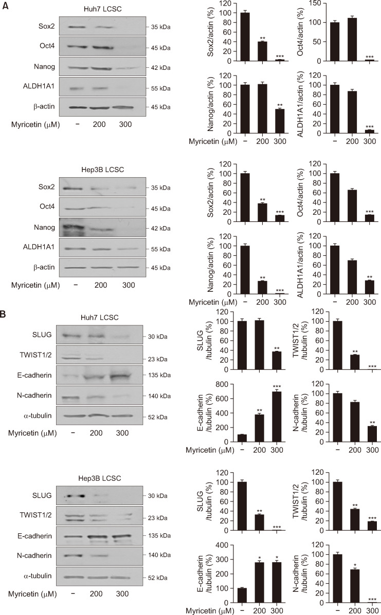
Liver cancer stem cells (LCSCs) play a significant role in the development, metastasis, treatment resistance, and recurrence of hepatocellular carcinoma (HCC). Targeting LCSCs offers a novel strategy to overcome treatment resistance in HCC. Myricetin, a flavonol from the flavonoid family, is known for its diverse biological activities, including anticancer effects. However, its potential for eradicating LCSCs had not been thoroughly investigated prior to this study. This study evaluated the effects of myricetin on LCSCs derived from Huh7 and Hep3B cell lines both in vitro and in vivo. LCSCs were treated with myricetin to assess cell proliferation, cell cycle arrest, apoptosis induction, autophagy regulation, stemness and EMT marker expression, and tumor growth suppression using a chicken embryo CAM model. Additionally, the combination therapy of myricetin with chloroquine, an autophagy inhibitor, was explored. Myricetin significantly inhibited the proliferation of Huh7- and Hep3B-derived LCSCs and suppressed tumor growth in the CAM model. It induced cell cycle arrest at the G0/G1 phase and triggered apoptosis through intrinsic and extrinsic pathways. Myricetin also stimulated autophagy by inhibiting the PI3K/AKT/mTOR pathway, reduced the expression of stemness markers, including Sox2, Oct4, Nanog, and ALDH1A1, and suppressed EMT. Combining myricetin with chloroquine enhanced apoptotic effects and further downregulated stemness markers by inhibiting STAT3 activation, demonstrating greater efficacy than myricetin alone. The findings establish myricetin, either as a standalone treatment or in combination with chloroquine, as a promising therapeutic candidate for targeting LCSC growth and overcoming chemotherapy resistance in HCC.

Keywords: Apoptosis; Autophagy; Chloroquine; Liver cancer stem cell; Myricetin.


Conflict of interest statement

CONFLICT OF INTEREST

All authors declare that there is no conflict of interest.

Figures

Fig. 1
Fig. 1
Effects of myricetin on the proliferation of Huh7- and Hep3B-derived LCSCs. (A) Huh7- and (B) Hep3B-derived LCSCs were exposed to the indicated concentrations of ampelopsin and myricetin for 0, 24, 48, and 72 h, and cell proliferation was assessed using a luciferase-based ATP luminescence assay. Tumorspheres were observed under an optical microscope after 72 h of ampelopsin and myricetin treatment, and cell images were captured at 20× magnification. Data are presented as mean ± SD (n=3). *p<0.05, **p<0.01, ***p<0.001 compared to the vehicle control. LCSC, liver cancer stem cell.
Fig. 2
Fig. 2
Effects of myricetin on in vivo tumor growth of Huh7-derived LCSCs in a CAM model. Huh7-derived LCSCs were combined with ECM gel, with or without myricetin (100, 200 µg/egg), and applied to the CAM surface of fertilized chick eggs. After 7 days of incubation, tumors were excised, and their formation rate, weight, and diameter were measured. Data are presented as mean ± SD (n=14). **p<0.01 compared to the vehicle control. LCSC, liver cancer stem cell; CAM, chicken embryo chorioallantoic membrane.
Fig. 3
Fig. 3
Effects of myricetin on cell cycle progression and apoptosis in Huh7- and Hep3B-derived LCSCs. Huh7- and Hep3B-derived LCSCs were treated with ampelopsin or myricetin at the indicated concentrations. (A) Cell cycle distribution was analyzed after 24 h of treatment using the Muse® Cell Cycle Kit. (B) Apoptosis was assessed after 72 h of treatment using the Muse® Annexin V & Dead Cell Kit. Flow cytometry analysis was performed using the Guava® Muse® Cell Analyzer. Data are presented as mean ± SD (n=3). *p<0.05, **p<0.01, ***p<0.001 compared to the vehicle control. LCSC, liver cancer stem cell.
Fig. 4
Fig. 4
Effects of myricetin on apoptotic characteristics in Huh7- and Hep3B-derived LCSCs. (A, B) Huh7- and Hep3B-derived LCSCs were treated with myricetin at the indicated concentrations for 24 h. (A) DAPI staining was used to observe nuclei under a fluorescence microscope, with nuclear condensation and fragmentation marked by white arrows. (B) Mitochondrial membrane potential was measured using TMRE, a fluorescent probe. Data are presented as mean ± SD (n=3). ***p<0.001 compared to the vehicle control. LCSC, liver cancer stem cell; DAPI, 4′,6-diamidino-2-phenylindole; TMRE, tetramethylrhodamine ethyl ester perchlorate.
Fig. 5
Fig. 5
Effects of myricetin on apoptosis and autophagy pathways in Huh7- and Hep3B-derived LCSCs. Huh7- and Hep3B-derived LCSCs were exposed to myricetin at the indicated concentrations for 24 h. The expression of apoptosis and autophagy markers was analyzed by western blotting and quantified by normalizing protein intensity to β-actin. Data are presented as mean ± SD (n=3). *p<0.05, **p<0.01, ***p<0.001 compared to the vehicle control. LCSC, liver cancer stem cell; DR5, death receptor 5; Bad, Bcl-2-associated death promoter; Bcl-2, B-cell lymphoma 2; Bax, Bcl-2-associated X protein; Atg5, autophagy-related gene 5; LC3B, microtubule-associated protein 1 light chain 3 beta; p62, sequestosome 1.
Fig. 6
Fig. 6
Effects of myricetin on the PI3K/AKT/mTOR pathway in Huh7- and Hep3B-derived LCSCs. Huh7- and Hep3B-derived LCSCs were treated with myricetin at the indicated concentrations for 24 h. Protein expression levels were analyzed by western blotting and normalized to β-actin. Data are presented as mean ± SD (n=3). *p<0.05, **p<0.01, ***p<0.001 compared to the vehicle control. LCSC, liver cancer stem cell; p-, phosphorylated; PI3K, phosphoinositide 3-kinase; AKT, protein kinase B; mTOR, mammalian target of rapamycin.
Fig. 7
Fig. 7
Effects of myricetin on stemness and EMT markers in Huh7- and Hep3B-derived LCSCs. (A, B) Huh7- and Hep3B-derived LCSCs were treated with myricetin at the indicated concentrations for 24 h. (A) Stemness and (B) EMT marker expression levels were evaluated by western blotting and normalized to β-actin or α-tubulin. Data are presented as mean ± SD (n=3). *p<0.05, **p<0.01, ***p<0.001 compared to the vehicle control. LCSC, liver cancer stem cell; Sox2, SRY-box transcription factor 2; Oct4, octamer-binding transcription factor 4; Nanog, Nanog homeobox; ALDH1A1, aldehyde dehydrogenase 1 family member A1; EMT, epithelial-mesenchymal transition; SLUG, Zinc finger protein SNAI2; TWIST1/2, twist-related protein 1/2; E-Cadherin, epithelial cadherin; N-cadherin, neural cadherin.
Fig. 8
Fig. 8
Chloroquine enhances the apoptotic effects of myricetin in Huh7- and Hep3B-derived LCSCs. (A-C) Huh7- and Hep3B-derived LCSCs were pretreated with chloroquine for 1 h, followed by myricetin treatment for 24 h. (A) Cell viability was assessed using a luciferase-based ATP luminescence assay after co-treatment with myricetin (50, 100, 200 μM) and chloroquine (50 μM). (B) Apoptotic cell death was analyzed by flow cytometry following treatment with myricetin (200 μM) and chloroquine (200 μM). (C) Expression of apoptosis markers was evaluated by western blotting after co-treatment with myricetin (100 μM) and chloroquine (200 μM). Data are presented as mean ± SD (n=3). *p<0.05, **p<0.01, ***p<0.001 compared to the myricetin-treated group. LCSC, liver cancer stem cell; NT, non-treated; CQ, chloroquine; Myri, myricetin.
Fig. 9
Fig. 9
Effects of co-treatment with chloroquine and myricetin on the JAK2/STAT3 pathway and stemness markers in Huh7- and Hep3B-derived LCSCs. Huh7- and Hep3B-derived LCSCs were pretreated with chloroquine (200 μM) for 1 h, followed by myricetin (100 μM) treatment for 24 h. Protein expression levels were determined by western blotting and are presented as the normalized ratio of each target protein (or phosphorylated protein) to β-actin or the corresponding total protein. Data are presented as mean ± SD (n=3). *p<0.05, **p<0.01 compared to the myricetin-treated group. LCSC, liver cancer stem cell; NT, non-treated; CQ, chloroquine; Myri, myricetin; p-, phosphorylated; JAK2, janus kinase 2; STAT3, signal transducer and activator of transcription 3; Sox2, SRY-box transcription factor 2; Oct4, octamer-binding transcription factor 4; Nanog, Nanog homeobox; ALDH1A1, aldehyde dehydrogenase 1 family member A1..
Fig. 10
Fig. 10
Proposed molecular mechanism of myricetin’s growth-inhibitory effects on Huh7- and Hep3B-derived LCSCs. Myricetin suppresses the growth of LCSCs through the regulation of apoptosis, autophagy, and stemness. p-, phosphorylated; PI3K, phosphoinositide 3-kinase; AKT, protein kinase B; mTOR, mammalian target of rapamycin; DR5, death receptor 5; Bad, Bcl-2-associated death promoter; Bcl-2, B-cell lymphoma 2; Bax, Bcl-2-associated X protein; Atg5, autophagy-related gene 5; LC3B, microtubule-associated protein 1 light chain 3 beta; p62, sequestosome 1; STAT3, signal transducer and activator of transcription 3; SLUG, Zinc finger protein SNAI2; TWIST1/2, twist-related protein 1/2; E-Cadherin, epithelial cadherin; N-cadherin, neural cadherin; Sox2, SRY-box transcription factor 2; Oct4, octamer-binding transcription factor 4; Nanog, Nanog homeobox; ALDH1A1, aldehyde dehydrogenase 1 family member A1.

Similar articles

References

    1. Agraharam G., Girigoswami A., Girigoswami K. Myricetin: a multifunctional flavonol in biomedicine. Curr. Pharmacol. Rep. 2022;8:48–61. doi: 10.1007/s40495-021-00269-2. - DOI - PMC - PubMed
    1. Аrtykov А. А., Belov D. A., Shipunova V. O., Trushina D. B., Deyev S. M., Dolgikh D. A., Kirpichnikov M. P., Gasparian M. E. Chemotherapeutic agents sensitize resistant cancer cells to the DR5-specific variant DR5-B more efficiently than to TRAIL by modulating the surface expression of death and decoy receptors. Cancers. 2020;12:1129. doi: 10.3390/cancers12051129. - DOI - PMC - PubMed
    1. Babaei G., Aziz S. G., Jaghi N. Z. Z. EMT, cancer stem cells and autophagy; the three main axes of metastasis. Biomed. Pharmacother. 2021;133:110909. doi: 10.1016/j.biopha.2020.110909. - DOI - PubMed
    1. Castelli V., Giordano A., Benedetti E., Giansanti F., Quintiliani M., Cimini A., Angelo M. The great escape: the power of cancer stem cells to evade programmed cell death. Cancers. 2021;13:328. doi: 10.3390/cancers13020328. - DOI - PMC - PubMed
    1. Cho H. J., Jung H. J. Cyclophilin A knockdown inhibits the proliferation and metastatic ability of AGS gastric cancer stem cells by downregulating CD147/STAT3/AKT/ERK and epithelial-mesenchymal transition. Mol. Med. Rep. 2025;31:14. doi: 10.3892/mmr.2024.13379. - DOI - PMC - PubMed

LinkOut - more resources