Review
. 2025 Aug;12(32):e03095.
doi: 10.1002/advs.202503095. Epub 2025 Jun 23.

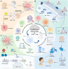
Mitochondrial Regulation of CD8⁺ T Cells: Mechanisms and Therapeutic Modulation

Affiliations
Review

Mitochondrial Regulation of CD8⁺ T Cells: Mechanisms and Therapeutic Modulation

Xu Chen et al. Adv Sci (Weinh). 2025 Aug.

Abstract

Mitochondria are integral to the regulation of CD8+ T cell function, critically influencing processes such as activation, differentiation, and long-term persistence during immune responses. Emerging evidence highlights the detrimental impact of mitochondrial dysfunction on CD8+ T cell activity, contributing to immune exhaustion and impairing both antitumor and antiviral immunity. This underscores the importance of understanding and modulating mitochondrial dynamics to optimize T cell-based immunotherapies. In this review, a comprehensive and in-depth analysis of the essential mitochondrial processes-including biogenesis, redox homeostasis, and metabolic reprogramming is provided-that govern CD8+ T cell function and are intricately linked to their therapeutic potential. The current strategies aimed at enhancing mitochondrial function in CD8+ T cells are also examined, focusing on both metabolic reprogramming and mitochondrial-targeted interventions. Despite these promising approaches, several significant challenges remain, such as achieving selective targeting, addressing mitochondrial plasticity, and mitigating off-target effects. Overcoming these obstacles will be crucial to improving the clinical efficacy and safety of mitochondrial modulation therapies. As the understanding of mitochondrial dynamics within CD8+ T cells continues to evolve, there is growing potential to leverage these insights to improve immune-based therapies across a range of diseases, including cancer and viral infections.

Keywords: CD8⁺ T cells; T cell exhaustion; immunotherapy; metabolic reprogramming; mitochondrial dynamics.


Conflict of interest statement

The authors declare no competing interests.

Figures

Figure 1
Figure 1
The crucial role of mitochondria in mediating CD8+ T function. A) TCR engagement induces PI3K‐Akt signaling, leading to mTORC1 activation via TSC1/2 and Rheb, thereby promoting metabolic reprogramming. Enhanced glycolysis and FAO support ATP production, driving clonal expansion, cytotoxicity, and survival of activated CD8⁺ T cells. B) 4‐1BB co‐stimulation activates LKB1‐AMPK signaling, facilitating FAO and ATP generation. Concurrently, NF‐κB activation promotes anti‐apoptotic pathways, while glucose metabolism supports cell cycle progression, ensuring sustained CD8⁺ T cell function. C) Memory CD8⁺ T cells rely on mitochondrial metabolism and the urea cycle, where CPS1, OTC, ASS1, ARG1, and NOS coordinate nitrogen metabolism. The conversion of aspartate to argininosuccinate supports NO production, contributing to enhanced memory development and longevity. D) Metabolic stress in the TME induces mitochondrial dysfunction in CD8⁺ T cells, leading to reduced mitochondrial content, altered morphology, and impaired oxidative metabolism, contributing to T cell exhaustion and dysfunction.
Figure 2
Figure 2
Mitochondrial dysfunction in CD8⁺ T cells under pathological conditions. A) Hypoxia in the nasopharyngeal microenvironment induces miR‐24, leading to c‐Myc suppression and reduced MFN1 expression, thereby impairing OXPHOS and ATP production. This metabolic dysregulation promotes CD8⁺ T cell exhaustion, while FGF11 activation partially supports cell growth. B) HIV‐1 infection disrupts mitochondrial function, exacerbated by glucose metabolism imbalance, leading to excessive ROS accumulation. Antioxidant mechanisms attempt to mitigate oxidative stress; however, mitochondrial dysfunction limits OXPHOS‐dependent ATP synthesis, impairing IL‐15‐mediated CD8⁺ T cell proliferation and leading to mitochondrial fragmentation. C) Chronic hepatitis B and inflammatory conditions drive mitochondrial dysfunction by disrupting MFN1 and OXPHOS, resulting in elevated ROS production. This oxidative stress contributes to mitochondrial fragmentation and restricts CD8⁺ T cell expansion. D) Mycobacterium tuberculosis infection induces T cell activation, yet sustained mitochondrial dysfunction promotes a metabolic shift toward glycolysis, driven by HIF‐1α. This metabolic reprogramming leads to lactate accumulation and AMPK activation, ultimately impairing IFN‐γ production and promoting CD8⁺ T cell exhaustion.
Figure 3
Figure 3
Mitochondrial regulation of CD8⁺ T cell function and exhaustion. A) Ncoa2 regulates PGC‐1α expression, enhancing OXPHOS and IFN‐γ production in CD8⁺ T cells, promoting antitumor immunity while preventing apoptosis. T cell differentiation progresses from naïve T cells to effector T cells, with aged CD8⁺ T cells exhibiting immunosenescence, impairing antitumor responses. B) OPA1 supports mitochondrial dynamics by maintaining FAO and OXPHOS, preventing apoptosis in effector T cells within the TME. Under hypoglycemic conditions, SIRT3‐SENP1‐YME1L1 signaling enhances mitochondrial fusion, sustaining CD8⁺ T cell function. However, PD‐1‐mediated AKT activation leads to mitochondrial elongation, contributing to T‐cell exhaustion. C) Hypoxia and persistent antigen stimulation induce ROS accumulation in CD8⁺ T cells, driving exhaustion through mitochondrial dysfunction. HIF‐1α‐mediated metabolic reprogramming downregulates MnSOD, promoting oxidative stress. Reduced EMP and ATP production further activate exhaustion‐associated genes, impairing T cell function. D) MFN2 regulates mitochondrial‐ER contacts, modulating Ca2⁺ homeostasis in T cells. CRAC channels facilitate Ca2⁺ influx, activating CaM‐CaN signaling and NFAT dephosphorylation, leading to T‐cell activation.
Figure 4
Figure 4
Mitochondrial metabolism in CD8⁺ T cell immune response and dysfunction. A) IL‐2 and IL‐21 orchestrate CD8⁺ T cell differentiation by modulating metabolic pathways. IL‐2 promotes glycolysis, while IL‐21 enhances OXPHOS, favoring memory CD8⁺ T cell formation and antitumor immunity. LDH downregulation shifts metabolism toward OXPHOS, supporting long‐term T‐cell persistence. B) TCR activation triggers metabolic reprogramming in CD8⁺ T cells, enhancing OXPHOS and ATP production through PDHK1‐mediated pyruvate metabolism, thereby promoting cytokine synthesis and proliferation. C) Memory CD8⁺ T cells rely on mitochondrial metabolism, with HK1‐VDAC interactions regulating glucose flux toward OXPHOS at the mitochondria‐ER junction. mTORC2‐AKT‐GSK3β signaling further enhances ATP generation and sustains T cell longevity. D) In melanoma, CD8⁺ TILs depend on OXPHOS for energy production. Enolase inhibition disrupts glycolysis, impairing metabolic flexibility and T‐cell function. E) In idiopathic pulmonary fibrosis, mtDNA leakage activates the cGAS‐STING‐IRF3 pathway, driving type I IFN responses in CD8⁺ T cells. (F) In metastatic pleural effusions, chronic antigen stimulation and inflammatory signals induce CD38 expression, leading to mitochondrial dysfunction, loss of mitochondrial membrane potential, and reduced IFN‐γ production, contributing to CD8⁺ T cell dysfunction.
Figure 5
Figure 5
Mitochondrial modulation mechanisms in CD8⁺ T cells and their implications for enhancing immunotherapy efficacy. Key small molecules—linoleic acid, bezafibrate, nicotinamide (NAM), SPD, and lithium carbonate—boost mitochondrial function, metabolism, and T cell activation. Linoleic acid promotes mitochondrial energetics and calcium signaling, preventing T‐cell exhaustion. Bezafibrate enhances FAO and OXPHOS, improving T cell survival and activation. NAM reduces mitochondrial content and ROS, enhancing T‐cell proliferation. SPD activates MTP to support FAO, especially in aged T cells. Lithium carbonate rescues mitochondrial oxidation of LA, improving T cell function. Natural compounds like resveratrol and oleuropein also enhance mitochondrial activity in chronic HBV infection.
Figure 6
Figure 6
Mitochondrial regulation of CD8⁺ T cell function and therapeutic interventions. A) Mitochondria‐targeted nanoparticle delivery modulates CD8⁺ T cell metabolism in the TME. These nanoparticles influence ATP production and TNF‐α signaling, potentially inducing CD8⁺ T cell apoptosis. Interactions between CD8⁺ T cells, DCs, Tregs, CAFs, and MDSCs shape mitochondrial responses. B) Heterogeneity in CD8⁺ T cell mitochondrial function drives functional diversity, including cytotoxic T cells, memory T cells, and exhausted T cells. Mitochondrial modulation strategies can influence Treg differentiation, with potential implications for immunotherapies. C) Hypoxia and metabolic constraints in the TME shift CD8⁺ T cell metabolism toward glycolysis, reinforcing the Warburg effect. LDH‐mediated lactate production under hypoxic conditions suppresses IFN responses, which can be counteracted using LDH inhibitors to restore mitochondrial function. D) Nanoparticle‐based immunotherapies require safety evaluation to assess pharmacological response, adverse effects, and long‐term toxicity. Preclinical animal testing and clinical trials are essential for validating therapeutic efficacy and safety.

Similar articles

References

    1. Guo Y., Xie Y. Q., Gao M., Zhao Y., Franco F., Wenes M., Siddiqui I., Bevilacqua A., Wang H., Yang H., Feng B., Xie X., Sabatel C. M., Tschumi B., Chaiboonchoe A., Wang Y., Li W., Xiao W., Held W., Romero P., Ho P. C., Tang L., Nat. Immunol. 2021, 22, 746. - PMC - PubMed
    1. Angajala A., Lim S., Phillips J. B., Kim J. H., Yates C., You Z., Tan M., Front. Immunol. 2018, 9, 1605. - PMC - PubMed
    1. Peters P. J., Borst J., Oorschot V., Fukuda M., Krähenbühl O., Tschopp J., Slot J. W., Geuze H J., J. Exp. Med. 1991, 173, 1099. - PMC - PubMed
    1. Nunnari J., Suomalainen A., Cell 2012, 148, 1145. - PMC - PubMed
    1. O'Sullivan D., Pearce E L., Trends Immunol. 2015, 36, 71. - PMC - PubMed

MeSH terms

LinkOut - more resources