. 2025 May 26;33(2):201006.
doi: 10.1016/j.omton.2025.201006. eCollection 2025 Jun 18.

Oncolytic adeno-immunotherapy improves allogeneic adoptive HER2.CAR-NK function against pancreatic ductal adenocarcinoma

Affiliations

Oncolytic adeno-immunotherapy improves allogeneic adoptive HER2.CAR-NK function against pancreatic ductal adenocarcinoma

Greyson Biegert et al. Mol Ther Oncol. .

Abstract

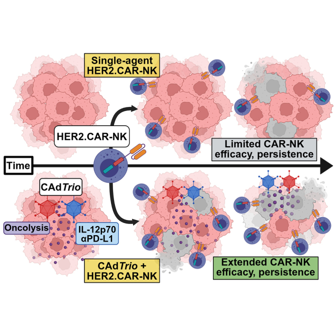
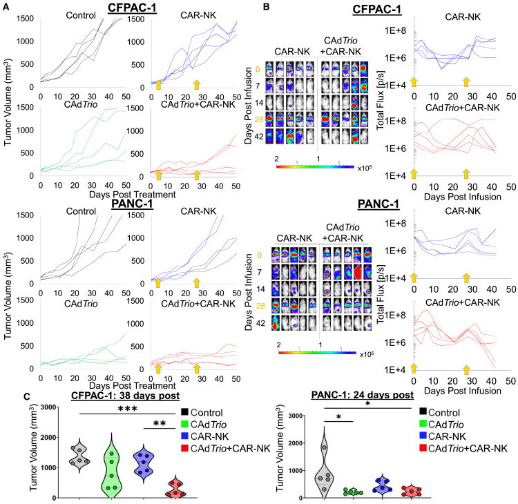
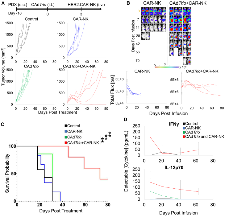
Pancreatic ductal adenocarcinoma (PDAC) responds poorly to conventional treatments and immunotherapy. We previously developed a binary oncolytic/helper-dependent adenovirus system (CAdTrio) that facilitated oncolysis and expressed the immunomodulatory molecule interleukin-12 and a programmed death ligand 1 (PD-L1) blocking mini-antibody. Given that CAdTrio enhanced endogenous natural killer (NK) cell anti-tumor activity in humanized mice bearing PDAC tumors and that NK cells can be adoptively transferred to patients safely in the allogeneic setting, we hypothesized that a combination of CAdTrio and allogeneic NK cells expressing a HER2-specific chimeric antigen receptor (HER2.CAR-NK) would be an effective, entirely "off-the-shelf" treatment against PDAC. We found that CAdTrio-derived immunomodulatory molecules prolonged HER2.CAR-NK persistence at tumor sites, allowing long-term tumor growth control and improved survival in both humanized mice and a heterogeneous PDAC patient-derived xenografts (PDX) model. This effect was based on CAdTrio-derived transgene support that shifted HER2.CAR-NK gene expression to that resembling an NK memory-like phenotype. Additionally, this allogeneic combination therapy was tolerated in humanized mice. Together, these data suggest that CAdTrio and HER2.CAR-NK cell combination immunotherapy may be a novel and effective option for the treatment for immunologically "cold" PDAC tumors.

Keywords: CAR-NK cell; MT: Regular Issue; PDX model; humanized mouse model; oncolytic viro-immuno therapy; pancreatic ductal adenocarcinoma.


Conflict of interest statement

M.S. received research funding from Tessa Therapeutic Ltd. and AstraZeneca. M.S. was a scientific consultant and C.P was a consultant for Tessa Therapeutic Ltd.

Figures

None
Graphical abstract
Figure 1
Figure 1
CAR modification improves NK cytotoxicity against PDAC (A) PBMC-derived NK cells from a single healthy donor were transduced with one of three different HER2.CAR constructs (CD3zeta.HER2.CAR [CD3z], CD28-CD3zeta.HER2.CAR [CD28z], or 41BB-CD3zeta.HER2.CAR [41BBz]). CAR transduction efficiency was determined using flow cytometry 72 h post transduction. (B) To measure cytotoxicity, non-transduced (nt) NKs and CAR-NKs were then cocultured with PDAC cell lines expressing GFP at an E:T of 1:5 in an IncuCyte for 72 h and measured total area of GFP+ target cells (measure of viable target cells) (n = 6). Killing efficacy was evaluated under increasingly stressful E:T conditions by measuring total area of GFP+ target cells at the 72 h time point (n = 6). (C) The killing capacity of 41BBz.HER2.CAR-NK cells from multiple donors was evaluated by coculturing non-transduced or HER2.CAR-NKs from three healthy donors with GFP expressing target cells for 48 h (E:T, 1:20; n = 4–6/donor). To test 41BBz.HER2.CAR-NK expansion, non-GFP expressing PDAC cell lines were cocultured with either non-transduced NKs or HER2.CAR.41BBz-NKs expressing GFP (E:T, 1:20; n = 4–6/donor). Data represent fold change expansion of non-transduced NKs or HER2.CAR-NKs. Data are presented as means ± SD, and p values were determined by mixed-effects analysis with Tukey multiple comparisons. ns, not significant, ∗p < 0.05, ∗∗p < 0.01, ∗∗∗p < 0.001, and ∗∗∗∗p < 0.0001.
Figure 2
Figure 2
CAdTrio-derived transgenes enhance HER2.CAR-NK activation and promote development (A) Total RNA was extracted from HER2.CAR-NKs after 24 h coculture with CFPAC-1 or PANC-1 infected with HD viruses: HDAd0 (no transgene), HDIL-12, HDPDL1, or HDTrio. Data represent 2-fold gene expression changes in HER2.CAR-NKs. Genes with increased expression are shown in red and decreased expression are shown in blue, while black are genes not substantially changed between comparators. (B) Total RNA was collected from HER2.CAR-NKs after coculture with CFPAC-1 cells infected with either HDAd0 or HDTrio for 24 h and converted to cDNA for PCR analysis (donor n = 5). Genes were quantified and normalized to human β-actin. p values were determined using two-tailed t test: IFNG t = 1.023; P2RX5 t = 1.011; LAG3 t = 1.876; TBX21 t = 6.719; TIGIT t = 0.3561; all df = 4. ns, not significant, ∗p < 0.05 and ∗∗p < 0.01.
Figure 3
Figure 3
CAdTrio combined with CAR-NK cells is superior to single agent HER2.CAR-NK therapy in NSG mouse (A) CFPAC-1 or PANC-1 cells were transplanted into the right flank of NSG mice (control [untreated], HER2.CAR-NK alone, CAdTrio alone, or CAdTrio+HER2.CAR-NK: n = 5). A total of 1 × 10⁷ vp of CAdTrio (OAd:HD = 1:1) were injected intratumorally, then 5 × 10⁶ HER2.CAR-NKs expressing GFP were administered via the tail vein 3 days after CAdTrio injection. Tumor volumes were monitored by caliper measurement at indicated time points, and data from individual animals are shown. (B) HER2.CAR-NK bioluminescence was monitored at indicated time points, and data from individual animals are shown. (C) Kaplan-Meier survival curve after CAdTrio administration (n = 5). p values were determined using the log rank Mantel-Cox test (dF = 3). ns, not significant, ∗p < 0.05 and ∗∗p < 0.01. Abbreviations are as follows: s.c., subcutaneous; i.t., intratumoral; i.v., intravenous.
Figure 4
Figure 4
CAdTrio and CAR-NK cell combination immunotherapy is both effective and well tolerated in humanized mouse models (A) CFPAC-1 or PANC-1 cells were transplanted into the right flank of humanized mice (control [untreated], HER2.CAR-NK alone, CAdTrio alone, or CAdTrio+HER2.CAR-NK) (n = 5). A total of 1 × 10⁷ vp of CAdTrio (OAd:HD = 1:1) were injected intratumorally, then 1 × 10⁶ HER2.CAR-NKs expressing GFP were systemically administered 3 days after CAdTrio injection, and then again 4 weeks later (yellow arrows). Tumor volumes were monitored by caliper measurement at indicated time points, and data from individual animals are shown. (B) HER2.CAR-NK bioluminescence was monitored at indicated time points, and data from individual animals are shown. (C) Tumor volumes at first endpoint event (CFPAC-1 day 38 [n = 5], PANC-1 day 24 [n = 5]). p values were determined by one-way ANOVA with Tukey multiple comparisons. ns, not significant, ∗p < 0.05, ∗∗p < 0.01, ∗∗∗p < 0.001, and ∗∗∗∗p < 0.0001. Abbreviations are as follows: s.c., subcutaneous; i.t., intratumoral; i.v., intravenous.
Figure 5
Figure 5
Combination immunotherapy controls heterogeneous PDX tumor growth and improves survival (A) PDAC PDX tumors were subcutaneously transplanted into the right flank of NSG mice (control [untreated], HER2.CAR-NK alone, CAdTrio alone, or CAdTrio+HER2.CAR-NK; n = 5–8 animals). A total of 1 × 10⁷ vp of CAdTrio (OAd:HD = 1:1) were injected intratumorally, and then 5 × 10⁶ HER2.CAR-NKs expressing GFP were systemically administered 3 days after CAdTrio injection. Tumor volumes were monitored by caliper measurement and (B) HER2.CAR-NK bioluminescence at indicated time points. Data from individual mice are shown. (C) Kaplan-Meier survival curve after CAdTrio administration. (D) Serum was collected at days 7, 21, 42, and 63 post HER2.CAR-NK infusion to measure IFN-γ and IL-12p70. p < 0.01. p values were determined using the log rank Mantel-Cox test (dF = 3). ns, not significant, ∗p < 0.05, ∗∗p < 0.01, ∗∗∗p < 0.001, and ∗∗∗∗p < 0.0001. Abbreviations are as follows: s.c., subcutaneous; i.t., intratumoral; i.v., intravenous.

Similar articles

References

    1. Halbrook C.J., Lyssiotis C.A., Pasca di Magliano M., Maitra A. Pancreatic cancer: Advances and challenges. Cell. 2023;186:1729–1754. doi: 10.1016/j.cell.2023.02.014. - DOI - PMC - PubMed
    1. Hu Z.I., O’Reilly E.M. Therapeutic developments in pancreatic cancer. Nat. Rev. Gastroenterol. Hepatol. 2024;21:7–24. doi: 10.1038/s41575-023-00840-w. - DOI - PubMed
    1. Orth M., Metzger P., Gerum S., Mayerle J., Schneider G., Belka C., Schnurr M., Lauber K. Pancreatic ductal adenocarcinoma: biological hallmarks, current status, and future perspectives of combined modality treatment approaches. Radiat. Oncol. 2019;14:141. doi: 10.1186/s13014-019-1345-6. - DOI - PMC - PubMed
    1. Harder J., Ihorst G., Heinemann V., Hofheinz R., Moehler M., Buechler P., Kloeppel G., Röcken C., Bitzer M., Boeck S., et al. Multicentre phase II trial of trastuzumab and capecitabine in patients with HER2 overexpressing metastatic pancreatic cancer. Br. J. Cancer. 2012;106:1033–1038. doi: 10.1038/bjc.2012.18. - DOI - PMC - PubMed
    1. Lin D., Shen Y., Liang T. Oncolytic virotherapy: basic principles, recent advances and future directions. Signal Transduct. Target. Ther. 2023;8:156. doi: 10.1038/s41392-023-01407-6. - DOI - PMC - PubMed

LinkOut - more resources