. 2025:12:5.
doi: 10.5281/zenodo.15531392. Epub 2025 May 27.

Advancing butterfly systematics through genomic analysis

Affiliations

Advancing butterfly systematics through genomic analysis

Jing Zhang et al. Taxon Rep Int Lepid Surv. 2025.

Abstract

Within the framework of an ongoing comparative genomic study of global butterfly diversity, we construct phylogenetic trees combining all protein-coding genes assembled from the whole genome shotgun data. When viewed in the context of current taxonomy and phenotypic knowledge, the genome-wide phylogeny points to further advances in butterfly systematics, which are presented here. We assign major clades with comparable levels of genetic divergence to the same taxonomic rank and apply criteria involving relative population divergence and gene flow to define species boundaries. As a result, one genus, 13 subgenera, 62 species, and five subspecies are proposed as new (type species in original combinations or type localities are given in parentheses): a genus Ajenorix Grishin, gen. n. (Rapala hypargyria Elwes, 1893) in Deudorigini Doherty, 1886, Lycaenidae [Leach], [1815]; subgenera: in Riodinidae Grote, 1895 (1827): Ouida Grishin, subgen. n. (Dodona ouida Hewitson, 1866) and Egeona Grishin, subgen. n. (Taxila egeon Westwood, 1851) of Dodona Hewitson, 1861, Locris Grishin, subgen. n. (Lasaia oileus Godman, 1903) of Lasaia H. Bates, 1868, Lucispila Grishin, subgen. n. (Hesperia lucianus Fabricius, 1793) of Parvospila J. Hall, 2018, Byzia Grishin, subgen. n. (Lemonias byzeres Hewitson, 1872) of Zelotaea H. Bates, 1868, Arichlosyne Grishin, subgen. n. (Apodemia ochracea Mengel, 1902) of Aricoris Westwood, 1851, and Lenca Grishin, subgen. n. (Lemonias lencates Hewitson, 1875) of Pachythone H. Bates, 1868; in Lycaenidae: Afrix Grishin, subgen. n. (Dipsas antalus Hopffer, 1855) of Capys Hewitson, 1865, Wacus Grishin, subgen. n. (Myrina epirus C. Felder, 1860) of Deudorix Hewitson, 1863, and Crates Grishin, subgen. n. (Hesperia isocrates Fabricius, 1793) of Virachola F. Moore, 1881; and in Hesperiidae Latreille, 1809: Ochloba Grishin, subgen. n. (Poanes batesi Bell, 1935), Ochlata Grishin, subgen. n. (Hesperia venata Bremer & Grey, 1853), and Ochluma Grishin, subgen. n. (Hesperia yuma W. H. Edwards, 1873) of Ochlodes Scudder, 1872; species: in Riodinidae: Lasaia cola Grishin, sp. n. (Mexico: Colima), Curvie wing Grishin, sp. n. (USA: Texas), Curvie westwing Grishin, sp. n. (Mexico: Sonora), Curvie chiapensis Grishin, sp. n. (Mexico: Chiapas), Emesis (Tenedia) tinia Grishin, sp. n. (Argentina), Emesis (Tenedia) guaya Grishin, sp. n. (Uruguay), Emesis (Aphacitis) bugaba Grishin, sp. n. (Panama: Chiriquí), and Synargis rectanga Grishin, sp. n. (Peru: San Martin) and in Hesperiidae: Phanus ecutinus Grishin, sp. n. (Ecuador: Pichincha), Entheus zeus Grishin, sp. n. (Brazil: Amazonas), Entheus guyaneus Grishin, sp. n. (Guyana), Entheus colombeus Grishin, sp. n. (eastern Colombia), Entheus proxemus Grishin, sp. n. (Brazil: Pará), Entheus peruveus Grishin, sp. n. (Peru: Madre de Dios), Entheus hyponota Grishin, sp. n. (Brazil: Amazonas), Entheus lina Grishin, sp. n. (Brazil: Pará), Entheus guato Grishin, sp. n. (Mexico: Chiapas), Entheus pano Grishin, sp. n. (Panama: Darién), Entheus venezuelius Grishin, sp. n. (Venezuela: Aragua), Entheus ecuadius Grishin, sp. n. (Ecuador: Napo), Entheus bogoteus Grishin, sp. n. (Colombia: Bogotá), Cecropterus (Thorybes) notochlorothrix Grishin, sp. n. (Brazil: Santa Catarina), Urbanus (Urbanoides) elma Grishin, sp. n. (Venezuela: Merida), Telegonus (Rhabdoides) missionus Grishin, sp. n. (USA: Texas), Telegonus (Rhabdoides) panavenus Grishin, sp. n. (Panama: Panamá), Telegonus (Rhabdoides) pacificus Grishin, sp. n. (Peru: Piura), Telegonus (Rhabdoides) amazonicus Grishin, sp. n. (Brazil: Rondônia), Telegonus (Rhabdoides) pallidus Grishin, sp. n. (Panama: Darién), Telegonus (Rhabdoides) subfuscus Grishin, sp. n. (Brazil: Santa Catarina), Telegonus (Rhabdoides) elorianus Grishin, sp. n. (likely Southeast or South Brazil), Telegonus (Rhabdoides) perumazon Grishin, sp. n. (Peru: Madre de Dios), Telegonus (Rhabdoides) steinhauseri Grishin, sp. n. (Mexico: Veracruz), Telegonus (Rhabdoides) chiapus Grishin, sp. n. (Mexico: Chiapas), Telegonus (Rhabdoides) colotrix Grishin, sp. n. (Colombia: Cauca), Telegonus (Rhabdoides) flavimargo Grishin, sp. n. (Costa Rica: Limón), Telegonus (Rhabdoides) sobrasus Grishin, sp. n. (Brazil: Santa Catarina), Telegonus (Rhabdoides) chuchuvianus Grishin, sp. n. (Ecuador: Esmeraldas), Telegonus (Rhabdoides) panamus Grishin, sp. n. (Panama: Panamá Oeste), Telegonus (Rhabdoides) tatus Grishin, sp. n. (Panama: Panamá), Telegonus (Rhabdoides) fulvimargo Grishin, sp. n. (Peru: Cuzco), Telegonus (Rhabdoides) alardinus Grishin, sp. n. (Brazil: Rio de Janeiro), Pellicia (Hemipteris) cina Grishin, sp. n. (Brazil: Rondônia), Gorgopas trochicuz Grishin, sp. n. (Peru: Cuzco), Gorgopas trocha Grishin, sp. n. (Colombia: Tolima), Gorgopas trochitango Grishin, sp. n. (Argentina: Salta), Perus (Perus) perus Grishin, sp. n. (Peru: Amazonas), Gomalia westafra Grishin, sp. n. (Ghana: Oti), Chirgus (Chirgus) argentinus Grishin, sp. n. (Argentina: Jujuy), Chirgus (Chirgus) teres Grishin, sp. n. (Peru: Junín), Chirgus (Chirgus) sombrus Grishin, sp. n. (Peru: Puno), Zopyrion (Zopyrion) xerxes Grishin, sp. n. (Honduras: San Pedro Sula), Anisochoria bacchoides Grishin, sp. n. (El Salvador: La Libertad), Onespa nuba Grishin, sp. n. (Mexico: Oaxaca), Vacerra tama Grishin, sp. n. (Mexico: Tamaulipas), Vacerra saltina Grishin, sp. n. (Argentina: Salta), Vacerra cuza Grishin, sp. n. (Peru: Cuzco), Oligoria (Oligoria) tinalandia Grishin, sp. n. (Ecuador: Santo Domingo de los Tsáchilas), Eutychide trombella Grishin, sp. n. (Costa Rica: Heredia), Talides hispina Grishin, sp. n. (Ecuador: Napo), Damas honduras Grishin, sp. n. (Honduras: San Pedro Sula), Damas kenos Grishin, sp. n. (Peru: Loreto), and Damas lavandas Grishin, sp. n. (Peru: Madre de Dios); and subspecies: in Nymphalidae Rafinesque, 1815: Erebia (Erebia) pawloskii bilibinia Grishin, ssp. n. (Russia: Chukotka) and in Hesperiidae: Telegonus (Rhabdoides) alector ecuadoricus Grishin, ssp. n. (Ecuador: Esmeraldas), Hesperia pahaska tehaska Grishin, ssp. n. (USA: Texas), Hesperia pahaska hidalgo Grishin, ssp. n. (Mexico: Hidalgo), and Hesperia pahaska bajanorta Grishin, ssp. n. (Mexico: Baja California Norte). The following are valid genera: Uraneis Bates, 1868, stat. rest. (not Thisbe Hübner, [1819]) and Virachola F. Moore, 1881, stat. rest. (not Deudorix Hewitson, 1863). The following are valid subgenera, not genera or synonyms: Balonca F. Moore, 1901, stat. rest. of Dodona Hewitson, 1861, Ariconias J. Hall & Harvey, 2002, stat. nov. of Aricoris Westwood, 1851, Thisbe Hübner, [1819], stat. nov. of Lemonias Hübner, [1807], and Pseudonymphidia Callaghan, 1985, stat. nov. of Pachythone H. Bates, 1868. The following are valid species, not subspecies or synonyms: Erebia (Erebia) pawloskii Ménétriés, 1859, stat. conf. (not Erebia (Erebia) theano (Tauscher, 1806) or Erebia (Erebia) stubbendorfii Ménétriés, 1847), Erebia (Erebia) demmia B. Warren, 1936, stat. nov. (not Erebia (Erebia) pawloskii Ménétriés, 1859), Lasaia oaxacensis Grishin, 2024, stat. nov. (not Lasaia sessilis Schaus, 1890), Curvie yucatanensis (Godman & Salvin, 1886), stat. rest. (not Curvie emesia (Hewitson, 1867)), Entheus talaus (Linnaeus, 1763), stat. rest. and Entheus pralina Evans, 1952, stat. nov. (not Entheus priassus (Linnaeus, 1758)), Entheus dius Mabille, 1898, stat. rest., Entheus aequatorius Mabille & Boullet, 1919, stat. rest., Entheus latifascius M. Hering, 1925, stat. rest., and Entheus marmato Salazar & Vargas, [2017], stat. nov. (not Entheus matho Godman & Salvin, 1879), Cecropterus (Thorybes) coxeyi (Williams, 1931), stat. rest. (not Cecropterus (Thorybes) egregius (Butler, 1870)), Cecropterus (Thorybes) chlorothrix (Röber, 1925), stat. rest. (not Cecropterus (Thorybes) virescens (Mabille, 1877)), Telegonus (Rhabdoides) hopfferi (Plötz, 1881), stat. rest. (not Telegonus (Rhabdoides) alector (C. Felder & R. Felder, 1867)), Telegonus (Rhabdoides) gilberti (H. Freeman, 1969), stat. rest. (not Telegonus (Rhabdoides) hopfferi (Plötz, 1881), stat. rest), Telegonus (Rhabdoides) bifascia (Herrich-Schäffer, 1869), stat. conf. and Telegonus (Rhabdoides) tinda (Evans, 1952), stat. conf. (not Telegonus (Rhabdoides) latimargo (Herrich-Schäffer, 1869)), Telegonus (Rhabdoides) parmenides (Stoll, 1781), stat. rest., Telegonus (Rhabdoides) crana (Evans, 1952), stat. nov., and Telegonus (Rhabdoides) cyprus (Evans, 1952), stat. nov. (not Telegonus (Rhabdoides) creteus (Cramer, 1780)), Telegonus (Rhabdoides) erana (Evans, 1952), stat. nov. and Telegonus (Rhabdoides) meretrix (Hewitson, 1876), stat. rest. (not Telegonus (Rhabdoides) chiriquensis Staudinger, 1875), Telegonus (Rhabdoides) grullus (Mabille, 1888), stat. rest. (not Telegonus (Rhabdoides) latimargo (Herrich-Schäffer, 1869)), Pellicia (Hemipteris) meno (Mabille, 1889), stat. rest. and Pellicia (Hemipteris) zamia (Plötz, 1882), stat. rest. (not Pellicia (Pellicia) dimidiata Herrich-Schäffer, 1870), Pellicia (Hemipteris) fumida Mabille, 1889, stat. rest., Pellicia (Hemipteris) aequatoria Williams & Bell, 1939, stat. rest., and Pellicia (Hemipteris) toza Evans, 1953, stat. nov. (not Pellicia (Hemipteris) tyana Plötz, 1882), Pellicia (Hemipteris) naja Steinhauser, 1989, stat. nov. (not Pellicia vecina Schaus, 1902, syn. nov.), Gomalia litoralis Swinhoe, 1885, stat. rest. (not Gomalia albofasciata F. Moore, 1879), Chirgus (Chirgus) biseriatus (Weymer, 1890), stat. rest. (not Chirgus (Chirgus) limbata (Erschoff, 1876)), Chirgus (Chirgus) trisignatus (Mabille, 1875), stat. rest. (not Chirgus (Chirgus) bocchoris (Hewitson, 1874)), Zopyrion (Zopyrion) thyas Evans, 1953, stat. nov. (not Zopyrion (Zopyrion) subvariegata Hayward, 1942), Vacerra cecropterus (Draudt, 1923), stat. rest. (not Vacerra hermesia (Hewitson, 1870)), and Damas corope (Herrich-Schäffer, 1869), stat. rest., Damas cervus (Möschler, 1877), stat. rest., and Damas angulis (Plötz, 1886), stat. rest. (not Damas clavus (Herrich-Schäffer, 1869). The following are valid subspecies, not species or synonyms: Erebia (Erebia) pawloskii sajana Staudinger, 1894 stat. rest. (not Erebia (Erebia) pawloskii pawloskii Ménétriés, 1859), Pellicia (Pellicia) dimidiata brasiliensis R. Williams & E. Bell, 1939, stat. rest. (not a synonym of Pellicia (Hemipteris) meno (Mabille, 1889), stat. rest.), Chirgus (Chirgus) biseriatus barrosi Ureta, 1956, stat. nov., Chirgus (Chirgus) bocchoris cuzcona Draudt, 1923, stat. conf., and Damas angulis ampyx (Mabille, 1891), stat. nov. (not a synonym of Damas clavus (Herrich-Schäffer, 1869)). Phareas serenus Plötz, 1883, syn. rev. is a junior objective synonym of Papilio talaus Linnaeus, 1763. The following are junior subjective synonyms, new or transferred between taxa: Esthemopheles Röber, 1903, syn. rev. of Uraneis Bates, 1868, stat. rest., (not of Thisbe Hübner, [1819]), Eudamus oenander Hewitson, 1876, syn. nov. of Aroma aroma (Hewitson, 1867), Aethilla weymeri Plötz, 1882, syn. rev. of Telegonus (Rhabdoides) chiriquensis Staudinger, 1875, Telegonus fabrici Ehrmann, 1918, syn. rev. of Telegonus (Rhabdoides) latimargo (Herrich-Schäffer, 1869) (not Telegonus (Rhabdoides) alardus (Stoll, 1790), Arteurotia demetrius Plötz, 1882, syn. nov. and Pellicia vecina Schaus, 1902, syn. nov. of Pellicia (Hemipteris) tyana Plötz, 1882, Carystus orope Capronnier, 1874, syn. rev. of Tigasis corope (Herrich-Schäffer, 1869) (not of Damas corope (Herrich-Schäffer, 1869)), Damas woldi Shuey, 2024, syn. nov. of Damas corope (Herrich-Schäffer, 1869), stat. rest., and Thracides polles Godman, 1901, syn. rev. and Perichares tripuncta Draudt, 1923, syn. rev. of Damas angulis ampyx (Mabille, 1891), stat. nov. (not of Damas corope (Herrich-Schäffer, 1869)). Pilodeudorix batikelides (W. Holland, 1920) (not Deudorix Hewitson, 1863) is a new genus-species combination and the following are new species-subspecies combinations: Telegonus (Rhabdoides) bifascia siges Mabille, 1903 (not Telegonus (Rhabdoides) creteus (Cramer, 1780)), Telegonus (Rhabdoides) latimargo aquila Evans, 1952 (not Telegonus (Rhabdoides) alardus (Stoll, 1790), and Gomalia jeanneli levana Benyamini, 1990, (not Gomalia elma (Trimen, 1862)). Lectotypes are designated for 24 taxa: Phareas serenus Plötz, 1883 (the Amazonian region), Peleus aeacus Swainson, 1831 (South America), Entheus matho Godman & Salvin, 1879 (Nicaragua), Eudamus hopfferi Plötz, 1881 (Mexico, likely south-central or southern), Telegonus bifascia (Herrich-Schäffer, 1869) (Brazil), Telegonus chiriquensis Staudinger, 1875 (Panama: Chiriquí), Aethilla weymeri Plötz, 1882 (likely Panama: Chiriquí), Pellicia zamia Plötz, 1882 (likely Venezuela), Pellicia tyana Plötz, 1882 (Brazil: likely São Paulo), Arteurotia demetrius Plötz, 1882 (Brazil: likely Rio de Janeiro), Pellicia violacea Mabille, 1891 (Brazil: likely Rio de Janeiro), Pellicia vecina Schaus, 1902 (Brazil: Rio de Janeiro), Pellicia bilinea Mabille, 1889 (Panama: Chiriquí), Pholisora clytius Godman & Salvin, 1897 (Mexico: Nayarit), Bolla semitincta Dyar, 1924 (Mexico: Colima), Carterocephalus biseriatus Weymer, 1890 (Bolivia), Zopyrion sandace Godman & Salvin, 1896 (Mexico: Guerrero), Goniloba clavus Herrich-Schäffer, 1869 (Southeast and South Brazil), Goniloba corope Herrich-Schäffer, 1869 (the Amazonian region, likely Suriname), Hesperia crataea Hewitson, 1876 (Brazil: Bahia), Proteides cervus Möschler, 1877 (Suriname), Proteides ampyx Mabille, 1891 (Panama: Chiriquí), Thracides polles Godman, 1901 (Panama: Chiriquí), and Carystus orope Capronnier, 1874 (Southeast or South Brazil). Neotypes are designated for six taxa: Papilio priassus Linnaeus, 1758 (Suriname), Papilio talaus Linnaeus, 1763 (the Amazonian region), Papilio peleus Linnaeus, 1763 (French Guiana), Peleus aeacus Swainson, 1831 (French Guiana), Eudamus blasius Plötz, 1881 (Southeast or South Brazil), and Hesperia angulis Plötz, 1886 (Panama: Panama). The type locality of Perichares tripuncta Draudt, 1923 is not in South Brazil, but likely in Panama: Chiriquí, as deduced by genomic comparison. Chlosyne flavula blackmorei Pelham, 2008 and Chlosyne palla sterope (W. H. Edwards, 1870) may be sympatric in British Columbia, Canada, and Lon co Grishin, 2023 is sympatric with Lon ma Grishin, 2023 in Monteverde, Costa Rica. Additional specimens of Cecropterus (Thorybes) viridissimus Grishin, 2023 confirm it as a species-level taxon, and Aethilla toxeus Plötz, 1882 is confirmed as a junior subjective synonym of Cecropterus (Murgaria) albociliatus albociliatus (Mabille, 1877) by further genomic sequencing. The holotype of Cecropterus (Thorybes) oaxacensis Grishin, 2023 is illustrated after being spread. Curiously, Onespa gala (Godman, 1900) and Onespa brockorum Austin & A. Warren, 2009 lack overall genetic differentiation typical of species-level taxa. Furthermore, preliminary taxonomic lists of Entheus Hübner, [1819] and Telegonus (Rhabdoides) Scudder, 1889 (from the clade analyzed in this work) are given. Finally, unless stated otherwise, all subgenera, species, subspecies, and synonyms of mentioned genera, subgenera, and species are transferred together with their parent taxa, and taxa not mentioned in this work remain as previously classified.

Keywords: biodiversity; classification; genomics; phylogeny; taxonomy.


Figures

Fig. 1.
Fig. 1.
Sequenced males of Chlosyne from Canada: British Columbia, Osoyoos [CNC], with their locality labels: a) C. flavula blackmorei NVG-24014H10 and b) C. palla sterope NVG-24015A09.
Fig. 2.
Fig. 2.
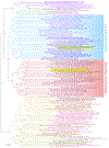
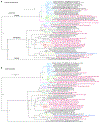
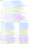
Phylogenetic trees of several Chlosyne species inferred from protein-coding regions in the nuclear genome (autosomes), based on 1,471,464 positions. Ultrafast bootstrap (Minh et al. 2013) values are shown at nodes. Different species are colored differently: C. damoetas (purple), C. whitneyi (green), C. palla (blue with C. palla sterope labeled in darker color), C. flavula (red with C flavula blackmorei labeled in darker color), C. acastus (orange with C. acastus dorothyi labeled in darker color), and C. gabbii (olive). Primary type specimens are labeled in magenta, and possibly sympatric specimens of C. flavula and C. palla shown in Fig. 1 are highlighted in yellow.
Fig. 3.
Fig. 3.
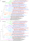
Phylogenetic trees of selected Erebia (Erebia) species constructed from protein-coding regions in: a) the nuclear genome (autosomes), based on 415,959 positions, and b) the mitochondrial genome. Different species and subspecies are colored differently: E. theano (brown), E. stubbendorfii (olive), E. pawloskii sajana stat. rest. (purple), E. pawloskii pawloskii (blue), E. pawloskii bilibinia (magenta), E. pawloskii alaskensis (green), E. pawloskii canadensis (dark blue), E. pawloskii ethela W. H. Edwards, 1891 (cyan), and E. demmia stat. nov. (red). Ultrafast bootstrap (Minh et al. 2013) values are shown at nodes. Gaps in terminal branches indicate that a segment of a tree was cut out to reduce its horizontal dimension (to allow an increase in the font size), i.e., a branch with a gap is longer than shown.
Fig. 4.
Fig. 4.
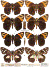
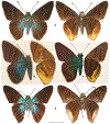
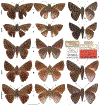
Erebia (Erebia) pawloskii bilibinia ssp. n. in dorsal (left) and ventral (right) views, data in text: a) holotype ♂ NVG-24041B06 and b) paratype ♀ NVG-24041B07.
Fig. 5.
Fig. 5.
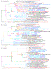
Phylogenetic trees of selected Dodona species constructed from protein-coding regions in: a) the nuclear genome (autosomes), based on 3,488,244 positions, and b) the mitochondrial genome. Different subgenera are colored differently: Ouida subgen. n. (red), Balonca (blue), Egeona subgen. n. (green), and Dodona (magenta). Ultrafast bootstrap (Minh et al. 2013) values are shown at nodes.
Fig. 6.
Fig. 6.
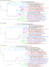
Phylogenetic trees of selected Lasaia species (L. sula species group) constructed from protein-coding regions in: a) the nuclear genome (autosomes), based on 4,871,172 positions, and b) the mitochondrial genome. Different species are colored differently: L. cola sp. n. (red), L. sula (blue), L. peninsularis (purple), and L. pallida Grishin, 2024 (green). Ultrafast bootstrap (Minh et al. 2013) values are shown at nodes.
Fig. 7.
Fig. 7.
Lasaia cola sp. n. holotype ♂ NVG-23103F05 in dorsal (left) and ventral (right) views, data in text.
Fig. 8.
Fig. 8.
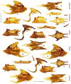
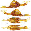
Male genitalia of Lasaia in left lateral (above each panel letter) and ventral (below each panel letter) views, data in text or below [MGCL]: a) L. sula NVG-24081A11 from Costa Rica, Guanacaste, 6 mi S and 6 mi W of Canas, Reserva Forestal Taboga, GPS 10.317, −85.150, 10-Jul-1968; b) L. cola sp. n. paratype NVG-24079H06 from Mexico: Colima; c) L. peninsulais NVG-25014D04 from USA: Texas, Hidalgo Co., Rio Rico Rd. near Relampago, 18-Nov-1998, E. C. Knudson leg. Green arrows point to characters useful for identification of these species, numbered 1 to 3, details in text.
Fig. 9.
Fig. 9.
Phylogenetic trees of Lasaia sessilis and relatives inferred from protein-coding regions in: a) the Z chromosome, based on 234,846 positions, and b) the mitochondrial genome. Different species are colored differently: L. sessilis (blue), L. oaxacensis stat. nov. (red), L. moeros (purple), and L. kennethi (green). Ultrafast bootstrap (Minh et al. 2013) values are shown at nodes.
Fig. 10.
Fig. 10.
Phylogenetic trees of Lasaia and relatives inferred from protein-coding regions in: a) the nuclear genome (autosomes), based on 6,309,384 positions, and b) the mitochondrial genome, showing subgenera Lasaia (blue) and Locris subgen. n. (red) labeled above corresponding branches. Ultrafast bootstrap (Minh et al. 2013) values are shown at nodes.
Fig. 11.
Fig. 11.
Phylogenetic trees of Curvie species constructed from protein-coding regions in: a) the nuclear genome (autosomes), based on 557,727 positions, and b) the mitochondrial genome. Ultrafast bootstrap (Minh et al. 2013) values are shown at nodes. Different species are colored differently: C. westwing sp. n. (cyan), C. wing sp. n. (red), C. yucatanensis (purple), C. emesia (blue), and C. chiapensis sp. n. (magenta).
Fig. 12.
Fig. 12.
Curvie wing sp. n. in dorsal (left) and ventral (right) views, data in text: a) holotype ♂ NVG-24103D07 and b) paratype ♀ NVG-24103D08. All Curvie specimens (Figs. 12, 14–15) are shown at the same scale to facilitate comparisons.
Fig. 13.
Fig. 13.
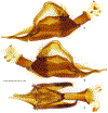
Male genitalia of Curvie in left lateral (left of the panel letter) and ventral (right of the panel letter) views, data in text or below: a) C. wing sp. n. paratype NVG-23112B10 from Mexico: Tamaulipas (tegumen with uncus and falces detached); b) C. westwing sp. n. paratype NVG-23111D09 from Mexico: Sinaloa; c) C. westwing sp. n. paratype NVG-23111D10 from Mexico: Colima; d) C. yucatanensis stat. rest. NVG-23112B07 Mexico: Yucatán, Chichén Itzá, E. C. Welling leg., genitalia NVG240817-04 [CMNH]; e) C. emesia NVG-23111D11 Guatemala: Zacapa, genitalia NVG240817-03 [CMNH]; f) C. emesia NVG-23115B03, 92-SRNP-4214 Costa Rica: Guanacaste Conservation Area, Cuesta Canyon Tigre, 270 m, eclosed 16-Aug-1992, genitalia NVG240817-06 [USNM].
Fig. 14.
Fig. 14.
Curvie westwing sp. n. holotype ♂ NVG-24087C09 in dorsal (left) and ventral (right) views, data in text.
Fig. 15.
Fig. 15.
Curvie chiapensis sp. n. in dorsal (left) and ventral (right) views, data in text. a) holotype ♂ NVG-23116C07 and b) paratype ♀ NVG-23116C08.
Fig. 16.
Fig. 16.
Phylogenetic trees of Emesis (Tenedia) species constructed from protein-coding regions in: a) the nuclear genome (autosomes), based on 7,184,877 positions, and b) the mitochondrial genome, showing the phylogenetic position of E. tinia sp. n. (magenta) and E. guaya sp. n. (green). Ultrafast bootstrap (Minh et al. 2013) values are shown at nodes.
Fig. 17.
Fig. 17.
Emesis (Tenedia) tinia sp. n. holotype ♂ NVG-24032C07 in dorsal (left) and ventral (right) views, data in text.
Fig. 18.
Fig. 18.
Emesis (Tenedia) guaya sp. n. holotype ♂ NVG-24032D02 in dorsal (left) and ventral (right) views, data in text.
Fig. 19.
Fig. 19.
Phylogenetic trees of selected Emesis (Aphacitis) species constructed from protein-coding regions in: a) the nuclear genome (autosomes), based on 3,086,919 positions, and b) the mitochondrial genome. Different species are colored differently: E. aurichica (brown), E. pruinapicalis (purple), E. auripana (cyan), E. aurimna (red), E. parvissima (blue), E. bugaba sp. n. (magenta), E. pallescens Grishin, 2024 (green), E. furvescens Grishin, 2024 (orange), and E. glaucescens Talbot, 1929 (olive). Ultrafast bootstrap (Minh et al. 2013) values are shown at nodes. A gap in a branch indicates that a segment of the branch was cut out to reduce its length (to allow an increase in the font size).
Fig. 20.
Fig. 20.
Emesis (Aphacitis) bugaba sp. n. in dorsal (left) and ventral (right) views, data in text: a) holotype ♂ NVG-18053H09 and b) paratype ♀ NVG-24031D06.
Fig. 21.
Fig. 21.
Phylogenetic trees of selected Synargis species constructed from protein-coding regions in: a) the nuclear genome (autosomes), based on 345,000 positions, and b) the mitochondrial genome. Different species are colored differently: S. maxidifa (green), S. rectanga sp. n. (magenta), S. tenebritorna Grishin, 2024 (blue), S. latidifa Grishin, 2024 (purple), and S. flavicauda Grishin, 2024 (cyan with S. flavicauda cosita Grishin, 2024 in olive). Ultrafast bootstrap (Minh et al. 2013) values are shown at nodes. Gaps in terminal branches indicate that a segment of a branch was cut out to reduce its length (to allow an increase in the font size), i.e., a branch with a gap is longer than shown.
Fig. 22.
Fig. 22.
Synargis specimens in dorsal (left) and ventral (right) views, data in text: a) S. rectanga sp. n. holotype ♀ NVG-23087C05 and b) S. maxidifa Grishin, 2024 holotype ♂ NVG-23103C10.
Fig. 23.
Fig. 23.
Phylogenetic trees of selected Nymphidiini constructed from protein-coding regions in the nuclear genome (autosomes), based on 4,956,768 positions. Different genera are colored differently, and different subgenera are labeled in different colors: Parvospila (brown with subgenus Lucispila subgen. n. labeled in magenta), Zelotaea (green with subgenus Byzia subgen. n. labeled in pink), Aricoris (purple with subgenera Arichlosyne subgen. n. and Ariconias stat. nov. labeled in orange and gray, respectively), Lemonias (cyan with subgenus Thisbe stat. nov. labeled in bright purple), Uraneis stat. nov. (red), and Pachythone (blue with subgenera Pixus Callaghan, 1982, Lamphiotes Callaghan, 1982, Lenca subgen. n., and Pseudonymphidia stat. nov. labeled in maroon, dark blue, aquamarine, and olive, respectively). Ultrafast bootstrap (Minh et al. 2013) values are shown at nodes.
Fig. 24.
Fig. 24.
Phylogenetic trees of selected Lycaenidae species constructed from protein-coding regions in: a) the nuclear genome (autosomes), based on 447,723 positions, and b) the mitochondrial genome; and c) a BioNJ (Gascuel 1997) dendrogram constructed from COI barcodes using the phylogeny.fr server (Dereeper et al. 2008). Different genera are colored differently: Ajenorix gen. n. (red), Bindahara (gray), Artipe (olive), Virachola stat. rest. (green with subgenus Crates subgen. n. in brown), Capys (purple with subgenus Afrix subgen. n. in magenta), Deudorix (blue with subgenus Wacus subgen. n. in dark blue), and Pilodeudorix (cyan with Pilodeudorix batikelides comb. nov. labeled in orange). Ultrafast bootstrap (Minh et al. 2013) values are shown in (a) and (b), and regular bootstrap values (as fractions) from 100 replicates are shown in (c). In the COI barcode dendrogram (c), species are added from the BOLD database (Ratnasingham and Hebert 2007) (identification not checked) and their BOLD Sample ID (not equal to 8 symbols) or GenBank accession (8 symbols starting from a letter) are given. Sequences obtained by us are denoted by the sample ID of 8 symbols starting from a number. Gaps in branches indicate where a vertical slice of the tree was removed to reduce its horizontal dimension (to allow an increase in the font size), i.e., branches with gaps are longer than shown.
Fig. 25.
Fig. 25.
Phylogenetic trees of Phanus species constructed from protein-coding regions in: a) the Z chromosome, based on 171,813 positions, and b) the mitochondrial genome: P. ecutinus sp. n. (magenta), P. ecitonorum (cyan), P. confusis Austin, 1993 (purple), P. rilma Evans, 1952 (green), and P. albiapicalis Austin, 1993 (blue). Ultrafast bootstrap (Minh et al. 2013) values are shown at nodes.
Fig. 26.
Fig. 26.
Phanus ecutinus sp. n. holotype ♀ NVG-24074D06 in dorsal (left) and ventral (right) views, data in text.
Fig. 27.
Fig. 27.
Genitalia of P. ecutinus sp. n. holotype ♀ NVG-24074D06 in ventral (left) and ventrolateral (right) views.
Fig. 28.
Fig. 28.
Phylogenetic trees of Entheus gentius group species constructed from protein-coding regions in: a) the nuclear genome (autosomes) and b) the mitochondrial genome: E. gentius (blue) and E. gentius sp. n. (red).
Fig. 29.
Fig. 29.
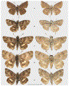
Entheus zeus sp. n. holotype ♂ NVG-22017H08 in dorsal (left) and ventral (right) views, data in text. The inset shows the hindtibial tuft enlarged two times compared to the specimen (scale not given).
Fig. 30.
Fig. 30.
Genitalia of Entheus zeus sp. n. paratype ♂, slide 488 (views): a) genitalia with valvae and aedeagus detached (left lateral); b, c) valvae (lateral); d) aedeagus (left lateral).
Fig. 31.
Fig. 31.
Phylogenetic trees of Entheus priassus group species constructed from protein-coding regions in: a) the nuclear genome (autosomes), based on 1,580,949 positions, and b) the mitochondrial genome. Primary and secondary type specimens are labeled in red and blue, respectively. Branches of new taxa are shown in red, and those with subspecies-to-species status change in blue. Ultrafast bootstrap (Minh et al. 2013) values are shown at nodes. Clades of more recently proposed species are colored: E. latebrosus (green), E. aureanota (purple), and E. curvus (cyan). The clades corresponding to the three species discussed in the text in detail are numbered 1, 2, and 3 with a yellow highlight.
Fig. 32.
Fig. 32.
Phylogenetic trees of Entheus priassus (blue), Entheus guyaneus sp. n. (red), and Entheus talaus (green) constructed from protein-coding regions in: a) the nuclear genome (autosomes), based on 2,495,064 positions, and b) the mitochondrial genome. Primary type specimens are labeled in magenta. Ultrafast bootstrap (Minh et al. 2013) values are shown at nodes.
Fig. 33.
Fig. 33.
Primary type specimens of Entheus designated in this work in dorsal (left) and ventral (right) views, data in text, insets show hindtibial tuft enlarged two times compared to specimens (scale not given): a) neotype of Papilio priassus ♂ NVG-18095F12; b) neotype of Papilio peleus ♂ NVG-23117A03; c) neotype of Peleus aeacus ♂ NVG-18038E04; d) lectotype of Phareas serenus ♀ NVG-22091A04 that is also the neotype of Papilio talaus, with its labels shown below and reduced by a quarter in size compared to specimens. The scale for labels is shown below them, and the scale for specimens is in the middle of the figure.
Fig. 34.
Fig. 34.
Female genitalia of Entheus primary types, data in text (ductus bursae and bursa copulatrix not shown): a–c) E. talaus neotype and, simultaneously, Phareas serenus lectotype NVG-22091A04; d–f) E. hyponota sp. n. holotype NVG-22091B03; g–i) E. lina sp. n. holotype NVG-15032C12; j–k) E. ecuadius sp. n. holotype NVG-14062C11 in different views: a, d, g, j) ventral; b, e, h) left ventrolateral; c, f, i, k) right ventrolateral.
Fig. 35.
Fig. 35.
Entheus guyaneus sp. n. in dorsal (left) and ventral (right) views, data in text: a) holotype ♂ NVG-14062D01, inset shows hindtibial tuft enlarged two times compared to specimens (scale not given); b) paratype ♀ NVG-14062D05.
Fig. 36.
Fig. 36.
Male genitalia of Entheus holotypes (unless indicated), data in text: a–d) E. guyaneus NVG-23119D12; e–g) E. proxemus NVG-24064A01; h–k) E. peruveus NVG-23119E01; l–o) E. pano NVG-23119E02; p–w) E. venezuelius NVG-15026F10; x–z) E. venezuelius paratype NVG-24028H11 in different views: a, e, h, l, s, u, w, x) left lateral; b, f, i, m, p, v, y) dorsal; c, j, n) posteroventral; d, g, k, o, r, z) posterior; q) ventral; t) right lateral. Complete genital capsule is shown, except p–s) with u, v) right and w) left valvae and t) aedeagus detached. Panel letters are on the lower right of each image.
Fig. 37.
Fig. 37.
Entheus colombeus sp. n. holotype ♂ NVG-15099C09 in dorsal (left) and ventral (right) views, data in text. The inset shows the hindtibial tuft enlarged two times compared to the specimen (scale not given).
Fig. 38.
Fig. 38.
Genitalia of Entheus colombeus sp. n. holotype ♂, slide 483 (views): a) genitalia with valvae and aedeagus detached (left lateral); b) left valva (right lateral); c) right valva (left lateral); d) aedeagus (left lateral). Dorsal tips of both harpes folded over during the slide mount.
Fig. 39.
Fig. 39.
Entheus proxemus sp. n. holotype ♂ NVG-23064B05 in dorsal (left) and ventral (right) views, data in text.
Fig. 40.
Fig. 40.
Entheus peruveus sp. n. in dorsal (left) and ventral (right) views, data in text: a) holotype ♂ NVG-14062B08, inset shows hindtibial tuft enlarged two times compared to specimens (scale not given); b) paratype ♀ NVG-14062B03.
Fig. 41.
Fig. 41.
Entheus hyponota sp. n. in dorsal (left) and ventral (right) views, data in text: a) holotype ♀ NVG-22091B03 with its labels below on the right and b) its illustration from Staudinger (1884–1888), identified as E. talaus at the time.
Fig. 42.
Fig. 42.
Entheus lina sp. n. holotype ♀ NVG-15032C12 in dorsal (left) and ventral (right) views, data in text.
Fig. 43.
Fig. 43.
Male genitalia of Entheus: a–c) E. matho lectotype minislide 108: a) genitalia with left valva detached; b) left valva, flipped (left-right inverted to facilitate comparison), interior view; c) illustration: fig. 29 on pl. 81 from Godman & Salvin (1894); d) E. guato sp. n. right valva, fig. 67 from Steinhauser (1989). a, b) © The Trustees of the Natural History Museum London and are made available under Creative Commons License 4.0 (https://creativecommons.org/licenses/by/4.0/).
Fig. 44.
Fig. 44.
Phylogenetic trees of Entheus matho group species inferred from protein-coding regions in: a) the nuclear genome (autosomes), based on 1,354,638 positions, b) the Z chromosome, based on 243,684 positions, and c) the mitochondrial genome. Ultrafast bootstrap (Minh et al. 2013) values are shown at nodes. Primary and secondary type specimens are labeled in red and blue, respectively. Branches of new taxa are shown in red and those with subspecies-to-species status change in blue.
Fig. 45.
Fig. 45.
Entheus guato sp. n. holotype ♂ NVG-15105A05 in dorsal (left) and ventral (right) views, data in text.
Fig. 46.
Fig. 46.
Entheus pano sp. n. holotype ♂ NVG-14062B12 in dorsal (left) and ventral (right) views, data in text. The inset shows the hindtibial tuft enlarged two times compared to the specimen (scale not given).
Fig. 47.
Fig. 47.
Entheus venezuelius sp. n. in dorsal (left) and ventral (right) views, data in text: a) holotype ♂ NVG-15026F10, inset shows hindtibial tuft enlarged two times compared to specimens (scale not given) and b) paratype ♀ NVG-15026F11.
Fig. 48.
Fig. 48.
Entheus ecuadius sp. n. holotype ♀ NVG-14062C11 in dorsal (left) and ventral (right) views, data in text.
Fig. 49.
Fig. 49.
Entheus bogoteus sp. n. holotype ♂ NVG-22042E08 in dorsal (left) and ventral (right) views, data in text. The inset shows the hindtibial tuft enlarged two times compared to the specimen (scale not given).
Fig. 50.
Fig. 50.
Phylogenetic trees of Entheus inferred from protein-coding regions in: a) the nuclear genome (autosomes), based on 3,486,282 positions, b) the Z chromosome, based on 279,462 positions, and c) the mitochondrial genome. Ultrafast bootstrap (Minh et al. 2013) values are shown at nodes. Different species groups and subgroups are colored: eumelus group (olive), gentius group (blue), priassus group (cyan, with telemus and pralina subgroups in purple and red, respectively), warreni group (magenta), and matho group (green). New taxa proposed in this work are labeled in red, and those with taxonomic changes, such as subspecies-to-species or synonym-to-species status (changes indicated in brackets), are labeled in blue.
Fig. 51.
Fig. 51.
Entheus specimens already illustrated above (data in text) shown life-size if printed on 8.5 by 11-inch paper for size comparison in dorsal (above each panel letter) and ventral (below) views: a) E. zeus sp. n. HT; b, c) E. priassus: b) NT, c) NT of = P. peleus; d) E. talaus stat. rest. NT & LT of = Ph. serenus; e, f) E. guyaneus sp. n.: e) HT, f) PT; g) E. lina sp. n. HT; h) E. colombeus sp. n. HT; i) E. proxemus sp. n. HT; j, k) E. peruveus sp. n.: j) PT, k) HT; l) E. hyponota sp. n. HT; m) E. guato sp. n. HT; n) E. pano sp. n. HT; o, p) E. venezuelius sp. n.: o) HT, p) PT; q) E. ecuadius sp. n. HT; r) E. bogoteus sp. n. HT.
Fig. 52.
Fig. 52.
Holotype of Cecropterus (Thorybes) oaxacensis ♂ NVG-19125B09 in dorsal (left) and ventral (right) views.
Fig. 53.
Fig. 53.
Phylogenetic trees of selected Cecropterus (Thorybes) species constructed from protein-coding regions in: a) the nuclear genome (autosomes), based on 11,630,790 positions, b) the Z chromosome, based on 338,631 positions, and c) the mitochondrial genome. Ultrafast bootstrap (Minh et al. 2013) values are shown at nodes. Different species are colored differently: C. egregius (blue), C. coxeyi stat. rest. (red), C. virescens (cyan), C. chlorothrix (purple), C. notochlorothrix sp. n. (orange), and C. viridissimus (green). Primary type specimens are labeled in magenta.
Fig. 54.
Fig. 54.
Cecropterus (Thorybes) notochlorothrix sp. n. holotype ♂ NVG-14111A02 in dorsal (left) and ventral (right) views.
Fig. 55.
Fig. 55.
Male genitalia of Cecropterus (Thorybes), data in text or below [MGCL]: a–b) C. (T.) notochlorothrix sp. n. paratype NVG-24124A07 Brazil, São Paulo, complete genital capsule and c–g) C. (T.) virescens NVG-24124A03 French Guiana, Saül, 8-Jun-1992, L. Sénécaux & A. Docquin leg., vial SRS-5288: c–d) genitalia with e) left and f) right valvae and g) aedeagus (vesica everted, cornutus below) detached and shown separately: in a, c, e–g) left lateral and b, d) dorsal views.
Fig. 56.
Fig. 56.
A pair of Cecropterus (Thorybes) viridissimus from Ecuador [SMF] in dorsal (left) and ventral (right) views: a) ♂ NVG-24021A04 Morona-Santiago, San Isidro, Macas, 1250 m, −2.12, −78.10, 17-Dec-2011, J.-C. Petit leg. And b) ♀ NVG-24021A09 Pastaza, Puyo, Mirador Condor, 1200 m, −1.28, −77.48, 7-Nov-2013 J.-C. Petit, E. & J. Brockmann leg.
Fig. 57.
Fig. 57.
Phylogenetic trees of selected Cecropterus (Murgaria) species constructed from protein-coding regions in: a) the nuclear genome (autosomes), based on 1,538,820 positions, and b) the mitochondrial genome. Ultrafast bootstrap (Minh et al. 2013) values are shown at nodes. Different species are colored differently: C. markwalkeri Grishin, 2023 (gray), C. albociliatus (blue, with the lectotype of A. toxeus in magenta), C. coyote (Skinner, 1892) (green), C. roeveri Grishin, 2025 (orange), C. nigrociliata (Mabille & Boullet, 1912) (purple), C. jalapus (Plötz, 1881) (cyan), and C. athesis (Hewitson, 1867) (olive). Gaps in branches indicate where vertical slices of the tree were removed to reduce its horizontal dimension (to allow an increase in the font size), i.e., branches with gaps are longer than shown.
Fig. 58.
Fig. 58.
Phylogenetic trees of selected Urbanus (Urbanoides) species (U. elma sp. n. in red and U. elmina in blue) constructed from protein-coding regions in: a) the Z chromosome, based on 615,213 positions, and b) the mitochondrial genome. Ultrafast bootstrap (Minh et al. 2013) values are shown at nodes.
Fig. 59.
Fig. 59.
Urbanus (Urbanoides) elma sp. n. in dorsal (left) and ventral (right) views, data in text: a) holotype ♀ NVG-19064C09 from Venezuela and b) paratype ♂ NVG-24019F07 from Colombia.
Fig. 60.
Fig. 60.
Eudamus hopfferi Plötz, 1881: a) the lectotype (designated herein) with its labels and b) illustrations from Draudt (1922: Pl. 167), which is likely a copy of an unpublished Plötz’s drawing t. 88, in dorsal (above) and ventral (below) views; c) an excerpt with the description of E. hopfferi from Plötz’s manuscript in ZSMC library dated 1876 that is an earlier version of his published works; d) a line for the No. 4969 in the MFNB collection catalog, written by Hopffer. Larger scale bar refers to the specimen, and smaller scale bar refers to labels, which are reduced by one-third compared to the specimen.
Fig. 61
Fig. 61
Phylogenetic trees of Telegonus specimens analyzed in this work inferred from protein-coding regions in: a) the nuclear genome (autosomes), based on 1,285,392 positions, b) the Z chromosome, based on 358,395 positions, and c) the mitochondrial genome. Ultrafast bootstrap (Minh et al. 2013) values are shown at nodes. Primary and secondary type specimens are labeled in red and blue, respectively. Branches of new taxa proposed in this work are shown in red and those with taxonomic changes, such as subspecies-to-species or synonym-to-species status or transfer of a subspecies between species (changes indicated in brackets) are shown in blue.
Fig. 61
Fig. 61
Phylogenetic trees of Telegonus specimens analyzed in this work inferred from protein-coding regions in: a) the nuclear genome (autosomes), based on 1,285,392 positions, b) the Z chromosome, based on 358,395 positions, and c) the mitochondrial genome. Ultrafast bootstrap (Minh et al. 2013) values are shown at nodes. Primary and secondary type specimens are labeled in red and blue, respectively. Branches of new taxa proposed in this work are shown in red and those with taxonomic changes, such as subspecies-to-species or synonym-to-species status or transfer of a subspecies between species (changes indicated in brackets) are shown in blue.
Fig. 62.
Fig. 62.
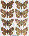
Telegonus (Rhabdoides) alector ecuadoricus ssp. n. holotype ♂ NVG-19071H10 in dorsal (left) and ventral (right) views.
Fig. 63.
Fig. 63.
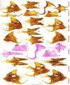
Male genitalia of Telegonus (Rhabdoides) holotypes (unless indicated), data in text or below: a–b) T. alector ecuadoricus ssp. n. NVG-19071H10; c–d) T. panavenus sp. n. paratype NVG-14111B09; e–f) T. pacificus sp. n. NVG-14111C02; g–h) T. amazonicus sp. n. NVG-14111C03; i–j) T. pallidus sp. n. NVG-14111D04; k–l) T. cyprus crilla comb. nov. specimen NVG-14111D07 from Peru, Huanuco, Tingo Maria, 800 m, May-Jun-1994 [USNM]; m–o) T. subfuscus sp. n. NVG-22078G12 in different views: a, d, e, h, i, l, o) left lateral; b, c, f, g, j, k, n) dorsal; m) right lateral. The complete genital capsule is shown. Beige arrows connect different views of the same genitalia.
Fig. 64.
Fig. 64.
Telegonus (Rhabdoides) missionus sp. n. holotype ♂ NVG-14111E04 in dorsal (left) and ventral (right) views.
Fig. 65.
Fig. 65.
Telegonus (Rhabdoides) panavenus sp. n. holotype ♂ NVG-14111B08 in dorsal (left) and ventral (right) views.
Fig. 66.
Fig. 66.
Telegonus (Rhabdoides) pacificus sp. n. holotype ♂ NVG-14111C02 in dorsal (left) and ventral (right) views.
Fig. 67.
Fig. 67.
Telegonus creteus and Telegonus parmenides stat. rest. in dorsal (left) and ventral (right) views: original drawings by Lambertz of type specimens of: a) Papilio creteus and b) Papilio parmenides used as models for published engravings; c) a male from Brazil in the Calkoen collection (known to include Cramer’s primary types) with its original labels [RMNH]. This specimen resembles the drawing of Papilio parmenides and is conspecific with the specimen from Guyana in MGCL that was chosen by Steinhauser as the neotype of P. parmenides, although this designation remains unpublished; d) a male we identify as conspecific with the Calkoen specimen shown in c), from Suriname, savanna forest adjacent to airport, Mar-2002, M. J. Simon leg., NVG-22078G06 [MGCL]. Images shown in a) and b) are © The Trustees of the Natural History Museum London and are made available under Creative Commons License 4.0 (https://creativecommons.org/licenses/by/4.0/).
Fig. 68.
Fig. 68.
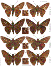
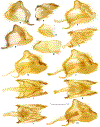
Male genitalia of Telegonus (Rhabdoides) non-type specimens [MGCL]: a–j) T. creteus: a–e) NVG-22078G05 Venezuela, Amazonas, Samariapo, Upper Orinoco, upstream from Maipures Rapids, 95–115 m, 5-Sep-1946, Rene Lichy leg., vial SRS-1799 and f–j) NVG-22078G04 Brazil, Pará, Fazenda Velna nr. Belém, 17-Nov-1973, C. Callaghan leg., vial SRS-853 and k–s) T. parmenides: k–p) NVG-24064B01 French Guiana, Maroni River, Oct-Nov-1903, ex coll. Le Moult, vial SRS-1786 and q–s) NVG-23063F08 Brazil, Amazonas, Manaus, km 26 AM-010, Reserva Ducke, GPS −2.9167, −59.9833, 13-Dec-1993, J. Bolling Sullivan & Roger W. Hutchings leg., vial SRS-4607 in different views: a, c–f, h–k, n–p, r) left lateral; b, g, l, s) dorsal; q) right lateral; and m) ventral: q–s) complete genital capsule and a, b, f, g, k–m) genitalia with c, h, n) aedeagus and d, i, o) right and e, j, p) left valvae detached and shown separately. Vesica is everted in c), cornuti pointing up at uncus in a). Beige arrows connect different views or parts of the same genitalia.
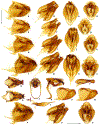
Fig. 69.
Fig. 69.
Telegonus (Rhabdoides) amazonicus sp. n. holotype ♂ NVG-14111C03 in dorsal (left) and ventral (right) views.
Fig. 70.
Fig. 70.
Telegonus (Rhabdoides) pallidus sp. n. holotype ♂ NVG-14111D04 in dorsal (left) and ventral (right) views.
Fig. 71.
Fig. 71.
Telegonus (Rhabdoides) subfuscus sp. n. holotype ♂ NVG-22078G12 in dorsal (left) and ventral (right) views.
Fig. 72.
Fig. 72.
Type specimens of Telegonus (Rhabdoides) in dorsal (top) and ventral (bottom) views, data in text: a) neotype of Eudamus blasius Plötz, 1881 ♂ NVG-24028D10 with its labels reduced to ¾ of the specimen scale (the scale for labels is below the handwritten label), and b) T. (R.) elorianus sp. n. holotype ♂ NVG-24028D11, no labels are shown for it.
Fig. 73.
Fig. 73.
Telegonus (Rhabdoides) perumazon sp. n. holotype ♂ NVG-14111C12 in dorsal (left) and ventral (right) views.
Fig. 74.
Fig. 74.
Male genitalia of Telegonus (Rhabdoides) holotypes (unless indicated), data in text: a–b) T. perumazon sp. n. NVG-14111C12; c–g) T. steinhauseri sp. n. NVG-23063E11; h–l) T. chiapus sp. n. NVG-23063G01; m–o) T. chiapus sp. n. paratype NVG-24064A06; p–t) T. colotrix sp. n. NVG-23063G02; u–v) T. sobrasus sp. n. NVG-19071H11 in different views: a,c, e, h, j, o, p, r, u) left lateral; b, d, i, n, q, v) dorsal; and f, g, k–m, s, t) right lateral: a, b, m–o, u, v) complete genital capsule and c, d, h, i, p, q) genitalia with e, j, r) aedeagus (vesica everted, cornuti on the right) and f, k, s) right and g, l, t) left valvae detached and shown separately. Beige arrows connect different views or parts of the same genitalia.
Fig. 75.
Fig. 75.
Telegonus chiriquensis and relatives in dorsal (left) and ventral (right) views: a) lectotype of Telegonus chiriquensis designated herein, NVG-24028C04, data in text; b) illustrations of T. chiriquensis from Draudt (1922); c) illustration of T. chiriquensis from Staudinger (1884–1888); d) a paralectotype ♂ of T. chiriquensis that is not conspecific with the lectotype and is T. grullus from Panama: Chiriquí, Ribbe leg., NVG-15031B10 [MFNB]; a) and d) photographed by Bernard Hermier.
Fig. 76.
Fig. 76.
Lectotype of Aethilla weymeri Plötz, 1882 NVG-24028C11 in dorsal (left) and ventral (right) views, data in text.
Fig. 77.
Fig. 77.
Telegonus (Rhabdoides) steinhauseri sp. n. holotype ♂ NVG-23063E11 in dorsal (left) and ventral (right) views.
Fig. 78.
Fig. 78.
Telegonus (Rhabdoides) chiapus sp. n. holotype ♂ NVG-23063G01 in dorsal (left) and ventral (right) views.
Fig. 79.
Fig. 79.
Telegonus (Rhabdoides) colotrix sp. n. holotype ♂ NVG-23063G02 in dorsal (left) and ventral (right) views.
Fig. 80.
Fig. 80.
Telegonus (Rhabdoides) flavimargo sp. n. holotype ♀ NVG-14105A05 in dorsal (left) and ventral (right) views.
Fig. 81.
Fig. 81.
Female genitalia of Telegonus (Rhabdoides) holotypes (unless indicated), data in text: a–b) T. colotrix sp. n. paratype NVG-14104A03; c–d) T. flavimargo sp. n. NVG-14105A05; and e–g) T. chuchuvianus sp. n. NVG-24086F12 in a, c, e, g) ventral; b, d) left ventrolateral; and f) right ventrolateral views: a, c, e, f) bursa copulatrix omitted and b, d, g) complete genitalia shown at 1/3 scale (indicated by smaller scale bars near them). Beige arrows connect different views and magnifications of the same genitalia.
Fig. 82.
Fig. 82.
Telegonus (Rhabdoides) sobrasus sp. n. holotype ♂ NVG-19071H11 in dorsal (left) and ventral (right) views.
Fig. 83.
Fig. 83.
Telegonus (Rhabdoides) chuchuvianus sp. n. holotype ♀ NVG-24086F12 in dorsal (left) and ventral (right) views.
Fig. 84.
Fig. 84.
Telegonus (Rhabdoides) panamus sp. n. holotype ♂ NVG-14111C10 in dorsal (left) and ventral (right) views.
Fig. 85.
Fig. 85.
Male genitalia of Telegonus (Rhabdoides) holotypes (unless indicated), data in text: a–b) T. panamus sp. n. NVG-14111C10; c–g) T. panamus sp. n. paratype NVG-23063F12; h–i) T. tatus sp. n. NVG-14111D05; j–k) T. fulvimargo sp. n. NVG-19075A12; l–n) T. fulvimargo sp. n. paratype NVG-24064B08; o–s) T. alardinus sp. n. NVG-23063G09; t–x) T. alardinus sp. n. paratypes: t–u) NVG-19075C11; v–x) NVG-24064B03 in different views: a, c, e, h, j, n, o, q, u, x) left lateral; b, d, i, k, m, p, t, w) dorsal; and f, g, l, r, s, v) right lateral: a, b, h–n, t–x) complete genital capsule and c, d, o, p) genitalia with e, q) aedeagus and f, s) right and g, r) left valvae detached and shown separately. Vesica is everted in e), cornuti on the right. Genitalia o–s) were stained using Double Stain, see text. Beige arrows connect different views or parts of the same genitalia.
Fig. 86.
Fig. 86.
Telegonus (Rhabdoides) tatus sp. n. holotype ♂ NVG-14111D05 in dorsal (left) and ventral (right) views.
Fig. 87.
Fig. 87.
Specimens of Telegonus (Rhabdoides) in dorsal (left) and ventral (right) views: a) T. (R.) fulvimargo sp. n. holotype ♂ NVG-19075A12 Peru: Cuzco, Cosñipata Valley, 22-X-2016. S. Kinyon [USMN] and b) T. (R.) meretrix non-type specimen ♂ NVG-24028D07 Ecuador: Pichincha, Santa Ines, old., R. Haensch S. [MFNB].
Fig. 88.
Fig. 88.
Telegonus (Rhabdoides) alardinus sp. n. holotype ♂ NVG-23063G09 in dorsal (left) and ventral (right) views.
Fig. 89.
Fig. 89.
Phylogenetic trees of Telegonus species analyzed in this study inferred from protein-coding regions in: a) the nuclear genome (autosomes), based on 9,184,581 positions, b) the Z chromosome, based on 319,194 positions, and c) the mitochondrial genome. Ultrafast bootstrap (Minh et al. 2013) values are shown at nodes. Different species groups are colored differently: alector group (green), elorus group (purple), creteus group (blue), parmenides group (orange), latimargo group (cyan), and galesus group (magenta). New taxa proposed in this work are labeled in red, and those with taxonomic changes, such as subspecies-to-species or synonym-to-species status or transfer of a subspecies between species (changes indicated in brackets) are labeled in blue.
Fig. 89.
Fig. 89.
Phylogenetic trees of Telegonus species analyzed in this study inferred from protein-coding regions in: a) the nuclear genome (autosomes), based on 9,184,581 positions, b) the Z chromosome, based on 319,194 positions, and c) the mitochondrial genome. Ultrafast bootstrap (Minh et al. 2013) values are shown at nodes. Different species groups are colored differently: alector group (green), elorus group (purple), creteus group (blue), parmenides group (orange), latimargo group (cyan), and galesus group (magenta). New taxa proposed in this work are labeled in red, and those with taxonomic changes, such as subspecies-to-species or synonym-to-species status or transfer of a subspecies between species (changes indicated in brackets) are labeled in blue.
Fig. 90.
Fig. 90.
Phylogenetic trees of selected Pellicia species inferred from protein-coding regions in: a) the nuclear genome (autosomes), b) the Z chromosome, and c) [see next page] the mitochondrial genome. Primary type specimens are labeled in red-purple, and a paralectotype of P. tyana (not conspecific with the lectotype) is labeled in blue. Different species mentioned in the text are shown in different colors. The sequence of SAMN18587826 is taken from the alignment provided in Kawahara et al. (2023).
Fig. 90.
Fig. 90.
Phylogenetic trees of selected Pellicia species inferred from protein-coding regions in: a) the nuclear genome (autosomes), b) the Z chromosome, and c) [see next page] the mitochondrial genome. Primary type specimens are labeled in red-purple, and a paralectotype of P. tyana (not conspecific with the lectotype) is labeled in blue. Different species mentioned in the text are shown in different colors. The sequence of SAMN18587826 is taken from the alignment provided in Kawahara et al. (2023).
Fig. 91.
Fig. 91.
Type specimens and illustrations of Pellicia species described by Plötz, data in text: a) P. theon lectotype NVG-15032E09; b) P. theon drawing t. 200; c) P. zamia lectotype NVG-15032E08; d) P. zamia drawing t. 201; e) P. toza stat. nov. specimen NVG-18056G09, which is a non-conspecific paralectotype of P. tyana, with its labels shown in f) and reduced by a quarter compared to specimens with the scale shown on the right; g) P. tyana lectotype NVG-15032D11; h) P. tyana drawing t. 202; i) Arteurotia demetrius syn. nov. of P. tyana, NVG-15032E12; j) A. demetrius drawing t. 205. The drawings are Godman’s copies of Plötz’s original illustrations (likely drawn by Horace Knight) (Godman 1907) and are in BMNH. Images b), d), h), and j) are © of the Trustees of the Natural History Museum London and are made available under Creative Commons License 4.0 (https://creativecommons.org/licenses/by/4.0/).
Fig. 92.
Fig. 92.
Pellicia (Hemipteris) cina sp. n. holotype ♂ NVG-23053D08 in dorsal (left) and ventral (right) views, data in text.
Fig. 93.
Fig. 93.
Male genitalia of Pellicia (Hemipteris) cina sp. n. holotype NVG-23053D08 (data in text) in different views: a) right lateral, b) left lateral, c) right posterolateral, d) left posterolateral, e) dorsal, and f) posterior tilted dorsad.
Fig. 94.
Fig. 94.
Phylogenetic trees of selected Gorgopas species constructed from protein-coding regions in: a) the Z chromosome, based on 336,096 positions, and b) the mitochondrial genome. Ultrafast bootstrap (Minh et al. 2013) values are shown at nodes. Different species are shown in different colors: G. trocha sp. n. (green), G. trochilus (blue), G. trochicuz sp. n. (red), and G. trochitango sp. n. (purple). Primary type specimens are labeled in magenta. The sequence of SAMN18587196 is taken from the alignment provided in Kawahara et al. (2023).
Fig. 95.
Fig. 95.
Gorgopas trochicuz sp. n. holotype ♂ NVG-7975 in dorsal (left) and ventral (right) views, data in text. All Gorgopas specimens (Figs. 95, 97, 99) are shown at the same scale to facilitate comparisons.
Fig. 96.
Fig. 96.
Genitalia of Gorgopas: a–c) G. trochicuz sp. n. holotype ♂ NVG-7975 data in text and d–f) G. trochilus ♂ NVG-23055H04 Ecuador, Napo, Misahuallí environs, 300 m, Oct-Nov-1978, N. Venedictoff leg., vial NVG250517-05 [MGCL] in different views: a, d) left lateral, b, e) right lateral, and c, f) dorsal.
Fig. 97.
Fig. 97.
Gorgopas trocha sp. n. holotype ♂ NVG-23055H03 in dorsal (left) and ventral (right) views, data in text.
Fig. 98.
Fig. 98.
Genitalia of Gorgopas trocha sp. n. holotype ♂ NVG-23055H03 in different views: a) left lateral, b) right lateral, c) dorsal.
Fig. 99.
Fig. 99.
Gorgopas trochitango sp. n. holotype ♂ NVG-23055H09 in dorsal (left) and ventral (right) views, data in text.
Fig. 100.
Fig. 100.
Genitalia of Gorgopas trochitango sp. n. holotype ♂ NVG-23055H09 in different views: a) left lateral, b) right lateral, c) dorsal.
Fig. 101.
Fig. 101.
Phylogenetic trees of Perus (Perus) cordillerae (blue) and Perus (Perus) perus sp. n. (red) constructed from protein-coding regions in: a) the Z chromosome, based on 315,390 positions, and b) the mitochondrial genome. Primary type specimens are labeled in magenta. Ultrafast bootstrap (Minh et al. 2013) values are shown at nodes.
Fig. 102.
Fig. 102.
Perus (Perus) perus sp. n. males in dorsal (left) and ventral (right) views, data in text: a) holotype NVG-7826 and b) paratype NVG-18058H06.
Fig. 103.
Fig. 103.
Male genitalia of Perus (Perus), data in text or below: a–d) P. (P.) perus sp. n.: a–b) holotype NVG-7826 and c–d) paratype NVG-18058H06 and e–k) P. (P.) cordillerae from Ecuador, Loja [MGCL]: e–f) NVG-25014A09 Vilcabamba, 1600 m, May-1974, R. de Lafebre leg., vial SRS-2023 and g–k) NVG-24065A08 km 28 of Loja–Catamayo Rd., 1700 m, 11-Sep-1975, S. S. Nicolay leg., vial SRS-1979 in a, c, e, h–k) left lateral and b, d, f, g) dorsal views: a–f) complete genitalia and g–h) genitalia with i) aedeagus and j) left and k) right valvae detached and shown separately.
Fig. 104.
Fig. 104.
Phylogenetic trees of Gomalia inferred from protein-coding regions in: a) the nuclear genome (autosomes), based on 1,176,192 positions, b) the Z chromosome, based on 116,643 positions, and c) the mitochondrial genome. Ultrafast bootstrap (Minh et al. 2013) values are shown at nodes. Primary types are labeled in red-purple. Different species are colored differently: G. jeanneli (purple), G. albofasciata (cyan), G. litoralis stat. rest. (red), G. elma (blue), and G. westafra sp. n. (green).
Fig. 105.
Fig. 105.
Specimens of Gomalia in dorsal (left) and ventral (right) views, additional data in text. a) G. jeanneli jeanneli ♂ NVG-21068F05 Ethiopia, Harari or Oromia Region, Erer River, 20-Aug-1955, S. Chojnacki leg. [MGCL], b) G. jeanneli levana ♂ NVG-24054D01, c–d) G. litoralis stat. rest. from Oman: c) ♂ NVG-24054B04 and d) ♀ NVG-24054B05, e) G. albofasciata ♂ NVG-22044B02 India, “Coimbatore Prov.” [Tamil Nadu], 9-Nov-1945, P. Susai Nathan leg. [CUIC].
Fig. 106.
Fig. 106.
Specimens of Gomalia in dorsal (left) and ventral (right) views, additional data are in text. Gomalia westafra sp. n.: a) holotype ♂ NVG-24066B03, b) paratype ♀ NVG-24054B03 and G. elma from South Africa: c) ♂ NVG-24066C07, UF FLMNH MGCL 1162207, “Transvaal Pienaar’s River” [Limpopo Province, Pienaarsrivier], ~1970, Wm. Henning leg., genitalia NVG241111-28 (Fig. 107b) [MGCL], d) ♀ NVG-19046G10 Pretoria, 6-Mar-1915 [AMNH].
Fig. 107.
Fig. 107.
Male genitalia of Gomalia in left lateral (above) and right dorsolateral (below, emphasizing the difference between species in the costa-ampulla area) views: a) G. westafra sp. n. paratype NVG-24066B02 (data in text), and b) G. elma NVG-24066C07 (specimen Fig. 106c, data in its legend).
Fig. 108.
Fig. 108.
Chirgus biseriatus and relatives in dorsal (right) and ventral (left) views: a) illustration of Carterocephalus biseriatus from Weymer and Maassen (1890); b) the lectotype of C. biseriatus designated herein, NVG-15033H08, data in text; c) a specimen of Chirgus nigella (NVG-15033H09) identified as C. biseriatus and placed next to the lectotype in the drawer; d) a specimen of Chirgus biseriatus stat. rest. from Peru: Arequipa Region, ca. 30 km NE of El Misti, 4100 m, 12-Oct-1983, E. S. Nielsen leg., NVG-22013C01 [RMNH]. All Chirgus specimens (Figs. 108, 110, 112, 114) are shown at the same scale to facilitate comparisons.
Fig. 109.
Fig. 109.
Phylogenetic trees of Chirgus (Chirgus) constructed from protein-coding regions in: a) autosomes, b) the Z chromosome, and c) the mitochondrial genome. Primary type specimens are labeled in red. Branches corresponding to different species are colored in different colors: C. biseriatus (purple), C. nigella (cyan), C. limbata (green), C argentinus sp. n. (orange), C. trisignatus (blue), C. teres sp. n. (red), C. sombrus sp. n. (magenta), and C. bocchoris (aquamarine).
Fig. 110.
Fig. 110.
Chirgus (Chirgus) argentinus sp. n. holotype ♂ NVG-15092G11 in dorsal (left) and ventral (right) views.
Fig. 111.
Fig. 111.
Male genitalia of Chirgus argentinus sp. n. holotype NVG-15092G11 in left lateral (left) and dorsal (right) views.
Fig. 112.
Fig. 112.
Chirgus (Chirgus) teres sp. n. holotype ♂ NVG-23058C10 in dorsal (left) and ventral (right) views, data in text.
Fig. 113.
Fig. 113.
Male genitalia of Chirgus (Chirgus) teres sp. n. holotype NVG-23058C10 in left lateral (left) and dorsal (right) views.
Fig. 114.
Fig. 114.
Chirgus (Chirgus) sombrus sp. n. holotype ♂ NVG-23058C12 in dorsal (left) and ventral (right) views, data in text.
Fig. 115.
Fig. 115.
Male genitalia of Chirgus sombrus sp. n. paratype NVG-17069B06 in left lateral (left) and dorsal (right) views.
Fig. 116.
Fig. 116.
Phylogenetic trees of Zopyrion species constructed from protein-coding regions in: a) the Z chromosome, based on 190,077 positions, and b) the mitochondrial genome: Z. subvariegata (purple), Zopyrion thyas stat. nov. (red), Z. sandace (blue), Z. xerxes sp. n. (magenta), and Z. satyrina (C. Felder & R. Felder, 1867) (green). Ultrafast bootstrap (Minh et al. 2013) values are shown at nodes.
Fig. 117.
Fig. 117.
Zopyrion (Zopyrion) xerxes sp. n. holotype ♂ NVG-19091F01 in dorsal (left) and ventral (right) views, data in text.
Fig. 118.
Fig. 118.
Male genitalia of Zopyrion (Zopyrion): a–c) Z. (Z.) xerxes sp. n. holotype NVG-19091F01, vial no. X-4380 J.M. Burns 1998 and d–f) Z. (Z.) sandace NVG-23124F07, vial no. X-4379 J.M. Burns 1998, Mexico, Oaxaca, 65 mi SE of Oaxaca, 15-Aug-1972, G. F. & S. Hevel leg. [USNM] in different views: a, d) left lateral, b, e) dorsal, and c, f) right dorsolateral.
Fig. 119.
Fig. 119.
Phylogenetic trees of selected Anisochoria species constructed from protein-coding regions in: a) the nuclear genome (autosomes), based on 1,278,153 positions, and b) the mitochondrial genome. Ultrafast bootstrap (Minh et al. 2013) values are shown at nodes. Different species are colored differently: A. bacchus (blue), A. bacchoides sp. n. (red), A. polysticta Mabille, 1877 (green), and A. pedaliodina (A. Butler, 1870) (purple).
Fig. 120.
Fig. 120.
Anisochoria bacchoides sp. n. holotype ♂ NVG-23054D06 in dorsal (left) and ventral (right) views, data in text.
Fig. 121.
Fig. 121.
Male genitalia of Anisochoria bacchoides sp. n. paratype NVG-20062H03 in a) left lateral, b) dorsal, and c) right posterolateral views.
Fig. 122.
Fig. 122.
Phylogenetic trees of Timochares fuscifasciata (blue) and Timochares ruptifasciata (red) inferred from protein-coding regions in: a) the nuclear genome (autosomes), based on 1,302,732 positions, and b) the mitochondrial genome. Primary type specimens are labeled in magenta.
Fig. 123.
Fig. 123.
Phylogenetic trees of Onespa species inferred from protein-coding regions in: a) the nuclear genome (autosomes), b) the Z chromosome, and c) the mitochondrial genome. Different species are shown in different colors: O. nuba sp. n. (red), O. nubis (blue), O. nakamura Austin & A. Warren, 2009 (black), O. brockorum (purple), and O. gala (green).
Fig. 124.
Fig. 124.
Onespa nuba sp. n. holotype ♂ NVG-18118E02 in dorsal (left) and ventral (right) views, data in text.
Fig. 125.
Fig. 125.
Male genitalia of Onespa nuba sp. n. paratype NVG-21107D04, X-2855 (data in text) in different views: a) left and b) right lateral, c) dorsal, and d) posterolateral, in the plane of three cornuti.
Fig. 126
Fig. 126
Phylogenetic trees of Hesperia pahaska specimens constructed from protein-coding regions in: a) the nuclear genome (autosomes), based on 4,407,714 positions, b) the Z chromosome, based on 341,715 positions, and c) the mitochondrial genome. Ultrafast bootstrap (Minh et al. 2013) values are shown at nodes. Different subspecies are colored differently: H. pahaska hannawackeri (blue), H. pahaska williamsi (olive), H. pahaska martini (cyan), H. pahaska tehaska sp. n. (purple), H. pahaska hidalgo sp. n. (red), H. pahaska bajanorta sp. n. (green), and H. pahaska pahaska (black, only two specimens shown). Holotypes are labeled in magenta. Gaps in branches indicate that a segment of a branch was cut out to reduce its length (to allow an increase in the font size), i.e., a branch with a gap is longer than shown.
Fig. 127.
Fig. 127.
Hesperia pahaska tehaska sp. n. holotype ♂ NVG-23049B09 in dorsal (left) and ventral (right) views, data in text. All Hesperia holotypes (Figs. 127, 129, 130) are shown at the same scale to facilitate comparisons.
Fig. 128.
Fig. 128.
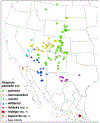
A map of sequenced specimens of Hesperia pahaska subspecies: pahaska (green squares), hannawackeri (yellow circles), martini (cyan inverted triangles), williamsi (blue triangles), tehaska ssp. n. (magenta ovals), hidalgo ssp. n. (red star), bajanorta ssp. n. (orange diamond). The type localities of subspecies are marked with tiny white circles inside symbols. Four U.S. subspecies converge near the NV–UT–AZ tripoint, where mixed populations occur, and subspecies assignment is currently tentative—if possible at all.
Fig. 129.
Fig. 129.
Hesperia pahaska hidalgo sp. n. holotype ♂ NVG-23049G08 in dorsal (left) and ventral (right) views, data in text.
Fig. 130.
Fig. 130.
Hesperia pahaska bajanorta sp. n. holotype ♂ NVG-23049G10 in dorsal (left) and ventral (right) views, data in text.
Fig. 131.
Fig. 131.
Phylogenetic trees of Ochlodes inferred from protein-coding regions in: a) the nuclear genome (autosomes), based on 13,712,217 positions, and b) the mitochondrial genome. Different subgenera are shown in different colors: Ochloba subgen. n. (red). Ochlata subgen. n. (purple), Ochluma subgen. n. (blue), and Ochlodes (green) and labeled above corresponding branches in the nuclear genome tree. Type species of subgenera are labeled in magenta.
Fig. 132.
Fig. 132.
Phylogenetic trees of three species of Lon inferred from protein-coding regions in: a) the nuclear genome (autosomes), based on 807,423 positions, and b) the mitochondrial genome. Specimens from Puntarenas Province, Costa Rica, are labeled in magenta. Ultrafast bootstrap (Minh et al. 2013) values are shown at nodes. Different species are in different colors: L. zabulon (blue), L. co (green), and L. ma (purple). The sequence of SAMN18587728 is taken from the alignment provided in Kawahara et al. (2023).
Fig. 133.
Fig. 133.
Lon males from Costa Rica: Puntarenas Prov., in dorsal (left half, a–f) and ventral (right half, g–l) views: L. co in the 1st and 3rd column (a–c, g–i) and L. ma in the 2nd and 4th column (d–f, j–l), the same specimen is shown in the same position in the left and right halves of the image. Green arrows point to characters useful for identification of these species, numbered 1 to 5, see text. Specimens were collected in Monteverde and are in MGCL, except as indicated. a, g) NVG-24065E10, Mar-1987, J. Brenner coll.; b, h) NVG-24065F01, 1280 m, 7-9-Sep-1988, P. F. Milner leg.; c, i) NVG-18115B07, USNMENT 01531555 PT, 1300 m, 18-May-1985, J. A. Chemsak leg. [USNM]; d, j) NVG-23048E11, Mar-1987, J. Brenner coll.; e, k) NVG-24065E11 Las Alturas, 1400 m, 5-Jul-1992 A. Sourakov leg.; f, l) NVG-24065E12, 1280 m, 7-9-Sep-1988, P. F. Milner leg.
Fig. 134.
Fig. 134.
Phylogenetic trees of Vacerra constructed from protein-coding regions in: a) the nuclear genome (autosomes) and b) the mitochondrial genome. Primary type specimens are labeled in magenta, and branches of selected species are colored differently: V. gayra (green), V. tama sp. n. (magenta), V. saltina sp. n. (purple), V. cecropterus stat. rest. (cyan), V. cuza sp.n. (red), and V. hermesia (blue).
Fig. 135.
Fig. 135.
Vacerra tama sp. n. males in dorsal (left) and ventral (right) views, data in text: a) holotype NVG-22056G03 and b) paratype NVG-24015D04.
Fig. 136.
Fig. 136.
Male genitalia of Vacerra tama sp. n. holotype NVG-22056G03 in different views: a) left lateral, b) right lateral, c) dorsal, and d) ventral.
Fig. 137.
Fig. 137.
Vacerra saltina sp. n. holotype ♂ NVG-23045D07 in dorsal (left) and ventral (right) views, data in text.
Fig. 138.
Fig. 138.
Male genitalia of Vacerra saltina sp. n. holotype NVG-23045D07 (data in text) in different views: a) left lateral, b) right lateral, and c) dorsal.
Fig. 139.
Fig. 139.
Vacerra cuza sp. n. holotype ♂ NVG-18128C01 in dorsal (left) and ventral (right) views, data in text.
Fig. 140.
Fig. 140.
Male genitalia of Vacerra cuza sp. n. holotype NVG-18128C01 (data in text) in different views: a) left lateral, b) right lateral, and c) dorsal.
Fig. 141.
Fig. 141.
Phylogenetic trees of Oligoria (Oligoria) species constructed from protein-coding regions in: a) the Z chromosome, based on 414,570 positions, and b) the mitochondrial genome: O. maculata (W. H. Edwards, 1865) (green), O. percosius (Godman, 1900) (red), O. rindgei (blue), O. tinalandia sp. n. (magenta), and O. lucifer (Hübner, [1831]) (cyan). Ultrafast bootstrap (Minh et al. 2013) values are shown at nodes.
Fig. 142.
Fig. 142.
Oligoria (Oligoria) tinalandia sp. n. holotype ♀ NVG-24065F10 in dorsal (left) and ventral (right) views.
Fig. 143.
Fig. 143.
Phylogenetic trees of Eutychide and its sister Dion Godman, 1901 constructed from protein-coding regions in: a) the nuclear genome (autosomes), based on 4,780,623 positions, and b) the mitochondrial genome. Different Eutychide species are colored differently: E. trombella sp. n. (magenta), E. paria (blue), E. ochus Godman, 1900 (orange), E. ochoides Grishin, 2023 (olive), E. rogersi (Kaye, 1914) (green), E. complana (Herrich-Schäffer, 1869) (purple), E. subcordata (Herrich-Schäffer, 1869) (brown), and E. physcella (Hewitson, 1866) (cyan). Ultrafast bootstrap (Minh et al. 2013) values are shown at nodes. Note the mitochondrial genome introgression among four species, which, as a result, cannot be identified using COI barcodes.
Fig. 144.
Fig. 144.
Eutychide trombella sp. n. holotype ♀ NVG-22109F02 in dorsal (left) and ventral (right) views, data in text.
Fig. 145.
Fig. 145.
Phylogenetic trees of Talides constructed from protein-coding regions in: a) the nuclear genome (autosomes) and b) the mitochondrial genome: T. hispina sp. n. (magenta), T. hispa (blue), T. alternata E. Bell, 1941 (purple), and T. laeta Grishin, 2023 (green).
Fig. 146.
Fig. 146.
Talides hispina sp. n. holotype ♂ NVG-23069C01 in dorsal (left) and ventral (right) views, data in text.
Fig. 147.
Fig. 147.
Male genitalia of Talides hispina sp. n. holotype NVG-23069C01 in different views, data in text: a) right lateral, b) anterodorsal, and c) dorsal.
Fig. 148.
Fig. 148.
Primary type specimens of Damas in dorsal (left) and ventral (right) views, data in text: a) lectotype of D. corope stat. rest., b) neotype of D. angulis stat. rest., c) holotype of D. honduras sp. n., and d) holotype of D. kenos sp. n. Insets show magnified (scale indicated by 0.5 cm bar) middle of forewing with digitally enhanced stigma of each specimen.
Fig. 149.
Fig. 149.
Male genitalia of Damas, data in text: a–d) lectotype of D. corope stat. rest., left valva detached, aedeagus not shown; e–g) specimen of D. angulis stat. rest. NVG-23123B02, left valva detached; h–i) paratype of D. honduras sp. n. NVG-23123A10; j–k) paratype of D. kenos sp. n. NVG-23123B06; and l–m) paratype of D. lavandas sp. n. NVG-24099C06. Views: b, e, h, j, m) left lateral, a, d, g) right lateral, c, f, i, k, l) dorsal, d) shows caudal part of the left valva.
Fig. 150.
Fig. 150.
Carystus orope in dorsal (left) and ventral (right) views: a) the lectotype of C. orope with it labels, labels are reduced by a third compared to the specimen: a smaller scale bar (placed vertically among labels) refers to labels; b) Godman’s copy of an unpublished illustration t. 533 by Plötz photographed by N.V.G., © The Trustees of the Natural History Museum London made available under Creative Commons License 4.0 (https://creativecommons.org/licenses/by/4.0/).
Fig. 151.
Fig. 151.
Phylogenetic trees of selected Tigasis species constructed from protein-coding regions in: a) the nuclear genome (autosomes), based on 934,104 positions, and b) the mitochondrial genome. Different species are colored differently: T. wellingi (H. Freeman, 1969) (green), T. arita (Schaus, 1902) (blue), and T. corope (red). Primary type specimens are labeled in magenta, and the lectotype of Carystus orope (which is simultaneously a female syntype of T. corope) is highlighted in yellow. Ultrafast bootstrap (Minh et al. 2013) values are shown at nodes. A gap in a branch indicates that a segment of the branch was cut out to reduce its length.
Fig. 152.
Fig. 152.
Phylogenetic trees of Damas inferred from protein-coding regions in: a) the nuclear genome (autosomes), b) the mitochondrial genome. Different taxa are shown in different colors: D. honduras sp. n. (green), D. angulis stat. rest. (purple), D. kenos sp. n. (orange), D. cervus stat. rest. (olive), D. corope stat. rest. (blue), D. lavandas sp. n. (red), and D. clavus (cyan). Primary type specimens are labeled in magenta.
Fig. 153.
Fig. 153.
DNA sequence alignment of three segments from the mitochondrial genome of Damas to illustrate identification of D. corope stat. rest. (lectotype in red font).
Fig. 154.
Fig. 154.
Damas lavandas sp. n. holotype ♂ NVG-23123B07 in dorsal (left) and ventral (right) views, data in text, at the same scale as Fig. 148. The inset displays a magnified view (scale indicated by 0.5 cm bar) of the middle of the forewing, highlighting digitally enhanced stigma.

References

    1. Anonymous. 2025. William Swainson (1789-1855) (https://www.museum.zoo.cam.ac.uk/collections-research/collections-uncove...). (Last Accessed 9 May 2025).
    1. Aurivillius POC 1882. Recensio critica Lepidopterorum Musei Ludovicæ Ulricæ qu descripsit Carolus a Linné. Kongliga svenska Vetenskaps-Akademiens Handlingar (Ny Följd) 19(5): 1–188, 1 pl.
    1. Austin GT 1993. A review of the Phanus vitreus group (Lepidoptera: Hesperiidae: Pyrginae). Tropical Lepidoptera 4(suppl. 2): 21–36.
    1. Austin GT 1997. Two new Entheus from Ecuador and Peru (Lepidoptera: Hesperiidae: Pyrginae). Tropical Lepidoptera 8(1): 18–21.
    1. Austin GT, and Mielke OHH. 1998. Hesperiidae of Rondônia, Brazil: Aguna Williams (Pyrginae), with a partial revision and descriptions of new species from Panama, Ecuador, and Brazil. Revista brasileira de Zoologia 14(4): 889–965.

LinkOut - more resources