. 2025 Jun 6:13:1564458.
doi: 10.3389/fcell.2025.1564458. eCollection 2025.

17β-estradiol maintains extracellular matrix homeostasis of nucleus pulposus cells by activating p70 S6K1 signaling pathway

Affiliations

17β-estradiol maintains extracellular matrix homeostasis of nucleus pulposus cells by activating p70 S6K1 signaling pathway

Tao Liu et al. Front Cell Dev Biol. .

Abstract

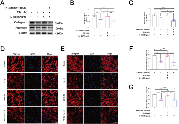
Background: Estrogen can inhibit the apoptosis of nucleus pulposus cells (NPCs) through the PI3K/AKT/mTOR signaling pathway. However, the downstream of mTOR signaling pathway remains elusive. This study investigates the effect of 17β-estradiol (E2) on intervertebral disc degeneration (IVDD) through the p70 S6K1 signaling pathway, downstream of mTOR.

Methods: The IVDD model of rats was established by needle puncture and bilateral ovariectomy. Fifteen Sprague-Dawley rats were randomly assigned to the following three groups: (A) Sham surgery group (Sham); (B) Bilateral ovariectomy, 21G needle puncture and carrier injection (OVX + veh); (C) Bilateral ovariectomy, 21G needle puncture, E2 supplementation (OVX + E2). The degree of IVDD was evaluated by X-ray, magnetic resonance imaging (MRI), hematoxylin and eosin (H&E), and Safranin O-Fast Green staining. The expression levels of target protein p70S6K1 and its phosphorylated products were detected by immunohistochemistry (IHC). Finally, Western blot analysis and immunofluorescence staining were used to investigate the effect of E2 on the p70 S6K1 signaling pathway in vitro.

Results: Histological staining and radiological results showed that E2 supplementation altered signaling, suggesting that it may have a protective effect against IVDD. IHC showed that compared with the Sham and OVX + E2 groups, the level of p70 S6K1 in the OVX + veh group was significantly increased while the expression of phosphorylated products (p-S6) was significantly decreased, suggesting that E2 could inhibit IVDD by activating p70 S6K1 signaling pathway, the downstream of mTOR. Furthermore, cellular immunofluorescence and Western blot showed that E2 can maintain extracellular matrix (ECM) balance and inhibits apoptosis of nucleus pulposus cells (NPCs) by activating the p70 S6K1 signaling pathway.

Conclusion: In summary, 17β-estradiol mitigates IVDD progression by maintaining ECM homeostasis and inhibiting NPCs apoptosis through activation of the p70 S6K1 signaling pathway downstream of mTOR.

Keywords: P70 S6K1; apoptosis; estradiol; intervertebral disc degeneration; mTOR; nucleus pulposus.


Conflict of interest statement

The authors declare that the research was conducted in the absence of any commercial or financial relationships that could be construed as a potential conflict of interest. The author(s) declared that they were an editorial board member of Frontiers, at the time of submission. This had no impact on the peer review process and the final decision.

Figures

FIGURE 1
FIGURE 1
Rat IVDD model induced by needle puncture and OVX and histological staining. Rat IVD sections shown from left to right: sham ovariectomy (Sham), needle puncture plus ovariectomy with vehicle injection (OVX + Veh), and needle puncture plus ovariectomy with estradiol hormone replacement injection (OVX + E2). (A, C) Representative X-ray images of rat coccygeal vertebra and measurement result of DHI changes. DHI% was calculated as: DHI = 2 × (D1 + D2 + D3)/(V1 + V2 + V3+ V4+ V5 + V6), DHI% = post-punctured DHI/pre-punctured DHI × 100%, where D indicates disc height and V indicates vertebra length. (B, D) MRI images of rat coccygeal IVDs from different treatment groups and the scores of IVDD evaluated by the modified Thompson classification. (E) There are low-magnification H&E sections and higher magnification images of boxed areas. (F) Low magnification Safranin O-Fast green sections and higher magnification images of boxed areas. The green components indicate an acidophilic matrix stained by Fast green, and the red components indicate a basophilic matrix stained by Safranin O. (G) Histological scores for rat IVDs in the three groups above. Values are mean ± SD (n = 5). ns: not significant; **p < 0.01; ***p < 0.001; ****p < 0.0001. E2, 17β-estradiol; AF, annulus fibrosus; IVD, intervertebral disc; IVDD, intervertebral disc degeneration; OVX, ovariectomy; veh, vehicle injection; DHI, disc height index; MRI, magnetic resonance imaging; H&E, hematoxylin and eosin staining; SOFG, Safranin O-Fast Green staining; SD, standard deviation.
FIGURE 2
FIGURE 2
E2 inhibits MMPs and cleaved caspase 3 expression and reduces the ECM degradation. The rat IVD sections shown from left to right: sham ovariectomy (Sham), needle puncture plus ovariectomy with vehicle injection (OVX + Veh), and needle puncture plus ovariectomy with estradiol hormone replacement injection (OVX + E2) groups. (A, C, D) The expression of aggrecan and collagen II, which are the main components of ECM in NP tissues stained by immunohistochemical staining. (B, E, F) The expression of MMP 3 and cleaved caspase 3 in NP tissues stained by immunohistochemical staining. Values are mean ± SD (n = 5). ns: not significant; *p < 0.05; **p < 0.01; ***p < 0.001; ****p < 0.0001. IVD, intervertebral disc; OVX, ovariectomy; veh, vehicle injection; E2, 17β-estradiol; MMP, matrix metalloproteinase; ECM, extracellular matrix; NP, nucleus pulposus; SD, standard deviation.
FIGURE 3
FIGURE 3
Immunohistochemistry showing that E2 mitigates IVDD by activating the p70 S6K1 signaling pathway downstream of mTOR in vivo. The rat IVD sections shown from left to right: sham ovariectomy (Sham), needle puncture plus ovariectomy with vehicle injection (OVX + Veh), and needle puncture plus ovariectomy with estradiol hormone replacement injection (OVX + E2) groups. (A) The levels of p70S6K1 and p-S6 in NP tissues of rat tails were detected using immunohistochemistry. (B, C) Data analysis of the immunohistochemistry. Values are mean ± SD (n = 5). ns: not significant; *p < 0.05; **p < 0.01; ***p < 0.001. IVD, intervertebral disc; IVDD, intervertebral disc degeneration; OVX, ovariectomy; veh, vehicle injection; E2, 17β-estradiol; NP, nucleus pulposus; p-S6, Phospho-S6 (the ribosomal protein S6); SD, standard deviation.
FIGURE 4
FIGURE 4
E2 activates the p70 S6K1 signaling pathway in vitro. (A–D) Western blotting was used to analyze the levels of p70 S6K1, p-S6K1 and p-S6 in human NPCs. (E, G) The level of p70 S6K1 in human NPCs by immunofluorescence staining. (F, H) The level of p-S6 in human NPCs by immunofluorescence staining. Values are mean ± SD (n = 3). ns: not significant; *p < 0.05; **p < 0.01; ***p < 0.001; ****p < 0.0001. E2, 17β-estradiol; NPCs, nucleus pulposus cells; IL-1β, interleukin-1β; PF, PF4708671 (an p70 S6K1 inhibitor); p-S6, Phospho-S6 (the ribosomal protein S6); SD, standard deviation.
FIGURE 5
FIGURE 5
E2 activation of p70 S6K1 signaling pathway improves ECM anabolism in vitro. (A–C) Western blot analysis was used to analyze the expression of Collagen II and Aggrecan in human NPCs. (D, F) The expression of Aggrecan in human NPCs by immunofluorescence staining. (E, G) The expression of Collagen II in human NPCs by immunofluorescence staining. Values are mean ± SD (n = 3). ns: not significant; *p < 0.05; **p < 0.01; ***p < 0.001; ****p < 0.0001. E2, 17β-estradiol; NPCs, nucleus pulposus cells; IL-1β, interleukin-1β; PF, PF4708671(an p70 S6K1 inhibitor); ECM, extracellular matrix; SD, standard deviation.
FIGURE 6
FIGURE 6
E2 activation of the p70 S6K1 signaling pathway inhibits MMP3 and cleaved caspase 3 in vitro. (A–D) Western blot analysis was used to analyze the expression of MMP3 and cleaved caspase 3. (E, F) The expression of MMP3 was determined by immunofluorescence staining. (G, H) The expression of cleaved caspase 3 was determined by immunofluorescence staining. Values are mean ± SD (n = 3). ns: not significant; *p < 0.05; **p < 0.01; ***p < 0.001. E2, 17β-estradiol; NPCs, nucleus pulposus cells; IL-1β, interleukin-1β; PF, PF4708671(an p70 S6K1 inhibitor); MMP3, matrix metalloproteinase 3; SD, standard deviation.

References

    1. Adams M. A., Dolan P., McNally D. S. (2009). The internal mechanical functioning of intervertebral discs and articular cartilage, and its relevance to matrix biology. Matrix Biol. 28 (7), 384–389. 10.1016/j.matbio.2009.06.004 - DOI - PubMed
    1. Andersson G. B. (1999). Epidemiological features of chronic low-back pain. Lancet 354 (9178), 581–585. 10.1016/S0140-6736(99)01312-4 - DOI - PubMed
    1. Bai X., Guo X., Zhang F., Zheng L., Ding W., Yang S. (2021). Resveratrol combined with 17β-estradiol prevents IL-1β induced apoptosis in human nucleus pulposus via the PI3K/AKT/mtor and PI3K/AKT/GSK-3β pathway. J. Invest Surg. 34 (8), 904–911. 10.1080/08941939.2019.1705941 - DOI - PubMed
    1. Chen J., Ma Y., Yang Z., Lan H., Liu G., Zhang Y., et al. (2020). Tnfaip3 ameliorates the degeneration of inflammatory human nucleus pulposus cells by inhibiting mtor signaling and promoting autophagy. Aging (Albany, NY.) 12 (23), 24242–24254. 10.18632/aging.104160 - DOI - PMC - PubMed
    1. Chen J., Xuan J., Gu Y., Shi K. S., Xie J. J., et al. (2017). Celastrol reduces il-1β induced matrix catabolism, oxidative stress and inflammation in human nucleus pulposus cells and attenuates rat intervertebral disc degeneration in vivo . Biomed. Pharmacother. 91, 208–219. 10.1016/j.biopha.2017.04.093 - DOI - PubMed

LinkOut - more resources