

Imaging Features of Primary Intraosseous Adenoid Cystic Carcinoma of the Mandible: A Case Report
- PMID: 40546616
- PMCID: PMC12181810
- DOI: 10.7759/cureus.84599
Imaging Features of Primary Intraosseous Adenoid Cystic Carcinoma of the Mandible: A Case Report
Abstract
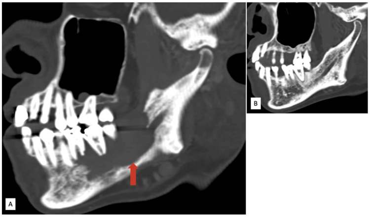
Intraosseous adenoid cystic carcinoma (ACC) of the mandible is an exceedingly rare malignancy. Its subtle onset and aggressive biological behavior, characterized by perineural invasion, present significant diagnostic and therapeutic challenges. We present a case involving a 68-year-old male who experienced persistent pain and swelling in the right hemimandible. Imaging studies using computed tomography (CT) revealed an ill-defined, osteolytic lesion affecting the right hemimandible, leading to cortical destruction. Magnetic resonance imaging (MRI) exhibited an enhancing mass in the right hemimandible, along with evidence of perineural spread along the inferior alveolar nerve. Histopathological analysis confirmed the diagnosis of adenoid cystic carcinoma, displaying a predominant cribriform pattern. The patient subsequently underwent segmental mandibulectomy, followed by adjuvant radiotherapy. This case highlights the essential roles of CT and MRI in the early detection, characterization, and surgical planning for intraosseous ACC. It is crucial for radiologists and clinicians to maintain a high index of suspicion when assessing destructive mandibular lesions, especially those that exhibit perineural spread. Timely diagnosis and a multidisciplinary management approach are vital for optimizing outcomes in this aggressive tumor.
Keywords: adenoid cystic carcinoma; ct; intraosseous; mandible; mri; perineural invasion.
Copyright © 2025, Munusamy et al.
Conflict of interest statement
Human subjects: Consent for treatment and open access publication was obtained or waived by all participants in this study. Conflicts of interest: In compliance with the ICMJE uniform disclosure form, all authors declare the following: Payment/services info: All authors have declared that no financial support was received from any organization for the submitted work. Financial relationships: All authors have declared that they have no financial relationships at present or within the previous three years with any organizations that might have an interest in the submitted work. Other relationships: All authors have declared that there are no other relationships or activities that could appear to have influenced the submitted work.
Figures


References
Publication types
LinkOut - more resources
Full Text Sources