Review
. 2025 Jun 17:20:7653-7683.
doi: 10.2147/IJN.S522198. eCollection 2025.

Extracellular Vesicle-Integrated Biomaterials in Bone Tissue Engineering Applications: Current Progress and Future Perspectives

Affiliations
Review

Extracellular Vesicle-Integrated Biomaterials in Bone Tissue Engineering Applications: Current Progress and Future Perspectives

Yan Huang et al. Int J Nanomedicine. .

Abstract

With an aging population and increased life expectancy, the clinical burden of bone-related disorders, especially large bone defects, continues to grow, underscoring the urgent need for effective regenerative strategies. Effective bone regeneration is essential not only for restoring skeletal structure and function but also for improving patients' quality of life and reducing the socioeconomic burden associated with prolonged recovery or surgical failure. Bone tissue engineering has emerged as a promising approach for healing large bone defects. Traditionally, stem cells, biomaterial scaffolds and growth factors have been considered the three essential elements of bone tissue engineering. However, stem cell-based therapies face several significant challenges, including ectopic tissue formation, malignant transformation, cell embolism, and immune rejection. In recent years, extracellular vesicles (EVs) have gained significant attention as an advanced alternative to stem cells and a novel cell-free therapy for bone regeneration due to their inherent advantages, such as low immune-rejection, excellent biocompatibility, significant bioactivity and high feasibility for carrying bioactive molecules or drugs. This review provides a comprehensive overview of the current state and future potential of EV-based strategies in bone tissue engineering. We first review the sources of parent cells for EVs applied in bone tissue engineering and the roles and potential mechanisms of EVs in bone regeneration. We then discuss the various modification strategies employed to enhance the therapeutic potential of EVs. Additionally, we summarize strategies for integrating EVs with various biomaterial scaffolds, with a specific focus on the latest advances in achieving controlled and sustained release of EVs from scaffolds at bone defect sites. Collectively, this review aims to offer key insights into the translational potential of EV-functionalized biomaterials and guide future directions in the development of next-generation bone regenerative therapies.

Keywords: biomaterials; bone tissue engineering; extracellular vesicles; mesenchymal stem cells.


Conflict of interest statement

The authors declare that they have no competing interests.

Figures
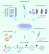
Figure 1
Figure 1
Schematic summary of strategies for endogenous engineering of EVs at the cellular level, including physical manipulation, pre-conditioning treatment (including osteogenic induction, hypoxic exposure and biochemical pretreatment) and genetic engineering of parent cells (such as lentivirus transduction and the EXPLOR technique). Adapted from Biomaterials, Volume 283, Tao SC, Li XR, Wei WJ, et al, Polymeric coating on beta-TCP scaffolds provides immobilization of small extracellular vesicles with surface-functionalization and ZEB1-Loading for bone defect repair in diabetes mellitus, Page 121465, Copyright 2022, with permission from Elsevier.
Figure 2
Figure 2
Schematic summary of strategies for EV surface modification. (A) Multivalent electrostatic interaction: positively charged polyethyleneimine (PEI) and negatively charged human BMP2 plasmids (phBMP2) were sequentially coated on the negatively charged membranes of EVs via electrostatic interactions, forming EVs–PEI/phBMP2 complexes. Used with permission of Royal Society of Chemistry, from Mesenchymal stem cell-derived microvesicles mediate BMP2 gene delivery and enhance bone regeneration, Liang Z, Luo Y, Lv Y, Volume 8, Edition 30, 2020; permission conveyed through Copyright Clearance Center, Inc. (B) Modification by anchoring CP05: The CP05 peptide specifically binds to the tetraspanin CD63 marker on EV membrane, while the titanium-binding peptide (TBP) specifically targets the titanium surface. EVs are directed to the titanium surface through a fusion peptide TBP-CP05. Used with permission from Royal Society of Chemistry, Synergetic osteogenesis of extracellular vesicles and loading RGD colonized on 3D-printed titanium implants, Ma S, Li X, Hu H, et al, Volume 10, Edition 17, 2022; permission conveyed through Copyright Clearance Center, Inc. (C) Hydrophobic interaction: the bone-targeting-peptide SDSSD, modified with DSPE-PEG-Mal, is effectively anchored to the EV membrane through hydrophobic interaction between the diacyllipid tail and the EV phospholipid layer. Adapted from Zou J, Shi M, Liu X, et al. Aptamer-functionalized exosomes: elucidating the cellular uptake mechanism and the potential for cancer-targeted chemotherapy. Anal Chem. 2019;91(3):2425–2430. Copyright 2019 American Chemical Society. (D) Aptamer-based surface modification: the 5′-end of a BMSC-specific aptamer is modified with an aldehyde group, allowing it to react with the amino groups of EV membrane proteins to produce aptamer-functionalized EVs. Used with permission from Royal Society of Chemistry, Aptamer-functionalized exosomes from bone marrow stromal cells target bone to promote bone regeneration, Luo ZW, Li FX, Liu YW, et al., Volume 11, Edition 43, 2019; permission conveyed through Copyright Clearance Center, Inc. (E) Click chemistry: alendronate (Ale) modified with an azide group (Ale-N3) is conjugated with EV membranes modified with an alkynyl group (EVs-DBCO) using click chemistry. Adapted from Wang Y, Yao J, Cai L, et al. Bone-targeted extracellular vesicles from mesenchymal stem cells for osteoporosis therapy. Int J Nanomed. 2020;15:7967–7977.
Figure 3
Figure 3
Schematic illustration of the procedures for fabricating self-assembled EV-functionalized biotin-doped polypyrrole titanium through the avidin-biotin system. First, a biotin-doped polypyrrole film (Bio-Ppy-Ti) is electrochemically deposited onto the titanium surface via an electrochemical potentiostatic method. Next, biotin-labeled EVs are immobilized on the surface of Bio-Ppy-Ti through the strepavidin-biotin interaction to form EV-Bio-Ppy-Ti. Finally, the osteodifferentiation of osteoblasts cultured on EV-Bio-Ppy-Ti is investigated to evaluate its osteoinductive properties. Reproduced from Chen L, Mou S, Li F, et al. Self-assembled human adipose-derived stem cell-derived extracellular vesicle-functionalized biotin-doped polypyrrole titanium with long-term stability and potential osteoinductive ability. ACS Appl Mater Interfaces. 2019;11(49):46183–46196. Copyright 2019 American Chemical Society.
Figure 4
Figure 4
Schematic illustration of the synthesis process for a polyethylene glycol (PEG)/DNA hybrid hydrogel system designed for the controlled release of DMSC-EVs in the treatment of diabetic bone defects. First, single DNA strands S1 and S2 are copolymerized with 8-arm vinyl sulfone (VS)-functionalized PEG to form PEG-DNA conjugates, designated as PS-1 and PS-2. These are then mixed in equal proportions with an MMP-9 aptamer-linker solution to form a stable hydrogel structure under suitable conditions, with EVs being introduced concurrently during the DNA hybridization process. Once the hydrogel is injected into the pathological defect site, the presence of MMP-9 triggers the separation of the DNA aptamer-linker from PS-1 and PS-2, leading to the disintegration of the DNA hydrogel and subsequent release of EVs. Reproduced from Jing X, Wang S, Tang H, et al. Dynamically bioresponsive DNA hydrogel incorporated with dual-functional stem cells from apical papilla-derived exosomes promotes diabetic bone regeneration. ACS Appl Mater Interfaces. 2022;14(14):16082–16099. Copyright 2022 American Chemical Society.

Similar articles

References

    1. Xue N, Ding X, Huang R, et al. Bone tissue engineering in the treatment of bone defects. Pharmaceuticals. 2022;15(7):879. doi: 10.3390/ph15070879 - DOI - PMC - PubMed
    1. Su N, Yang J, Xie Y, et al. Bone function, dysfunction and its role in diseases including critical illness. Int J Biol Sci. 2019;15(4):776–787. doi: 10.7150/ijbs.27063 - DOI - PMC - PubMed
    1. Qi J, Yu T, Hu B, Wu H, Ouyang H. Current biomaterial-based bone tissue engineering and translational medicine. Int J Mol Sci. 2021;22(19):10233. doi: 10.3390/ijms221910233 - DOI - PMC - PubMed
    1. Bauso LV, La Fauci V, Longo C, Calabrese G. Bone tissue engineering and nanotechnology: a promising combination for bone regeneration. Biology. 2024;13(4):237. doi: 10.3390/biology13040237 - DOI - PMC - PubMed
    1. Shang F, Yu Y, Liu S, et al. Advancing application of mesenchymal stem cell-based bone tissue regeneration. Bioact Mater. 2021;6(3):666–683. doi: 10.1016/j.bioactmat.2020.08.014 - DOI - PMC - PubMed

Substances

LinkOut - more resources