Review
. 2025 Jun 16:20:7685-7714.
doi: 10.2147/IJN.S488026. eCollection 2025.

Advancements, Challenges, and Future Prospects of Nanotechnology in Sepsis Therapy

Affiliations
Review

Advancements, Challenges, and Future Prospects of Nanotechnology in Sepsis Therapy

Yukun Liu et al. Int J Nanomedicine. .

Abstract

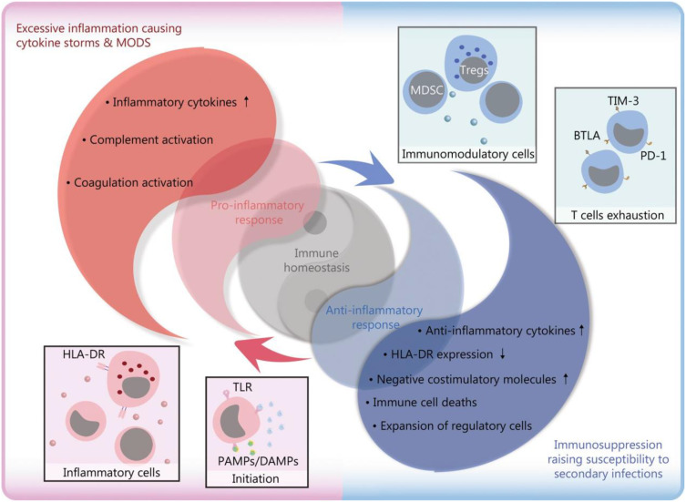
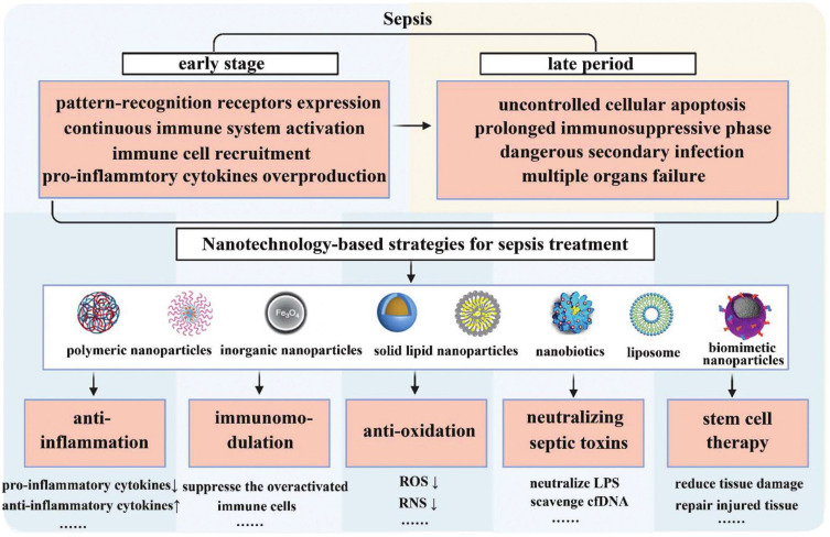
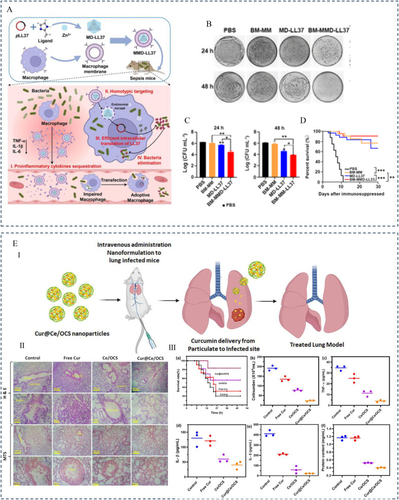
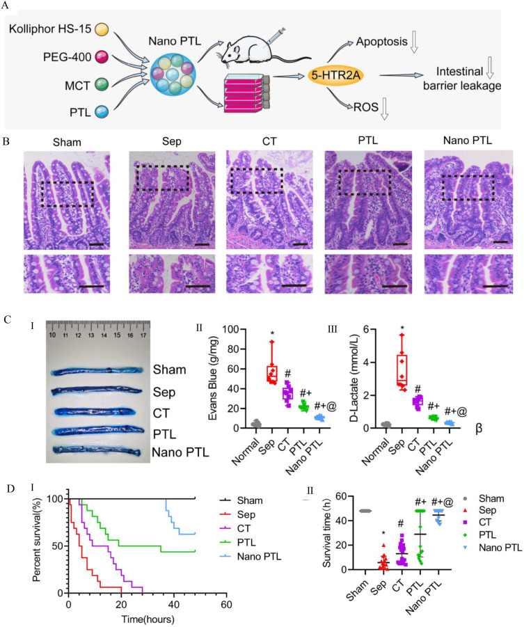
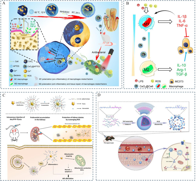
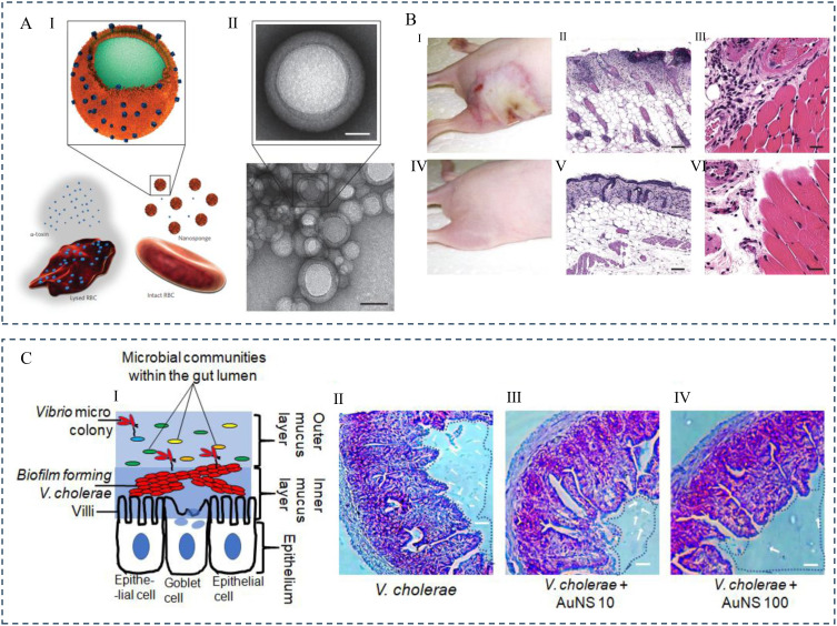
Sepsis is a life-threatening systemic inflammatory syndrome, typically triggered by infection, that can lead to multi-organ failure and high mortality rates. Traditional treatments for sepsis often have limited efficacy and significant side effects, necessitating the exploration of innovative therapeutic strategies. In recent years, the application of nanotechnology in sepsis therapy has garnered widespread attention due to its potential to modulate immune responses, reduce inflammation and oxidative stress, and eliminate bacterial toxins. This review aims to provide an overview of the latest advancements, challenges, and future prospects of nanotechnology in sepsis treatment. By analyzing recent developments in anti-inflammatory, immunomodulatory, antioxidant, and detoxification applications of nanotechnology, key findings and therapeutic potential are summarized, including the use of nanocarriers, biomimetic nanoparticles, and self-assembled nanomaterials. Furthermore, this review addresses the challenges in clinical translation, such as drug targeting, long-term safety, and biocompatibility. Future research will require large-scale clinical trials and interdisciplinary collaboration to validate the efficacy of nanotechnology in sepsis treatment and facilitate its integration into clinical practice. Overall, nanotechnology presents unprecedented opportunities for sepsis management, and this review seeks to offer insights into ongoing research while promoting further advancements in this field.

Keywords: MODS; nanoimmunotherapy; nanomedicine delivery methods; nanotechnology; sepsis.


Conflict of interest statement

The authors declare that the research was conducted in the absence of any commercial or financial relationships that could be construed as a potential conflict of interest.

Figures

Figure 1
Figure 1
Pathophysiological changes caused by infection-induced sepsis. Reproduced from Liu D, Huang SY, Sun JH, et al. Sepsis-induced immunosuppression: mechanisms, diagnosis and current treatment options. Mil Med Res. 2022;9(1):56. Copyright 2022, Springer Nature. This article is licensed under a Creative Commons Attribution 4.0 International License.
Figure 2
Figure 2
Overview of the pathophysiological process of sepsis and nanotechnology-based sepsis treatment strategies. Reproduced from Zhao Y, Pu M, Zhang J, et al. Recent advancements of nanomaterial-based therapeutic strategies toward sepsis: bacterial eradication, anti-inflammation, and immunomodulation. Nanoscale. 2021;13(24):10726–10747. © Royal Society of Chemistry 2021.
Figure 3
Figure 3
(A) I. Schematic illustration of the synthesis of SNP. II. Functionalization with aminopropyl groups. III. Functionalization with propyl methyl phosphonate groups. IV. Functionalization with octadecyl methyl groups. Reproduced from Ndayishimiye J, Cao Y, Kumeria T, Blaskovich MAT, Falconer JR, Popat A. Engineering mesoporous silica nanoparticles towards oral delivery of vancomycin. J Mater Chem B. 2021;9(35):7145–7166. © The Royal Society of Chemistry 2021. (B) I. Design and bio-functionalization of IME-responsive nanoparticles (NPs) for targeted drug delivery. II. Schematic of multifunctional NP design. III. Drug-loaded NPs-anti-ICAM-1 specifically target activated endothelial cells at infection sites after intravenous injection, binding to them, crossing blood vessels, and releasing drugs in response to local infection signals. IV. Structure and pH/enzyme-sensitive amphiphilic block copolymers. V. Coarse-grained simulation of CIP-PMs self-assembly using DPD methods. Reproduced from Zhang CY, Gao J, Wang Z. Bioresponsive nanoparticles targeted to infectious microenvironments for sepsis management. Adv Mater. 2018;30(43):e1803618. © 2018 WILEY‐VCH Verlag GmbH & Co. KGaA, Weinheim. (C) γ3 peptide grafted onto nanoparticle (NP) surfaces enhances uptake by activated endothelial cells (ECs) and specifically targets the lungs to alleviate inflammation. I. Schematic of the preparation of γ3 peptide-modified PLGA NPs loaded with SFX and TAC. II. Schematic of targeted therapy in mice with lung infection via intravenous injection of NPs. III. Digital images of lungs from mice with acute lung injury (ALI) infection treated with different formulations. IV. H&E sections of lungs from ALI mice receiving different treatments. V. Digital images of plated lungs from mice with acute lung infection caused by Pseudomonas aeruginosa receiving different treatments. VI. Colony-forming unit (CFU) counts. VII. Survival rate of different groups. I: PBS, II: free SFX, III: free TAC, IV: free S + T, V: PLGA/S + T, VI: γ3-PLGA/S + T. Data are shown as mean ± SD (n = 3). *p < 0.05, **p < 0.01, ***p < 0.001. Reproduced from Yang Y, Ding Y, Fan B, et al. Inflammation-targeting polymeric nanoparticles deliver sparfloxacin and tacrolimus for combating acute lung sepsis. J Control Release. 2020;321:463–474. Copyright © 2020 Elsevier B.V. All rights reserved.
Figure 4
Figure 4
(A) Schematic presentation of the antibacterial mechanism of AMPs. Reproduced from Zhang R, Xu L, Dong C. Antimicrobial peptides: an overview of their structure, function and mechanism of action. Protein Pept Lett. 2022;29(8):641–650. Copyright© Bentham Science Publishers. (B) I. Illustration of a passive nanosystem, demonstrating antimicrobial peptide delivery into an infected cell. II. Illustration of an active nano system demonstrating antimicrobial peptide delivery into an infected cell. Reproduced from Biswaro LS, da Costa Sousa MG, Rezende TMB, Dias SC, Franco OL. Antimicrobial peptides and nanotechnology, recent advances and challenges. Front Microbiol. 2018;9:855. Copyright © 2018 Biswaro, da Costa Sousa, Rezende, Dias and Franco. This is an open-access article distributed under the terms of the Creative Commons Attribution License (CC BY). (C) I. Synthesis of SNAPPs by ring-opening polymerization of lysine and valine N-carboxyanhydrides (NCAs), with poly(amidoamine) (PAMAM) dendrimer terminal amines as initiators. II. Comparison of the antibacterial mechanisms of typical membrane-disrupting cationic antimicrobial peptides (AMPs) and SNAPPs against Gram-negative bacteria. a. Cationic AMPs bind to the outer membrane (OM) of Gram-negative bacteria via electrostatic interactions, penetrate the OM through membrane destabilization, and disrupt the cytoplasmic membrane (CM) physical integrity via “barrel-stave”, “toroidal pore”, or “carpet” mechanisms (not shown). b. SNAPPs, whether aggregated or not, interact with the OM, peptidoglycan (PG), and CM layers of Gram-negative bacteria via electrostatic attraction, destabilizing/disrupting the OM and potentially the CM, leading to uncontrolled ion movement and cell death through apoptosis-like pathways (not shown). Reproduced from Lam SJ, O’Brien-Simpson NM, Pantarat N, et al. Combating multidrug-resistant Gram-negative bacteria with structurally nanoengineered antimicrobial peptide polymers. Nat Microbiol. 2016;1(11):16162. Copyright © 2016, Macmillan Publishers Limited. (D) I. Schematic of Ts-LPs-LEV preparation. II. Schematic of Ts-LPs-LEV bactericidal process. III. Kaplan–Meier survival curve representing mortality data of levofloxacin formulations in a multidrug-resistant (MDR) clinical isolate-induced septic shock model. Reproduced from Fan X, Fan J, Wang X, Wu P, Wu G. S-thanatin functionalized liposome potentially targeting on Klebsiella pneumoniae and its application in sepsis mouse model. Front Pharmacol. 2015;6:249. Copyright © 2015 Fan, Fan, Wang, Wu and Wu. This is an open-access article distributed under the terms of the Creative Commons Attribution License (CC BY).
Figure 5
Figure 5
(A) Summary of natural extracts from various natural sources, including plants, animals, or microbial species, for antibacterial applications. Reproduced from Chen C, Chen L, Mao C, et al. Natural extracts for antibacterial applications. Small. 2024;20(9):e2306553. © 2023 Wiley‐VCH GmbH. (B) Schematic of the synthesis of metal nanoparticles/nanocellulose composite materials. I. Simple mixing of cellulose source with prepared inorganic nanoparticles. II. Synthesis of nanoparticles using an external reducing agent. III. Synthesis of nanoparticles through surface modification of nanocellulose. IV. Synthesis of nanoparticles using nanocellulose itself without an external reducing agent. (C) Illustration of the synthesis of CNC/Ag composite nanoparticles. Reproduced from Oun AA, Shankar S, Rhim JW. Multifunctional nanocellulose/metal and metal oxide nanoparticle hybrid nanomaterials. Crit Rev Food Sci Nutr. 2020;60(3):435–460. Rights managed by Taylor & Francis. (D) I. Schematic of surface modification of Curcumin and SNP-immobilized Cu-MSN and their possible antibacterial mechanism. II. TEM images of Cur-Cu-MSN-SNP samples, scale bars: (a) 50 nm and (b) 200 nm. Reproduced from Kuthati Y, Kankala RK, Busa P, et al. Phototherapeutic spectrum expansion through synergistic effect of mesoporous silica trio-nanohybrids against antibiotic-resistant gram-negative bacterium. J Photochem Photobiol B. 2017;169:124–133. opyright © 2017 Elsevier B.V. All rights reserved.
Figure 6
Figure 6
(A) I. Approaches for the design and fabrication of diverse stimuli-responsive biomaterials and their utilization in the formulation of stimuli-responsive nano-delivery systems. R, S, and COO- are ROS-responsive moieties. II. Strategies for designing and fabricating biomimetic nanoparticles targeting sepsis microenvironments. Ismail EA, Devnarain N, Govender T, Omolo CA. Stimuli-responsive and biomimetic delivery systems for sepsis and related complications. J Control Release. 2022;352:1048–1070. Copyright © 2022 Elsevier B.V. All rights reserved. (B) I. Nano-antibody production process using phage display technology. II. Effect of administration times on survival rate of rats. Reproduced from Liao S, Liu S, Zhang Y. Preparation of anti toll-like receptor-4 nano-antibody and its effect on gram negative sepsis. J Nanosci Nanotechnol. 2021;21(2):1048–1053. Copyright 2021, American Scientific Publishers.
Figure 7
Figure 7
(A) Synthesis and characterization of immunomodulatory particles. I. The particles are formulated from polymers with different molecular weights (low or high), compositions (glycolic acid (GA) and lactic acid (LA)), or surfactants (PVA or PEMA). II. Measurements of particle size and zeta potential. III. The interaction kinetics between particles and bone marrow-derived macrophages (BMMØs) depend on the emulsifying surfactant. (B) Particles regulate innate inflammatory responses. I. Schematic of cytokine inhibition assay. II. Inhibition of inflammatory cytokine production in BMMØs treated with particles (300 μg/mL) and stimulated with 100 ng/mL LPS. III. Particle-induced inhibition of BMMØ cytokine production is dependent on particle and LPS concentration. IV. Evaluation of live cell surface marker expression in BMMØs by flow cytometry. (C) Intravenous injection of PLA particles alters cytokine responses of splenocytes to LPS and CpG-ODN stimulation in mice. I. Evaluation of inflammatory cytokine secretion by splenocytes. II. Cytokine secretion after in vitro stimulation with LPS (100 ng/mL). III. Cytokine secretion after in vitro stimulation with CpG-ODN (100 ng/mL). Cytokines were measured by ELISA after 2 days of culture. Reproduced from Casey LM, Kakade S, Decker JT, et al. Cargo-less nanoparticles program innate immune cell responses to toll-like receptor activation. Biomaterials. 2019;218:119333. © 2019 Elsevier Ltd. All rights reserved.
Figure 8
Figure 8
(A) Interaction of nanoparticles (NPs) with macrophages and the regulation of macrophage pro/anti-inflammatory functions by NPs. (I) Upon entering the body, NPs bind to plasma proteins and are internalized by macrophages. NPs are endocytosed into endosomes, degraded, and then released extracellularly to exert their active effects; other endosomes fuse with lysosomes to form endolysosomes, exerting intracellular effects. (II) First, NPs can eliminate macrophage activation by phagocytosis and restriction of pathogen-associated molecular patterns (PAMPs); second, they inhibit the interaction between PAMPs and pattern recognition receptors (PRRs); third, NPs that enter the cytoplasm inhibit the transmission of inflammatory signaling pathways; finally, NPs inhibit the release of active products from inflammatory pathways, controlling cell and tissue damage caused by overactivated macrophages. (III) NPs modulate the pro-inflammatory activity of macrophages. NPs can enhance PRR activation to initiate macrophage inflammatory responses. Once in the cytoplasm, NPs activate downstream pathways and inflammasomes to induce the production of pro-inflammatory factors. NPs, nanoparticles; PAMPs, pathogen-associated molecular patterns; ROS, reactive oxygen species; ILs, interleukins; TNFs, tumor necrosis factors; MAPK, mitogen-activated protein kinase; NF-kB, nuclear factor-κB. (B) Regulation of macrophage polarization by nanoparticles (NPs). M1-type macrophages typically function during the cytokine storm phase of sepsis, releasing large amounts of pro-inflammatory mediators, including reactive oxygen species (ROS), interferons (IFNs), interleukins (ILs), and tumor necrosis factors (TNFs). In contrast, M2-type macrophages usually appear during the immune paralysis phase, secreting anti-inflammatory mediators, most notably IL-10. During excessive inflammatory stimulation, the continuous release of pro-inflammatory mediators by M1-type macrophages causes damage to the body, whereas, during immune paralysis, excessive activation of M2-type macrophages increases the risk of secondary infections. NPs can modulate macrophage polarization at different stages of sepsis to improve prognosis. NPs, nanoparticles; ROS, reactive oxygen species; ILs, interleukins; IFNs, interferons; TNFs, tumor necrosis factors. (C) Interference of macrophage pyroptosis by nanoparticles (NPs). NPs can block the activation of caspase-1 and caspase-4/5/11, reducing the release of DAMPs (damage-associated molecular patterns) and preventing unnecessary tissue and cell damage. NPs, nanoparticles; LPS, lipopolysaccharide; IL-1b, interleukin-1b; DAMPs, damage-associated molecular patterns; GSDMD, gasdermin D. Reproduced from Song C, Xu J, Gao C, Zhang W, Fang X, Shang Y. Nanomaterials targeting macrophages in sepsis: a promising approach for sepsis management. Front Immunol. 2022;13:1026173. Copyright © 2022 Song, Xu, Gao, Zhang, Fang and Shang. This is an open-access article distributed under the terms of the Creative Commons Attribution License (CC BY).
Figure 9
Figure 9
(A) Design, synthesis, and hypothesized mechanism of macrophage membrane-coated MOFs (MMD-LL37) for delivering pLL37 in the treatment of sepsis. (I) MMD-LL37 inhibits potential inflammatory responses in sepsis by adsorbing pro-inflammatory cytokines. (II) Homologous targeting and intracellular delivery of antibacterial plasmids. (III) Transfection with the LL37 plasmid induces macrophages to act as “factories” for continuous LL37 production. (IV) Systematic elimination of bacteria hidden within macrophages and circulating in the bloodstream. (B) Representative images of blood plate colonies at 24 hours and 48 hours post-treatment. (C) Bacterial load in the blood at 24 hours and 48 hours post-administration. (D) Survival rate of septic mice under different treatment regimens. Reproduced from Cao H, Gao Y, Jia H, et al. Macrophage-membrane-camouflaged nonviral gene vectors for the treatment of multidrug-resistant bacterial sepsis. Nano Lett. 2022;22(19):7882–7891. Copyright © 2022 American Chemical Society. (E) (I) Design and schematic illustration elaborating the formulation of curcumin and CeO2-loaded nanoformulations for the treatment of lung infectious sepsis. (II) Histopathological observations of lung tissue with H&E and MTS staining 20 hours post-treatment following Pseudomonas aeruginosa infection. (III) Observations of the therapeutic potential of curcumin-loaded Ce/OCS nanoparticles in lung sections of induced bacterial sepsis; (a) Survival rate of mice in the peritonitis-induced sepsis model after treatment with different nanoformulations, (b) white blood cell count, (c) TNF-α, (d) IL-1β, (e) IL-6, and (f) protein content in peritoneal fluid after treatment with different formulations (including control (PBS), free curcumin, Ce/OCS, and Cur@Ce/OCS nanoparticles). Reproduced from Teng L, Zhang Y, Chen L, Shi G. Fabrication of a curcumin encapsulated bioengineered nano-cocktail formulation for stimuli-responsive targeted therapeutic delivery to enhance anti-inflammatory, anti-oxidant, and anti-bacterial properties in sepsis management. J Biomater Sci Polym Ed. 2023;34(12):1716–1740. Rights managed by Taylor & Francis.
Figure 10
Figure 10
(A) The schematic diagram illustrating the mechanism of the therapeutic effect of Nano PTL on intestinal barrier function after sepsis. (B) Representative microscopic images of H&E-stained intestinal sections (scale bar, 100 µm, n=8 per group). (C) (I and II) Intestinal Evans Blue (EB) leakage after treatment with Nano PTL (n=8 per group). (III) D-lactate levels in septic rats. (D) Protective effect of Nano PTL on septic rats. (I) Survival rate. (II) Survival time of each group. Reproduced from Guo NK, She H, Tan L, et al. Nano parthenolide improves intestinal barrier function of sepsis by inhibiting apoptosis and ROS via 5-HTR2A. Int J Nanomed. 2023;18:693–709. © 2023 Guo et al. Dove Medical press, Creative Commons Attribution – Non Commercial.
Figure 11
Figure 11
(A) Schematic illustration of the synthesis, antibacterial mechanism, and regulation of macrophage polarization by CeO2@Ce6 nanocomposites for the treatment of periodontal disease. The enhanced antibacterial efficiency of CeO2@Ce6 may rely on ROS produced by aPDT and the inherent antibacterial activity of CeO2. Nanocomposites containing CeO2 exhibit SOD- and CAT-mimetic activities, capable of scavenging excess ROS by switching between Ce(III) and Ce(IV) states. CeO2@Ce6 nanocomposites can actively modulate macrophage polarization from the pro-inflammatory M1 phenotype to the anti-inflammatory (regenerative) M2 phenotype. Ce6: chlorin e6; aPDT: antimicrobial photodynamic therapy; ROS: reactive oxygen species; SOD: superoxide dismutase; CAT: catalase. (B) Schematic images illustrating the shift of macrophage polarization between the M1 phenotype and the M2 phenotype with the regulation of regenerative functions. Zhou X, Zhou Q, He Z, et al. ROS balance autoregulating core-shell CeO(2)@ZIF-8/Au nanoplatform for wound repair. Nanomicro Lett. 2024;16(1):156. Copyright © 2024, The Author(s). Creative Commons CC BY license. (C) Schematic diagram of Atv/PTP-TCeria NPs used for acute kidney injury (AKI). Atv/PTP-TCeria NPs can be passively targeted to the kidney and release the drug in response to high levels of ROS, while TCeria NPs target mitochondria to scavenge excess ROS, thereby improving AKI. Reproduced from Yu H, Jin F, Liu D, et al. ROS-responsive nano-drug delivery system combining mitochondria-targeting ceria nanoparticles with atorvastatin for acute kidney injury. Theranostics. 2020;10(5):2342–2357. Creative Commons Attribution License. (D) Schematic illustration depicting the on-demand delivery of melatonin to the liver during sepsis and its mechanism for reducing excess ROS and inflammation. Reproduced from Chen G, Deng H, Song X, et al. Reactive oxygen species-responsive polymeric nanoparticles for alleviating sepsis-induced acute liver injury in mice. Biomaterials. 2017;144:30–41. Copyright © 2017 Elsevier Ltd. All rights reserved.
Figure 12
Figure 12
(A) (I) Schematic and the actual structure of the system. Schematic representation of the structure of toxin nanosponges and their mechanism for neutralizing pore-forming toxins (PFTs). (II) TEM (transmission electron microscopy) images of nanosponges mixed with α-toxin (scale bar, 80 nm) and an enlarged view of a single toxin-adsorbed nanosponge (scale bar, 20 nm). Samples were negatively stained with uranyl acetate before TEM imaging. (B) In vivo toxin neutralization. (I–III) Skin lesions in mice three days after injection with α-toxin. (IV–VI) No skin lesions in mice injected with α-toxin/nanosponge mixture. Each group had n = 6 mice. Reproduced from Hu CM, Fang RH, Copp J, Luk BT, Zhang L. A biomimetic nanosponge that absorbs pore-forming toxins. Nat Nanotechnol. 2013;8(5):336–340. Copyright © 2013, Springer Nature Limited. (C) Antibacterial effect of AuNS100 on biofilms of V. cholerae adhering to epithelial surfaces. (I) Schematic illustration of V. cholerae interacting with villi in the inner mucus layer to form an intestinal biofilm. (II–IV) Microscopic images of small intestine sections from mice infected with V. cholerae before and after treatment. Reproduced from Chatterjee T, Saha T, Sarkar P, Hoque KM, Chatterjee BK, Chakrabarti P. The gold nanoparticle reduces Vibrio cholerae pathogenesis by inhibition of biofilm formation and disruption of the production and structure of cholera toxin. Colloids Surf B Biointerfaces. 2021;204:111811. Copyright © 2021. Published by Elsevier B.V.
Figure 13
Figure 13
(A) (I) Schematic illustration demonstrating the ability of human red blood cell-derived nanosponges (hRBC-NS) to capture β-H/C, thereby reducing cytotoxic damage to lung epithelial cells and mitigating macrophage inflammasome activation, leading to inhibited IL-1β production (II). Reproduced from Koo J, Escajadillo T, Zhang L, Nizet V, Lawrence SM. Erythrocyte-coated nanoparticles block cytotoxic effects of group B streptococcus beta-hemolysin/cytolysin. Front Pediatr. 2019;7:410. Copyright © 2019 Koo, Escajadillo, Zhang, Nizet and Lawrence. Creative Commons Attribution License (CC BY). (B) (I) Schematic diagram showing the preparation and characterization of red blood cell nanosponges (RBC-NS) and their use in treating mice subjected to MRSA toxin-induced toxic shock. (II) The schematic includes the survival rate of mice within 24 hours of treatment (n = 6 per cohort). Reproduced from Chen Y, Zhang Y, Chen M, et al. Biomimetic nanosponges suppress in vivo lethality induced by the whole secreted proteins of pathogenic bacteria. Small. 2019;15(6):e1804994. © 2019 WILEY‐VCH Verlag GmbH & Co. KGaA, Weinheim. (C) (I) Schematic illustration of using macrophage-derived nanoparticles (MΦ-NPs) to neutralize endotoxins and pro-inflammatory cytokines as a two-step process for sepsis management. (II) Levels of pro-inflammatory cytokines, including TNF-α and IL-6, in plasma (n = 6). (III) Survival rate (n = 10). Reproduced from Thamphiwatana S, Angsantikul P, Escajadillo T, et al. Macrophage-like nanoparticles concurrently absorbing endotoxins and proinflammatory cytokines for sepsis management. Proc Natl Acad Sci U S A. 2017;114(43):11488–11493. (D) (I) Schematic representation and detoxification process of Fe3O4@MMs. (II) Schematic structure of Fe3O4@MMs and the mechanism by which they neutralize LPS. (III) Transmission electron microscopy (TEM) image of PEI-modified Fe3O4 nanoparticles. (IV) TEM image of Fe3O4@MMs nanoparticles. (V) Enlarged view of a single Fe3O4@MMs nanoparticle. Reproduced from Shen S, Han F, Yuan A, et al. Engineered nanoparticles disguised as macrophages for trapping lipopolysaccharide and preventing endotoxemia. Biomaterials. 2019;189:60–68. Copyright © 2018 Elsevier Ltd. All rights reserved.

Similar articles

References

    1. Singer M, Deutschman CS, Seymour CW, et al. The third international consensus definitions for sepsis and septic shock (Sepsis-3). JAMA. 2016;315(8):801–810. doi: 10.1001/jama.2016.0287 - DOI - PMC - PubMed
    1. Sartelli M, Kluger Y, Ansaloni L, et al. Raising concerns about the Sepsis-3 definitions. World J Emerg Surg. 2018;13(1):6. doi: 10.1186/s13017-018-0165-6 - DOI - PMC - PubMed
    1. Rudd KE, Johnson SC, Agesa KM, et al. Global, regional, and national sepsis incidence and mortality, 1990-2017: analysis for the global burden of disease study. Lancet. 2020;395(10219):200–211. doi: 10.1016/S0140-6736(19)32989-7 - DOI - PMC - PubMed
    1. Kumar S, Tripathy S, Jyoti A, Singh SG. Recent advances in biosensors for diagnosis and detection of sepsis: a comprehensive review. Biosens Bioelectron. 2019;124-125:205–215. doi: 10.1016/j.bios.2018.10.034 - DOI - PubMed
    1. Gotts JE, Matthay MA. Sepsis: pathophysiology and clinical management. BMJ. 2016;353:i1585. doi: 10.1136/bmj.i1585 - DOI - PubMed

LinkOut - more resources