. 2025 May 26;28(6):112741.
doi: 10.1016/j.isci.2025.112741. eCollection 2025 Jun 20.

PTEN protein phosphatase activity regulates metastasis by targeting PKCδ

Affiliations

PTEN protein phosphatase activity regulates metastasis by targeting PKCδ

Yanlin Yu et al. iScience. .

Abstract

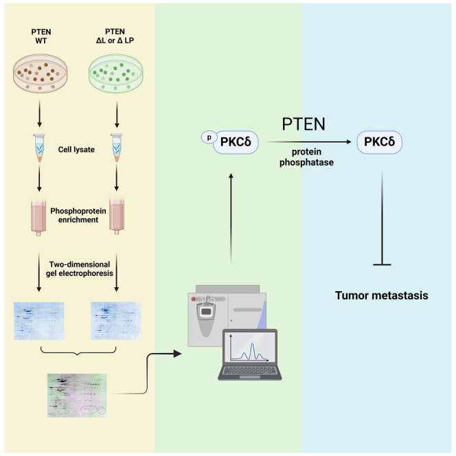
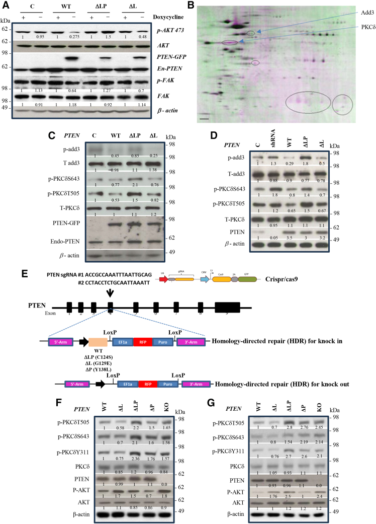
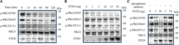
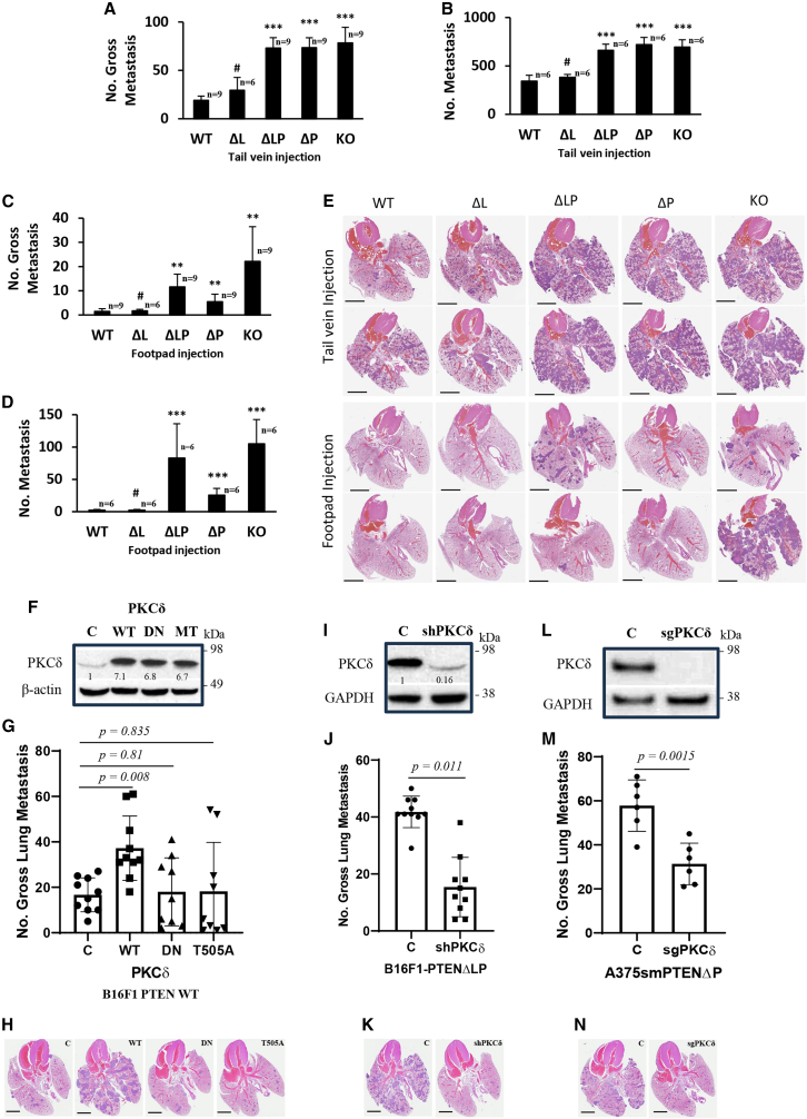
PTEN acts as a tumor suppressor through its lipid and protein phosphatase activities. We previously reported that PTEN phosphatase inhibits metastasis independent of its lipid phosphatase. To determine PTEN phosphatase downstream substrates and their role precisely in metastatic suppression, we used proteomic approaches to identify PKCδ as PTEN protein phosphatase substrates. We show that the inactivation of PTEN protein phosphatase activity causes loss of the capability to dephosphorylate PKCδ at S643, T505, and Y311, but wild-type or PTEN lipid phosphatase deficient mutants can maintain. We then established knock-in and knock-out models to confirm that PTEN protein phosphatase is required to inhibit PKCδ phosphorylation and necessary to suppress tumor metastasis. Notably, we found that PKCδ could promote metastasis of melanoma cells with wild-type PTEN. Still, the knockdown of PKCδ abrogated the metastatic potential of PTEN phosphatase-deficient melanoma cells, linking PTEN metastasis suppressor function to PTEN protein phosphatase and its substrate PKCδ.

Keywords: Proteomics; cancer.


Conflict of interest statement

The authors declare that they have no competing interests.

Figures

None
Graphical abstract
Figure 1
Figure 1
Identification of candidate substrates of PTEN protein phosphatase (A) The western blotting showed induced expression of various PTEN mutants and protein levels of p-AKT, AKT, p-FAK, and FAK in mouse melanoma B16F1 cells transfected with various PTEN mutant forms in tet-off inducible expressing vectors, which exogenous gene expression is controlled by the absence of doxycycline. C, empty vector; WT, PTEN wild type; ΔLP, PTEN phosphatase dead mutant (C124S); ΔL, PTEN lipid phosphatase deficient (G129E). The protein level of β-actin is the loading control. The expression of transfected various PTEN mutant forms was induced by taking off doxycycline for 24 h. +, with doxycycline; −, without doxycycline. The value of western blot band density was quantified by ImageJ analysis. After normalization with the value of β-actin, the fold change was presented to compare with no exogenous PTEN expression (+, with doxycycline). (B) Representative identification of different phosphoproteins in B16F1 cells with various PTEN mutants by Two-dimensional gel electrophoresis (2DE). The combination of two images from 2D gel electrophoresis of phosphoproteins purified from wildtype PTEN (green) and phosphatase dead mutant PTEN (magenta) B16F1 cells. Several phosphoprotein spots that were differentially represented are circled. The differentiated spots (phosphoproteins) were collected for mass spectrometry (MS) protein identification analysis. Using 2D gel electrophoresis and mass-spec technology, we identified 14 candidates, including AKT, IRS1, Add3, and PKCδ. The scale bar, 10 mM. (C) Western blot showed p-Add3, Add3, p-PKCδS643, p-PKCδT505, total PKCδ, and PTEN protein levels from B16F1 cells transfected with PTEN various mutant forms. C, empty vector; WT, PTEN wild type; ΔLP, PTEN phosphatase dead mutant; ΔL, PTEN lipid phosphatase deficient. The protein level of β-actin is the loading control. The value of western blot band density was quantified by ImageJ analysis. After normalization with the value of β-actin, the fold changes were presented to compare with control (C). The related protein levels were quantified with three repeated western blots (see Figure S2). (D) Western blot analysis of indicated proteins from A375sm transfectants harboring empty vector (C), PTEN wildtype (WT), and PTEN phosphatase dead mutant (ΔLP) and PTEN knockdown by shRNA. The value of western blot band density was quantified by ImageJ analysis. After normalization with the value of β-actin, the fold changes were presented to compare with control (C). The related protein levels were quantified with three repeated western blots (see Figure S3). (E) The schematic of CRISPR/Cas9 editing technology with a homology-directed repair (HDR) system for PTEN various mutant forms knock-in or knock-out specifically at PTEN exon 4 local. WT, wildtype PTEN; ΔL, PTEN lipid deficient mutant; ΔLP, PTEN phosphatase dead mutant; ΔP, PTEN protein deficient mutant. The human melanoma A375sm or A375p cells were cotransfected with the CRISPR/Cas9 expressed sgRNA for PTEN plasmid and a homology-directed repair (HDR) vector with various PTEN forms to insert the various PTEN forms into PTEN exon 4, or with a homology-directed repair (HDR) with RFP and puromycin genes into PTEN exon 4, therefore disrupted the PTEN gene. (F and G) PTEN protein phosphatase activity is required for dephosphorylating PKCδ. Western blots showed the indicated protein level from the melanoma cell lines A375sm (F) and A375p (G) with knocked-in PTEN wild type (WT), lipid phosphatase deficient mutant (ΔL), protein phosphatase deficient mutant (ΔP), PTEN phosphatase dead mutant (ΔLP), or total loss of PTEN (KO). The value of western blot band density was quantified by ImageJ analysis. After normalization with the value of β-actin, the fold changes were presented to compare with knocked-in PTEN wildtype (WT).
Figure 2
Figure 2
PTEN is a protein phosphatase for PKCδ in vitro (A) Purified PTEN proteins were used in a phosphatase assay in vitro using PKCδ as a substrate. The reaction was run for the described time interval. Changes in PKCδ phosphorylation at Y311, T505, and S643 were detected by immunoblotting. (B) Western blot showed PKCδ phosphorylation levels in a phosphatase assay in vitro using PKCδ as a substrate following the increased PTEN. The value of western blot band density was quantified by ImageJ analysis. After normalization with the value of PKCδ, the fold changes were presented to compare with 0 time point (0). (C) Western blot showed PKCδ phosphorylation levels in a phosphatase assay in vitro using PKCδ as a substrate following the increased PTEN with/without a phosphatase inhibitor. The value of western blot band density was quantified by ImageJ analysis. After normalization with the value of PKCδ, the fold change was presented to compare with 0 ng PTEN (0).
Figure 3
Figure 3
PTEN protein phosphatase activity is required to suppress metastasis in A375sm human melanoma cells (A and B) Gross pulmonary metastases (A) and micrometastases (B) from A375sm cells knock-in PTEN wild type (WT), PTEN lipid phosphatase deficient mutant (ΔL), PTEN protein phosphatase deficient mutant (ΔP), PTEN phosphatase dead mutant (ΔLP) and PTEN knockout (KO) in the experimental metastasis assay by tail vein injection. (C and D) Gross pulmonary metastases (C) and micrometastases (D) from A375sm cells knock-in PTEN wild type (WT), PTEN lipid phosphatase deficient mutant (ΔL), PTEN protein phosphatase deficient mutant (ΔP), PTEN phosphatase dead mutant (ΔLP) and PTEN knockout (KO) in the orthotopic metastasis assay by footpad transplantation. The p values were presented from an unpaired t-test analysis (two-tailed) compared with PTEN wild type (WT). #, no statistical difference; ∗p < 0.05; ∗∗p < 0.01; ∗∗∗p < 0.001. Data are represented as mean ± SEM. (E) Representative histopathology (H&E staining) of lung sections with metastases from mice bearing A375sm cells knock-in PTEN wild type (WT), PTEN lipid phosphatase deficient mutant (ΔL), PTEN protein phosphatase deficient mutant (ΔP), PTEN phosphatase dead mutant (ΔLP) and PTEN knockout (KO) in the experimental metastasis assay by tail vein injection or orthotopic metastasis assay by footpad transplantation. The scale bar = 5 mM. (F–N) PKCδ functions in PTEN-mediated metastatic regulation. (F, G, and H) Overexpression of PKCδ in B16F1 cells expressing PTEN wild type enhances metastasis in vivo by tail vein injection. Western blot analysis of PKCδ wildtype (WT), dominant negative (DN), and T505A mutant (MT) forms expressing level (F), number of metastatic nodes on the surface of the lung (G) and representative histopathology (H&E staining) of lung sections with metastases from mice (H). C, the cells with empty vector as control; WT, the cells with PKCδ wildtype expression; DN, the cells with PKCδ dominant negative mutant form expression; MT, the cells with PKCδ T505A mutant form expression. The value of western blot band density was quantified by ImageJ analysis. After normalization with the value of β-actin, the fold changes were presented to compare with control (C). Data are represented as mean ± SEM. The scale bar, 5 mM. (I–K) Knockdown of PKCδ in B16F1 cells expressing PTENΔLP inhibits metastasis in vivo. Western blot analysis (I), number of lung metastasis (J) and representative histopathology (H&E staining) of lung sections with metastases from mice (K). C, the cells with shRNA scramble control; shPCKδ, the cells with shRNA expression for PKCδ; GAPDH was used as a control. The value of western blot band density was quantified by ImageJ analysis. After normalization with the value of GAPDH, the fold changes were presented to compare with control (C). Data are represented as mean ± SEM. The scale bar, 5 mM. (L–N) Knockout of PKCδ in A375sm cells expressing PTENΔP inhibits metastasis in vivo. Western blot analysis (L), number of lung metastasis (M) and representative histopathology (H&E staining) of lung sections with metastases from mice (N). C, the cells with sgRNA scramble control; sgPCKδ, the cells with sgRNA expression for PKCδ; GAPDH was used as a control. The p values were presented from an unpaired t test analysis (two-tailed) compared with the control-empty vector (c). Data are represented as mean ± SEM. The scale bar, 5 mM.
Figure 4
Figure 4
Treatment of PKCδ inhibitors blocks the metastatic potential of melanoma (A) Pretreatment of PKCδ inhibitors reduces the metastatic potential of melanoma B16F1. DMSO, a mock control; Cal C, 100 nM of calphostin C; rottlerin, 5 μM of rottlerin; BMI, 20 nM of bisindoylmaleimide I; β inhibitor, 20 nM of PKCβ inhibitor I; Go 6976, 3 nM of PKCα inhibitor. (B) Pretreatment of PKCδ promoter PMA (100 nM of phorbol 12-myristatel-13-acetate) promotes the metastasis of melanoma B16F1 expressing PTEN. (C) Pretreatment of PKCδ inhibitors reduces the metastatic potential of melanoma B16F1 cells expressing PTENΔLP. DMSO, a mock control; Cal C, 100 nM of calphostin C; rottlerin, 5μM of rottlerin; BMI, 20 nM of bisindoylmaleimide I; β inhibitor, 20 nM of PKCβ inhibitor; Go 6976, 3 nM of PKCα inhibitor. The p values were presented from an unpaired t test analysis (two-tailed) compared with the control—DMSO (DMSO). Data are represented as mean ± SEM. (D) Western blot analysis of indicated proteins from B16F1 cells (B16) pretreated with indicated inhibitors. DMSO, as a control. (E) Western blot analysis of indicated proteins from B16F1 cells expressing PTEN (B16 PTEN) pretreated with PKC promoter PMA (100 nM of phorbol 12-myristatel-13-acetate). DMSO, as a control. (F) Western blot analysis of indicated proteins from B16F1 cells expressing PTENΔLP (B16 PTENΔLP) pretreated with indicated inhibitors. DMSO, as a control. The value of western blot band density was quantified by ImageJ analysis. After normalization with the value of GAPDH, the fold changes were presented to compare with the control (DMSO). (G) Gross pulmonary metastases from mice transplanted with B16F1 PTENΔLP cells treated with two doses of PKCδ inhibitor calphostin C. Mock, as a control; 0.7 mg/kg; 1.4 mg/kg. Data are represented as mean ± SEM. The p values were presented from an unpaired t test analysis (two-tailed) compared with the control (Mock). (H) Representative histopathology (H&E staining) of lung sections with metastases from mice bearing B16F1 PTENΔLP cells treated with two doses of PKCδ inhibitor calphostin C (0.7 mg/kg or 1.4 mg/kg) and mock control (Mock). The scale bar, 5 mM. (I) Gross pulmonary metastases from mice transplanted with B16F1 PTENΔLP cells treated with 1 mg/kg of PKCδ inhibitor KAI-9803. Mock, as a control; KAI-9803, 1 mg/kg of KAI-9803. Data are represented as mean ± SEM. The p values were presented from an unpaired t test analysis (two-tailed) compared with the control (Mock). (J) Representative histopathology (H&E staining) of lung sections with metastases from mice bearing B16F1 PTENΔLP cells treated with 1 mg/kg of PKCδ inhibitor KAI-9803 (KAI-9803) and mock control (Mock). The scale bar, 5 mM. (K and L) Representative immunohistochemistry staining with indicated PKCδ antibodies of lung sections with metastases from mice bearing B16F1 PTENΔLP cells treated with PKCδ inhibitor calphostin C (K) or KAI-9803 (L) and mock control (Mock). The scale bar, 200 μM.

Similar articles

References

    1. Megino-Luque C., Bravo-Cordero J.J. Metastasis suppressor genes and their role in the tumor microenvironment. Cancer Metastasis Rev. 2023;42:1147–1154. doi: 10.1007/s10555-023-10155-6. - DOI - PMC - PubMed
    1. Dankort D., Curley D.P., Cartlidge R.A., Nelson B., Karnezis A.N., Damsky W.E., Jr., You M.J., DePinho R.A., McMahon M., Bosenberg M. Braf(V600E) cooperates with Pten loss to induce metastatic melanoma. Nat. Genet. 2009;41:544–552. doi: 10.1038/ng.356. - DOI - PMC - PubMed
    1. Valastyan S., Weinberg R.A. Tumor metastasis: molecular insights and evolving paradigms. Cell. 2011;147:275–292. doi: 10.1016/j.cell.2011.09.024. - DOI - PMC - PubMed
    1. Yu Y., Dai M., Huang L., Chen W., Yu E., Mendoza A., Michael H., Khanna C., Bosenberg M., McMahon M., Merlino G. PTEN phosphatase inhibits metastasis by negatively regulating the Entpd5/IGF1R pathway through ATF6. iScience. 2023;26 doi: 10.1016/j.isci.2023.106070. - DOI - PMC - PubMed
    1. Liu A., Zhu Y., Chen W., Merlino G., Yu Y. PTEN Dual Lipid- and Protein-Phosphatase Function in Tumor Progression. Cancers (Basel) 2022;14 doi: 10.3390/cancers14153666. - DOI - PMC - PubMed

LinkOut - more resources