Review
. 2025 Jun 20;10(2):e24.00249.
doi: 10.2106/JBJS.OA.24.00249. eCollection 2025 Apr-Jun.

Long-Segment Ventral Spinal Epidural Abscesses Caused by Mycobacterium Tuberculosis: A Report of 2 Cases and Review

Affiliations
Review

Long-Segment Ventral Spinal Epidural Abscesses Caused by Mycobacterium Tuberculosis: A Report of 2 Cases and Review

Shiyong Le et al. JB JS Open Access. .

Abstract

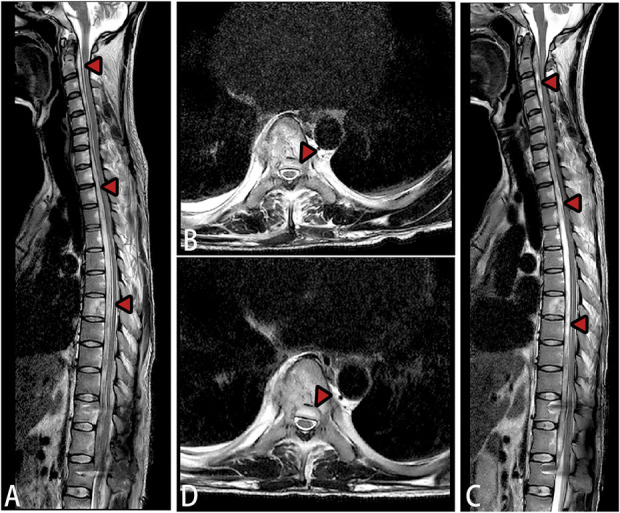
» Abscesses caused by spinal tuberculosis typically occur in the anterior and lateral regions of the vertebral bodies, often extending to the paraspinal muscles as cold abscesses. Although intraspinal abscesses due to spinal tuberculosis are very common, cases with longitudinally extensive abscesses in the ventral epidural space of the spinal cord are not well-documented. The disease is characterized by insidious onset, rapid progression, and a high risk of irreversible neurological damage, highlighting the necessity for early diagnosis and prompt treatment. » This review presents 2 rare cases of long-segment tuberculous spinal epidural abscesses, with both patients achieving significant symptom improvement after precise surgical intervention and standardized antituberculosis therapy. A long-segment tuberculous spinal epidural abscess (SEA) is an extremely rare complication that may cause severe neurological impairment, and magnetic resonance imaging revealed extensive spinal epidural abscesses. Clinical manifestations, such as neck stiffness, can easily be mistaken for tuberculous encephalopathy; therefore, contrast-enhanced MRI is recommended to differentiate a SEA from tuberculous encephalopathy. » Local decompression with catheter drainage can help preserve neurological function, establish a definitive etiological diagnosis, and guide subsequent treatment. Early, adequate, and comprehensive antituberculosis therapy is crucial for successful management.


Conflict of interest statement

All patients consented to their data being used anonymously. The Clinical Ethics Committee of our hospital approved this retrospective study (2024-ER-076). The author(s) declared no potential conflicts of interest with respect to the research, authorship, and/or publication of this article. Disclosure: The Disclosure of Potential Conflicts of Interest forms are provided with the online version of the article (http://links.lww.com/JBJSOA/A818).

Figures

Fig. 1
Fig. 1
Imaging findings of long-segment tuberculous epidural abscesses in the cervical spine. Fig. 1-A Sagittal T2-weighted image shows abnormal high signal intensity (red arrows) within the ventral epidural space at the C2-T1 levels, with evident spinal cord compression. Additional abnormal high signal intensity (blue arrows) is observed at the posterior margins of the C5 and C7 vertebrae. Fig. 1-B Sagittal fat-suppressed sequence demonstrates uneven, strip-shaped high signal intensity within the C2-T1 spinal canal, with a low-signal margin around the lesion. Fig. 1-C Axial T1-weighted image displays abnormal high signal intensity within the ventral epidural space at the C2 level, with significant spinal cord compression and a surrounding ring of high signal intensity. Fig. 1-D Cervical and thoracic CT image illustrates a circular, low-density lesion posterior to the C4 vertebra. No significant bone destruction was observed in the cervical vertebrae. Fig. 1-E Chest CT image shows patchy high-density areas in the upper lobe of the right lung. CT = computed tomography.
Fig. 2
Fig. 2
Postoperative imaging findings of long-segment tuberculous epidural abscesses in the cervical spine. Figs. 2-A, 2-B, and 2-C Images obtained on postoperative day 3; (Figs. 2-D and 2-E) images obtained at 1 week after surgery. Figs. 2-A and 2-D Sagittal T2-weighted images demonstrate a significant reduction in high signal intensity within the ventral spinal epidural space at the C2-T1 levels, in comparison with preoperative images. Fig. 2-B Cervical CT image illustrates the placement of a CVC within the epidural abscess cavity during the operation. Figs. 2-C and 2-E Axial T2-weighted images show a reduction in abnormal high signal intensity within the ventral epidural space at the C2 level, and spinal cord compression has significantly improved. CT = computed tomography and CVC = central venous catheter.
Fig. 3
Fig. 3
Imaging findings of long-segment tuberculous epidural abscesses in the cervical and thoracic regions. Fig. 3-A Sagittal T2-weighted image displays a strip-shaped area of high signal intensity within the ventral epidural space at the C1-T12 levels, with evident spinal cord compression. Fig. 3-B Sagittal fat-suppressed sequence demonstrates uneven, strip-shaped high signal intensity within the C1-T12 spinal canal with a low-signal margin around the lesion. Fig. 3-C Cervical and thoracic CT image reveals irregular osteolytic destruction of the T7 and T11 vertebrae. Fig. 3-D Axial T2-weighted image shows agglomerate or flake-like high signal intensity (blue arrows) at the right margin of the T7 vertebral body, abnormal high signal intensity (red arrows) within the ventral epidural space at the T7 level, indicating severe spinal cord compression, and a surrounding ring of low signal intensity. Fig. 3-E Chest CT image shows multiple areas of abnormal density in both lungs. CT = computed tomography.
Fig. 4
Fig. 4
Postoperative imaging findings of long-segment tuberculous epidural abscesses in the cervical and thoracic spine. Figs. 4-A and 4-B Images obtained on postoperative day 3; (Figs. 4-C and 4-D) images obtained on postoperative day 10. Figs. 4-A and 4-C Sagittal T2-weighted images indicate a reduction in abnormal high signal intensity within the spinal epidural area at C1-T12 levels, in comparison with preoperative images. Figs. 4-B and 4-D Axial T2-weighted images show a reduction in high signal intensity within the ventral epidural space at the C7 level, and the spinal cord compression was relieved postoperatively.
Fig. 5
Fig. 5
Imaging findings of long-segment spinal epidural tuberculous abscesses in the cervical and thoracic regions. Figs. 5-A, 5-B, and 5-C Images obtained on 6 months postoperatively; (Figs. 5-D, 5-E, and 5-F) images obtained on 10 months postoperatively. Figs. 5-A and 5-B Sagittal T2-weighted images at 4 months show a reduction in abnormal high signal intensity within the spinal epidural area at C1-T12 levels, in comparison with preoperative images. Figs. 5-D and 5-E Sagittal T2-weighted images at 8 months demonstrate a further decrease in abnormal high signal intensity within the same region, compared with the 4-month findings. Figs. 5-C and 5-F Axial T2-weighted images reveal a significant reduction in ventral epidural high signal intensity within the spinal canal, with substantial alleviation of spinal cord compression, compared with preoperative findings.

Similar articles

References

    1. Hasan GA, Kani SM, Alqatub A. Tuberculous lumbar spinal epidural abscess in a young adult (case report). SICOT J. 2018;4:5. - PMC - PubMed
    1. Tetsuka S, Suzuki T, Ogawa T, Hashimoto R, Kato H. Spinal epidural abscess: a review highlighting early diagnosis and management. JMA J. 2020;3(1):29-40. - PMC - PubMed
    1. Darouiche RO. Spinal epidural abscess. N Engl J Med. 2006;355(19):2012-20. - PubMed
    1. Reihsaus E, Waldbaur H, Seeling W. Spinal epidural abscess: a meta-analysis of 915 patients. Neurosurg Rev. 2000;23(4):175-205. - PubMed
    1. Manoharan SR, Leitao J, Emberton P, Quraishi NA. A large tuberculosis abscess causing spinal cord compression of the cervico-thoracic region in a young child. Eur Spine J. 2013;22(7):1459-63. - PMC - PubMed