. 2025 May 12;4(3):100251.
doi: 10.1016/j.cellin.2025.100251. eCollection 2025 Jun.

Rapid and direct discovery of functional tumor specific neoantigens by high resolution mass spectrometry and novel algorithm prediction

Affiliations

Rapid and direct discovery of functional tumor specific neoantigens by high resolution mass spectrometry and novel algorithm prediction

Huajian Tian et al. Cell Insight. .

Abstract

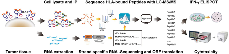
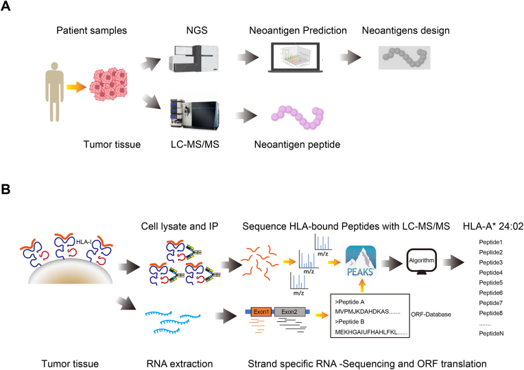
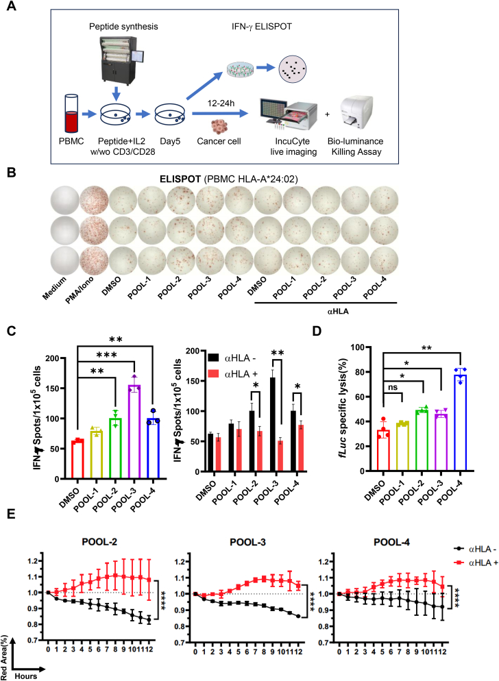
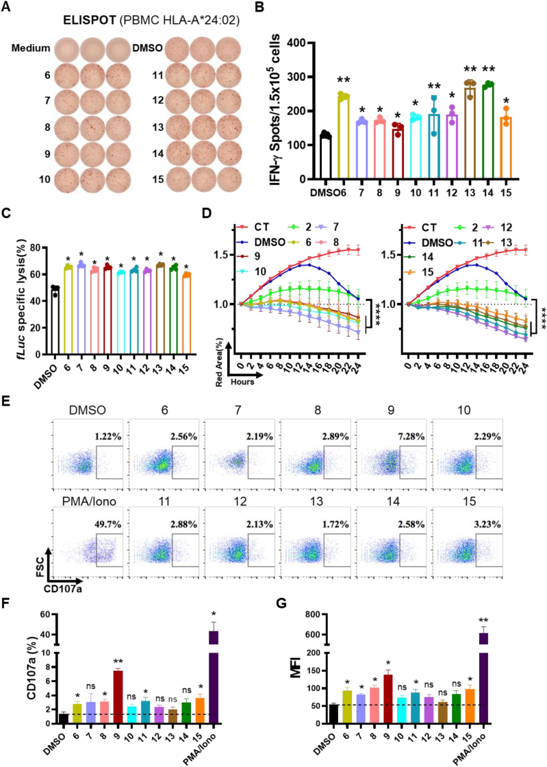
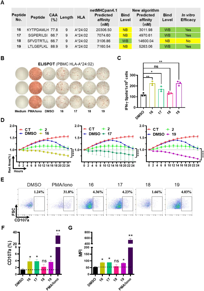
While immune cell therapies have transformed cancer treatment, achieving comparable success in solid tumors remains a significant challenge compared to hematologic malignancies like non-Hodgkin lymphoma (NHL) and multiple myeloma (MM). Over the past four decades, various immunotherapeutic strategies, including tumor vaccines, tumor-infiltrating lymphocyte (TIL) therapies, and T cell receptor (TCR) therapies, have demonstrated clinical efficacy in select solid tumors, suggesting potential advantages over CAR-T and CAR-NK cell therapies in specific contexts. The dynamic nature of the cancer-immunity cycle, characterized by the continuous evolution of tumor-specific neoantigens, enables tumors to evade immune surveillance. This highlights the urgent need for rapid and accurate identification of functional tumor neoantigens to inform the design of personalized tumor vaccines. These vaccines can be based on mRNA, dendritic cells (DCs), or synthetic peptides. In this study, we established a novel platform integrating immunoprecipitation-mass spectrometry (IP-MS) for efficient and direct identification of tumor-specific neoantigen peptides. By combining this approach with our proprietary AI-based prediction algorithm and high-throughput in vitro functional validation, we can generate patient-specific neoantigen candidates within six weeks, accelerating personalized tumor vaccine development.

Keywords: Mass spectrometry; Neoantigen; New algorithm; Tumor vaccine.


Conflict of interest statement

The authors declare that there are no conflicts of interest related to this manuscript.

Figures

Image 1
Graphical abstract
Fig. 1
Fig. 1
Comparison between traditional neoantigen identification methods and the technical approach in this study. (A) Current methods for discovering tumor neoantigens. (B) Schematic diagram of technical methods used in this research.
Fig. 2
Fig. 2
Immunoprecipitation pMHC complex of Xenograft tumor tissue. (A) Identify W6/32 antibody purity by SDS-PAGE with non-reducing or reducing loading buffer. N, non-reducing; R, reducing; FL, Full Length; HC, heavy chain; LC, light chain. (B) Quality Controlling the binding affinity of W6/32 with MHC complex using T2 cell line or negative controlled CHO-S cell line. (C) Immunoprecipitant the refold pMHC complex with W6/32. All protein samples were loaded with reduced buffer. The result was presented by Coomassie staining, right panel. HC, heavy chain; LC, light chain. Schematic of pMHC refold in biochemical condition, left panel. (D) Immunoprecipitant the MIA PaCa-2 Xenograft tumor lysate with W6/32. The result was shown by Western Blot.
Fig. 3
Fig. 3
The analysis of MS result in PEAKS Online. (A) Overview of data acquisition strategies, analysis approaches, and core algorithms of PEAKS Online. (B) An example of integrated workflow for DDA data analysis. DDA:Data-dependent acquisition. (C) An example of ORF-DB calibrated the error of de novo sequencing. The de novo sequencing mistake was marked in red while the accurate peptide sequence in ORF-DB was marked in green. ORF-DB: a peptide database of open reading frame (ORF) translations based on RNA-seq data. (D) Peptides length distribution and Sequence logos of IP(UniProt) and IP(ORF-DB). (E) The peptides overlapping of IP(UniProt) vs. IP(ORF-DB), IP(UniProt) vs. IP(de novo) and IP(ORF-DB) vs. IP(de novo); CT(UniProt)/CT(ORF-DB)/CT(de novo) was a control group to filter the non-specific binding peptides; IP: immunoprecipitated with W6/32; CT: immunoprecipitated with mIgG.
Fig. 4
Fig. 4
Neoantigens POOLs activated the Immune responses and cytotoxicity. (A) Schematic diagram of in vitro functional assays of neoantigens, HLA-A∗24:02 PBMCs were used for IFN-γ ELISPOT and cytotoxic assays. w/wo = with or without. (B) PBMCs stimulated by neoantigen pools were used for IFN-γ ELISPOT detection; anti-HLA(W6/32) was used to block T cell recognition; (C) Number of ELISPOTs was calculated. Student two-tailed test. Data represent mean ± SEM from 3 independent replicates (∗P < 0.05, ∗∗P < 0.01, ∗∗∗P < 0.001). αHLA: anti-HLA antibody(W6/32). (D) Detection of neoantigen pools stimulated cytotoxic effects on target cells by using bio-luminance assay. Student two-tailed test. Data represent mean ± SEM from 3 independent replicates (∗P < 0.05, ∗∗P < 0.01; ns, not significant). fLuc: Firefly luciferase (E) IncuCyte analysis the inhibitory effects of PBMCs against target cells. Anti-HLA(W6/32) was used to block T cell recognition. Student two-tailed test. Data represent mean ± SEM from 3 independent replicates (∗∗∗∗P < 0.0001). αHLA: anti-HLA antibody(W6/32). Red area (%) indicates the live cells percentage.
Fig. 5
Fig. 5
Screening neoantigens by measuring immune responses and tumor killing effects. (A) PBMCs induced by No.1-15 peptides were used for IFN-γ ELISPOT detection. (B) Number of ELISPOTs was calculated. Student two-tailed test. Data represent mean ± SEM from 3 independent replicates (∗P < 0.05, ∗∗P < 0.01). (C) Detection of cytotoxic effects on target cells induced by No.1-15 peptides via using bio-luminance assay. Student two-tailed test. Data represent mean ± SEM from 3 independent replicates (∗P < 0.05). fLuc: Firefly luciferase (D) IncuCyte analysis of the inhibitory effects of PBMCs against target cells. Student two-tailed test. Data represent mean ± SEM from 3 independent replicates (∗∗∗∗P < 0.0001); the CT group was only without PBMCs. Peptide No.2 was a negative control. The red area (%) indicated live cells percentage. (E–G) CD8+T cells are activated by neoantigens in vitro. CD8+T cells were stimulated with peptides or control (DMSO) and the efficacy was evaluated by T cell degranulation. CD107a positive percentage and mean fluorescent intensity were summarized. Data represent mean ± SEM from 3 independent replicates (∗P < 0.05, ∗∗P < 0.01; ns, not significant). PMA-Iono: PMA-Ionomycin; MFI: Mean fluorescence intensity.
Fig. 6
Fig. 6
Measurement of immune responses and tumor killing effects induced byNo.16-19peptides. (A) Comparison of the differences in affinity prediction results for peptides between the new algorithm and netMHCpan4.1. (B) PBMCs induced by No.16-19 peptides were used for IFN-γ ELISPOT assay. PMA-Iono: PMA-Ionomycin; (C) Number of ELISPOTs was calculated. Student two-tailed test. Data represent mean ± SEM from 3 independent replicates (∗P < 0.05, ∗∗P < 0.01; ns, not significant). (D) IncuCyte analysis of the inhibitory effects of PBMCs against target cells. Student two-tailed test. Data represent mean ± SEM from 3 independent replicates (∗∗∗∗P < 0.0001); the CT group was only MIA PaCa-2 without PBMCs. Peptide No.2 was a negative control. (E)–(G) CD8+T cells activated by neoantigens in vitro. CD8+T cells were stimulated with peptides or control (DMSO) and the efficacy was evaluated by T cell degranulation. CD107a positive percentage and mean fluorescent intensity were summarized. MFI: Mean fluorescence intensity. Data represent mean ± SEM from 3 independent replicates (∗P < 0.05, ∗∗P < 0.01; ns, not significant).
Fig. 7
Fig. 7
No.19 peptide loaded pMHC triggers T cell activation via the specific TCR interaction. (A) Schematic of scTCR Jurkat-NFAT-GFP activated upon binding with T2 loaded with No.19 peptide. (B) 10 μg/mL FITC conjugated No.19 Peptide was loaded onto empty T2 cells for 2 h at 37 °C, T2 cells were then rinsed twice and checked via FACS and controlled by T2 cells without any peptide loading. (C) Jurkat NFAT-GFP were stably transduced with the lentivirus containing scTCR-V5-BBZ. The positive transduced cells were polyclonal sorted by Flow cytometer. The sorted cells were maintained and the scTCR expression was checked before co-culture. (D) 24 h post of the co-culture of No.19 peptide loaded T2 cells and the Jurkat NFAT-GFP cells. High-content imaging analysis at 10 × magnification was applied to evaluate the NFAT-GFP reporter signals (Left). Besides, the Mean fluorescence intensity (MFI) of GFP (right) was quantified by the imaging software. Data represent mean ± SEM from 3 independent replicates (∗P < 0.05). (E)–(H) After High-content imaging analysis, the co-culture cells were harvested and stained with viability dye, CD3, and CD69 antibodies. Flow cytometry was applied to compare the NFAT-GFP reporter signals ((E)–(F)) as well as the CD69 expression stands for the T cell activation ((G)-(H)). Data represent mean ± SEM from 3 independent replicates (∗∗P < 0.01, ∗∗∗P < 0.001).

Similar articles

References

    1. Abdollahi P., Norseth H.M., Schjesvold F. Advances and challenges in anti-cancer vaccines for multiple myeloma. Frontiers in Immunology. 2024;15 - PMC - PubMed
    1. Borno H., Moses K.A., Monk P., Fitch K., Harmon M., Flanders S., Mckay R.R. Sipuleucel-T (sip-T) and oral androgen axis inhibitors in black men being treated for metastatic castration-resistance prostate cancer (mCRPC): Observations from the Medicare Fee for Service (FFS) population. Journal of Clinical Oncology. 2020;38
    1. Buonaguro L., Tagliamonte M. Peptide-based vaccine for cancer therapies. Frontiers in Immunology. 2023;14 - PMC - PubMed
    1. Chervin A.S., et al. 2021. Bispecific binding molecules. WO2021163366A1.
    1. Chervin A.S., Stone J.D., Konieczna I., Calabrese K.M., Wang N., Haribhai D., Dong F., White M.K., Rodriguez L.E., Bukofzer G.T., Ellis P.A., Cosgrove C., Hecquet C., Clarin J.D., Palma J.P., Reilly E.B. ABBV-184: A novel survivin-specific TCR/CD3 bispecific T-cell engager is active against both solid tumor and hematologic malignancies. Molecular Cancer Therapeutics. 2023;22:903–912. - PubMed

LinkOut - more resources