

Ultrasound in intensive care unit patients: applications, observations, and comparison of two established ultrasound methods
- PMID: 40547216
- PMCID: PMC12182898
- DOI: 10.15557/jou.2025.0016
Ultrasound in intensive care unit patients: applications, observations, and comparison of two established ultrasound methods
Abstract
Aim: To evaluate the benefit of abdominal ultrasonography performed routinely and thus independently of symptomatology in patients in the intensive care unit, and to assess the value of a portable ultrasound device. Diagnostic yield and documented results with clinical consequences were considered and compared with findings obtained using a high-end ultrasound device.
Material and methods: A total of 120 patients of an internal medicine intensive care unit were included over 12 months. The investigator had limited experience in sonography (approximately 300 abdominal sonographies performed). The abdomen and basal portions of the thorax were examined.
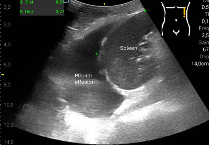
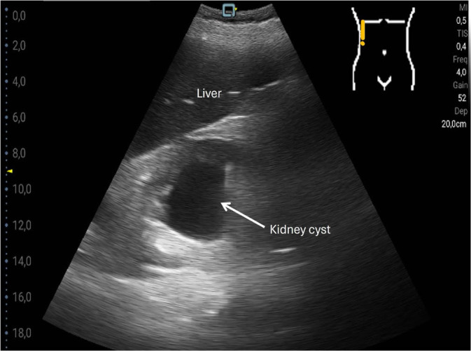
Results: The most common pathological findings were renal cysts in 34/120 (28.3%), left-sided or right-sided pleural effusions in 33/120 (27.5%) and 29/120 (24.2%) patients, respectively, dilatation of the vena cava in 24/120 (20.0%), and urinary retention in 14/120 (11.7%) patients. In 13/120 (10.8%) patients, the sonographic examination resulted in a diagnostic consequence, while in 38/120 (31.7%) patients in a therapeutic consequence. Among the false-negative findings using the hand-held ultrasound device, no finding was of therapeutic relevance. Four findings that were missed by the hand-held ultrasound device were diagnostically significant: two lesions of the kidney, one lesion of the liver, and one case of urinary stasis kidney.
Conclusions: With the hand-held ultrasound device, only 33 of 52 focal lesions were detected. Thus, a high-end ultrasound device cannot be replaced by a hand-held ultrasound device for this purpose, but certain clinical questions can be answered reliably with a hand-held ultrasound device (such as the presence of a puncture-worthy pleural effusion in patients with dyspnea, or verification of the volume status based on the diameter of the vena cava).
Keywords: abdomen; hand-held ultrasound device; high-end ultrasound; intensive care unit; liver lesion.
© 2025 André Ignee et al., published by Sciendo.
Conflict of interest statement
Conflict of interest The authors do not report any financial or personal connections with other persons or organizations which might negatively affect the contents of this publication and/or claim authorship rights to this publication.
Figures
Similar articles
-
Signs and symptoms to determine if a patient presenting in primary care or hospital outpatient settings has COVID-19.Cochrane Database Syst Rev. 2022 May 20;5(5):CD013665. doi: 10.1002/14651858.CD013665.pub3. Cochrane Database Syst Rev. 2022. PMID: 35593186 Free PMC article.
-
Intravenous magnesium sulphate and sotalol for prevention of atrial fibrillation after coronary artery bypass surgery: a systematic review and economic evaluation.Health Technol Assess. 2008 Jun;12(28):iii-iv, ix-95. doi: 10.3310/hta12280. Health Technol Assess. 2008. PMID: 18547499
-
Contrast-enhanced ultrasound for the diagnosis of hepatocellular carcinoma in adults with chronic liver disease.Cochrane Database Syst Rev. 2022 Sep 2;9(9):CD013483. doi: 10.1002/14651858.CD013483.pub2. Cochrane Database Syst Rev. 2022. PMID: 36053210 Free PMC article.
-
Magnetic resonance imaging for the diagnosis of hepatocellular carcinoma in adults with chronic liver disease.Cochrane Database Syst Rev. 2022 May 6;5(5):CD014798. doi: 10.1002/14651858.CD014798.pub2. Cochrane Database Syst Rev. 2022. PMID: 35521901 Free PMC article.
-
Comparison of the effectiveness of inhaler devices in asthma and chronic obstructive airways disease: a systematic review of the literature.Health Technol Assess. 2001;5(26):1-149. doi: 10.3310/hta5260. Health Technol Assess. 2001. PMID: 11701099
References
-
- Dietrich CF, Nolsøe CP, Barr RG, Berzigotti A, Burns PN, Cantisani V. et al. Guidelines and Good Clinical Practice Recommendations for Contrast-Enhanced Ultrasound (CEUS) in the Liver-Update 2020 WFUMB in Cooperation with EFSUMB, AFSUMB, AIUM, and FLAUS. Ultrasound Med Biol. 2020;46:2579–2604. doi: 10.1016/j.ultrasmedbio.2020.04.030. - DOI - PubMed
-
- Sidhu PS, Cantisani V, Dietrich CF, Gilja OH, Saftoiu A, Bartels E. et al. The EFSUMB Guidelines and Recommendations for the Clinical Practice of Contrast-Enhanced Ultrasound (CEUS) in Non-Hepatic Applications: Update 2017 (Long Version) Ultraschall Med. 2018;39:e2–e44. doi: 10.1055/a-0586-1107. English. - DOI - PubMed
LinkOut - more resources
Full Text Sources