. 2025 Jun 10:52:271-286.
doi: 10.1016/j.bioactmat.2025.06.001. eCollection 2025 Oct.

One-step strategy for fabricating icariin-encapsulated biomimetic Scaffold: Orchestrating immune, angiogenic, and osteogenic cascade for enhanced bone regeneration

Affiliations

One-step strategy for fabricating icariin-encapsulated biomimetic Scaffold: Orchestrating immune, angiogenic, and osteogenic cascade for enhanced bone regeneration

Fengxin Zhao et al. Bioact Mater. .

Abstract

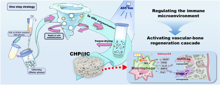
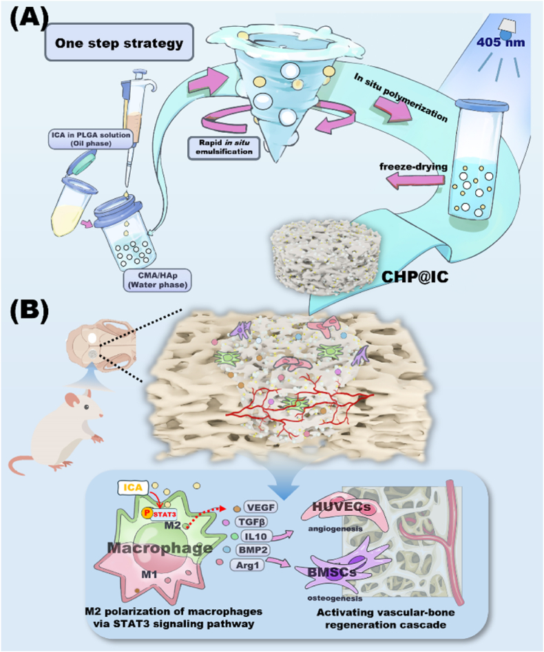
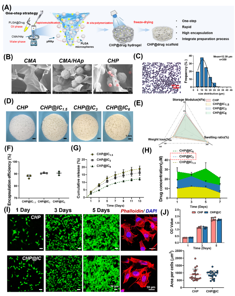
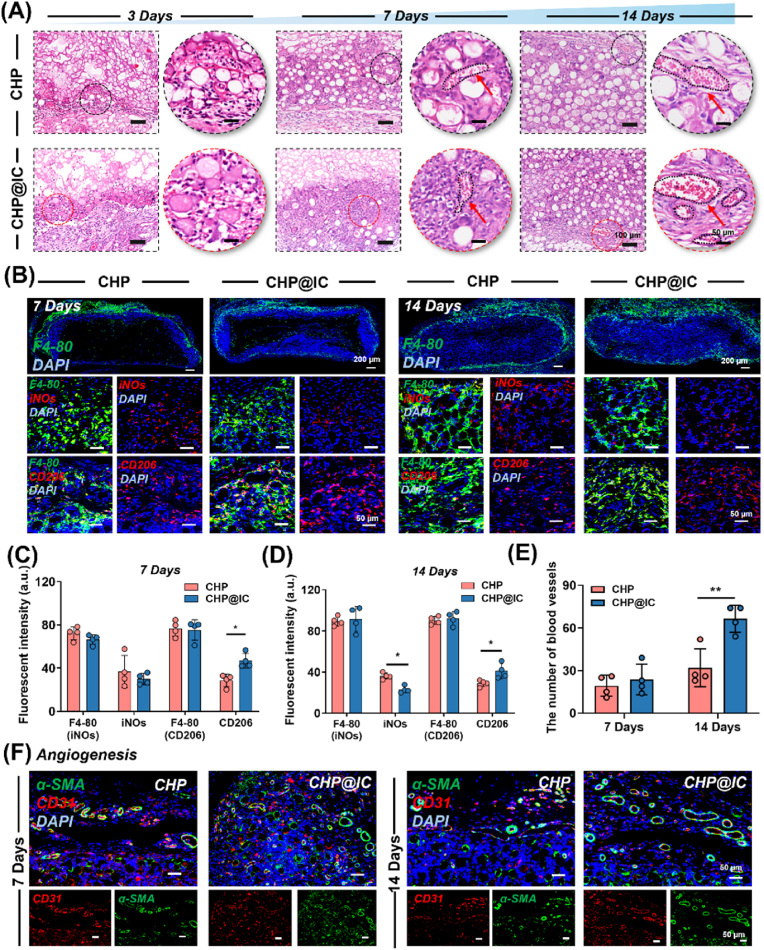
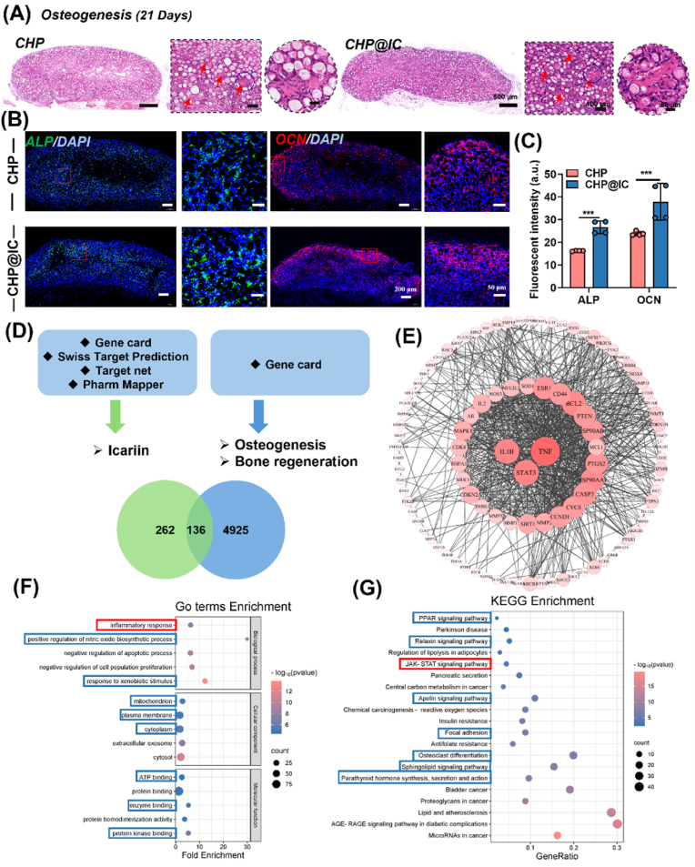
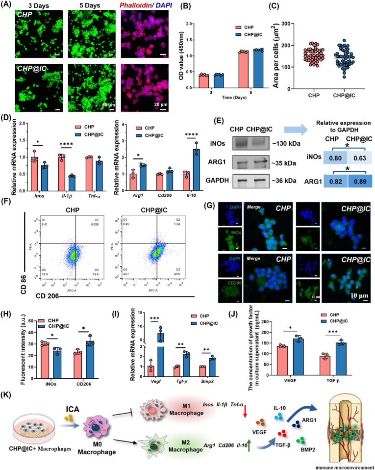
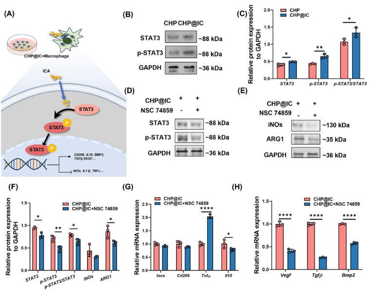
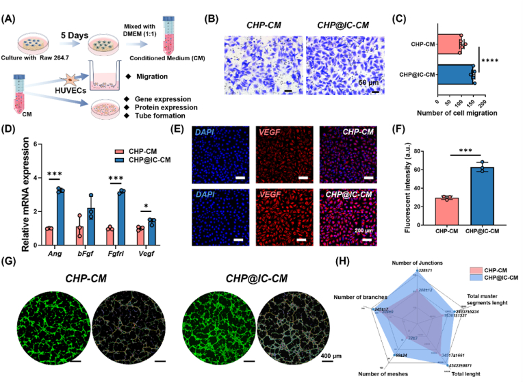
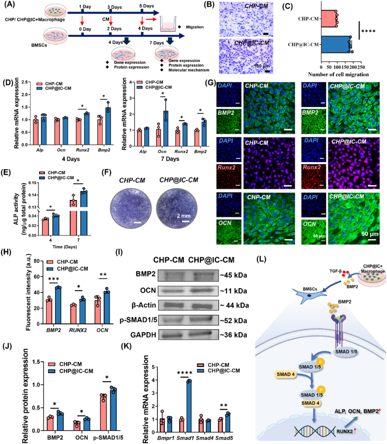
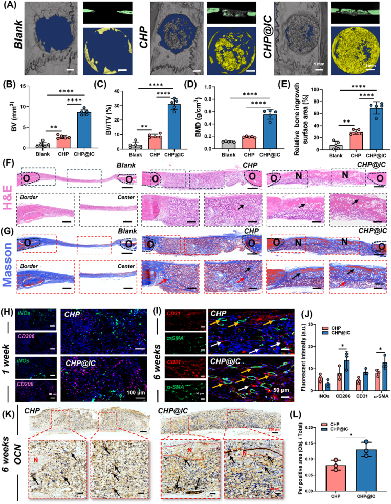
The repair of bone defects relies on the intricate coordination of inflammation, angiogenesis, and osteogenesis. However, scaffolds capable of integrating osteo-immunomodulation and vascular-bone coupling to cascade-activate these processes remain a challenge. Here, a biomimetic scaffold (CHP@IC) with in situ PLGA@icariin (PLGA@IC) microspheres encapsulation was successfully fabricated using a one-step emulsification and polymerization strategy. This approach not only simplifies the fabrication process but also ensures high encapsulation efficiency and sustained release of IC through PLGA@IC microspheres. The findings from subcutaneous implantation, network pharmacology-predicted molecular targets, and in vitro studies collectively reveal that the CHP@IC-induced M2 polarization of macrophages via STAT3 signaling pathway triggers the sequential activation of inflammation, angiogenesis, and osteogenesis to enhance bone regeneration. The CHP@IC scaffold exhibited a significant osteogenic advantage in cranial defect repair, yielding new bone volumes approximately 3-fold and 10-fold greater than those in the CHP group and blank control group, respectively. This study not only elucidates the mechanism of IC in promoting regeneration of bone but also provides a novel method for designing scaffolds aimed at the efficient repair of bone defects.

Keywords: Biomimetic scaffold; Bone regeneration; Cascade activation; Icariin delivery; One-step strategy.


Conflict of interest statement

Kai Zhang is an editorial board member for Bioactive Materials and was not involved in the editorial review or the decision to publish this article. All authors declare that there are no competing interests.

Figures

Image 1
Graphical abstract
Scheme 1
Scheme 1
(A) One-step strategy prepared CHP@IC scaffold containing PLGA@IC microspheres. (B) CHP@IC activates immune-mediated vascular-bone regeneration cascade via STAT3 signaling pathway.
Fig. 1
Fig. 1
Preparation and characterization of CHP and CHP@IC scaffolds. (A) Design conception of the CHP@Drug scaffold. (B) Network structure of scaffolds and PLGA microsphere morphology (pink color) in biomimetic scaffolds from SEM images. (C) The gross view and particle size distribution of the in situ prepared PLGA microspheres (The transparent white balls are PLGA microspheres, and the opaque black balls are HAp in CHP). (D and E) The gross view and physicochemical properties of CHP@IC scaffolds. (F–H) The encapsulation efficiency, the cumulative release and the real-time concentrations of ICA in CHP@IC scaffolds. (I and J) The FDA/PI staining, phalloidin staining, CCK8 result and cell spread area of BMSCs cultured on the surface of CHP and CHP@IC scaffolds.
Fig. 2
Fig. 2
Scaffold-induced biological responses at 3, 7, 14 days. (A) The H&E staining for subcutaneous implantation of CHP and CHP@IC scaffolds at 3, 7, 14 days (red arrow: blood vessels). (B–D) The IF staining and quantitative analysis of F4-80, iNOs and CD206 at 7, 14 days (n = 4). (E) The number of blood vessels per scaffold (n = 4). (F) The IF staining of and CD31 and α-SMA at 7, 14 days (n = 4).
Fig. 3
Fig. 3
Scaffold-induced biological responses at 21 Days and network pharmacology analysis (A) The H&E staining for subcutaneous implantation of CHP and CHP@IC scaffolds at 21 days (Red arrow: bone-like matrix). (B) The IF staining and quantitative analysis of ALP and OCN in CHP and CHP@IC scaffolds at 21 days (n = 4). (D) The target of the ICA, the bone regeneration related gene and the intersecting genes of ICA and bone regeneration. (E) The PPI network of intersecting genes. (F) The GO enrichment analysis of intersecting genes. (G) The KEGG cluster enrichment analysis of intersecting genes.
Fig. 4
Fig. 4
CHP@IC directs macrophage M2 polarization to regulate the immune microenvironment. (A–C) The FDA/PI staining, phalloidin staining, CCK8 result, and cell spread area of Raw264.7 cultured on the surface of CHP and CHP@IC scaffolds. (D) The RT-qPCR results of macrophage phenotypic transition at 5 days. (E)The WB results and the relative protein expression of iNOs, and ARG1 at 5 days. (F) The FC result of CD86 and CD206 at 5 days. (G) The IF staining of iNOs and CD206 at 5 days. (H) Quantitative analysis of iNOs, and CD206 fluorescent intensity (n = 3). (I) The RT-qPCR results of the cytokine in immune microenvironment at 5 days. (J) ELISA results of the cytokine at 5 days. (I) Schematic representation of the CHP@IC-mediated shift in the immune microenvironment.
Fig. 5
Fig. 5
CHP@IC activates the STAT3 signaling pathway. (A) Schematic representation of CHP@IC activation of the STAT3 signaling pathway. (B–C) The WB results and the relative protein expression of STAT3, p-STAT3 and p-STAT3/STAT3 treated with CHP@IC scaffold. (D) The WB results of STAT3, p-STAT3 and p-STAT3/STAT3 treated with/without NSC 74859. (E) The WB results of iNOs and ARG1 treated with/without NSC 74859. (F)The quantitative analysis of protein expression with/without NSC 74859. (G–H) The RT-qPCR results of macrophages treated with/without NSC 74859.
Fig. 6
Fig. 6
CHP@IC mediated macrophages to improve the angiogenesis of HUVECs. (A) The illustration of experimental design. (B–C) Trans-well assay images and the quantitative analysis of migrated HUVECs treated with CHP-CM and CHP@IC-CM (n = 5). (D) The RT-qPCR results of angiogenesis (n = 3). (E–F) The IF staining and quantitative analysis of VEGF treated with CHP-CM and CHP@IC-CM (n = 3). (G–H) The tube forming image of HUVECs and quantitative analysis(n = 3).
Fig. 7
Fig. 7
CHP@IC mediated macrophages to promote osteogenic differentiation of BMSCs (A) The illustration of experimental design. (B–C) Trans-well assay images and the quantitative analysis of migrated BMSCs treated with CHP-CM and CHP@IC-CM (n = 5). (D) The RT-qPCR results of osteogenic differentiation (n = 3). (E–F) The ALP staining and quantitative analysis treated with CHP-CM and CHP@IC-CM (n = 3). (G–H) The IF staining and quantitative analysis treated with CHP-CM and CHP@IC-CM (n = 3). (I–J) The WB results and the relative protein expression of BMP2, OCN and p-SAMD1/5 treated with CHP@IC scaffold (n = 3). (K) The RT-qPCR results about the mechanisms of osteogenic differentiation (n = 3). (L) Schematic representation about the mechanism of osteogenic differentiation treated with CHP@IC scaffold.
Fig. 8
Fig. 8
CHP@IC accelerated bone defect repair. (A) Micro-CT analysis of bone defects after 6 weeks. (B–E) Newly formed bone volume (BV), bone mineral density (BMD),bone volume fraction (BV/TV), and relative bone ingrowth surface area in the cranial defects after 6 weeks (n = 5). (F–G) H&E and Masson staining of the defect sites after 6 weeks. (O: old bone, N: new bone, black arrow: new bone, red arrow: immature bone matrix). (H) The IF staining of iNOs and CD206 in the defect sites after 1 week (n = 3) (I)The IF staining of CD31 and α-SMA in the defect sites after 6 weeks (white arrow: immature vessel, yellow arrow: mature vessel) (n = 3). (J) the quantitative analysis of IF staining. (K–L) The IHC staining and quantitative analysis of OCN in the defect sites after 6 weeks (black arrow: OCN positive area, N: new bone) (n = 3).

Similar articles

References

    1. Ho-Shui-Ling A., Bolander J., Rustom L.E., Johnson A.W., Luyten F.P., Picart C. Bone regeneration strategies: engineered scaffolds, bioactive molecules and stem cells current stage and future perspectives. Biomaterials. 2018;180:143–162. doi: 10.1016/j.biomaterials.2018.07.017. - DOI - PMC - PubMed
    1. Tang D., Tare R.S., Yang L.-Y., Williams D.F., Ou K.-L., Oreffo R.O.C. Biofabrication of bone tissue: approaches, challenges and translation for bone regeneration. Biomaterials. 2016;83:363–382. doi: 10.1016/j.biomaterials.2016.01.024. - DOI - PubMed
    1. Salhotra A., Shah H.N., Levi B., Longaker M.T. Mechanisms of bone development and repair. Nat. Rev. Mol. Cell Biol. 2020;21:696–711. doi: 10.1038/s41580-020-00279-w. - DOI - PMC - PubMed
    1. Wang W., Yeung K.W.K. Bone grafts and biomaterials substitutes for bone defect repair: a review. Bioact. Mater. 2017;2(4):224–247. doi: 10.1016/j.bioactmat.2017.05.007. - DOI - PMC - PubMed
    1. Xue X., Hu Y., Deng Y., Su J. Recent advances in design of functional biocompatible hydrogels for bone tissue engineering. Adv. Funct. Mater. 2021;31(19) doi: 10.1002/adfm.202009432. - DOI

LinkOut - more resources