Misdiagnosis of sacroiliac joint gout as ankylosing spondylitis: Solving the diagnostic dilemma with dual-energy computed tomography
- PMID: 40547413
- PMCID: PMC12181733
- DOI: 10.1177/2050313X251351769
Misdiagnosis of sacroiliac joint gout as ankylosing spondylitis: Solving the diagnostic dilemma with dual-energy computed tomography
Abstract
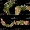
Gout rarely affects the axial joints, and sacroiliac joint involvement is exceptionally uncommon.1 This report describes the case of a 30-year-old female with a family history of gout who had recurrent knee swelling and low back pain for 2 years, initially misdiagnosed with ankylosing spondylitis. Laboratory findings showed episodic hyperuricemia and elevated inflammatory markers, while MRI revealed bilateral sacroiliitis and bone island in the right sacroiliac joint. HLA-B27 was negative, and no family history of psoriasis or ankylosing spondylitis was noted. The atypical presentation of inflammatory low back pain, along with episodic joint redness, swelling, and pain, prompted further investigation. Dual-energy computed tomography confirmed urate crystal deposition in the sacroiliac joint and knees, accompanied by bone erosion, leading to a final diagnosis of primary sacroiliac joint gout. The patient's symptoms improved significantly after being treated with diclofenac and benzbromarone. This case emphasizes dual-energy computed tomography's diagnostic utility in differentiating gouty arthritis from inflammatory sacroiliitis, especially in patients with atypical presentations, family history of gout, or hyperuricemia. Although rare, axial joint gout should be considered a differential diagnosis for axial and large joint pain. Dual-energy computed tomography provides critical insights, allowing the accurate localization of urate deposits and preventing misdiagnosis or delayed treatment. This case highlights the need for increased clinical awareness and appropriate imaging for rare presentations of gout.
Keywords: DECT; ankylosing spondylitis; gout; misdiagnosis; sacroiliac joint; urate crystal deposition.
© The Author(s) 2025.
Conflict of interest statement
The author(s) declared no potential conflicts of interest with respect to the research, authorship, and/or publication of this article.
Figures
Similar articles
-
Sulfasalazine for ankylosing spondylitis.Cochrane Database Syst Rev. 2005 Apr 18;(2):CD004800. doi: 10.1002/14651858.CD004800.pub2. Cochrane Database Syst Rev. 2005. Update in: Cochrane Database Syst Rev. 2014 Nov 27;(11):CD004800. doi: 10.1002/14651858.CD004800.pub3. PMID: 15846731 Updated.
-
Colchicine for acute gout.Cochrane Database Syst Rev. 2021 Aug 26;8(8):CD006190. doi: 10.1002/14651858.CD006190.pub3. Cochrane Database Syst Rev. 2021. PMID: 34438469 Free PMC article.
-
Sacral joint infection caused by Salmonella: a post-gastroenteritis complication-a case report.J Med Case Rep. 2025 Jul 4;19(1):315. doi: 10.1186/s13256-025-05144-y. J Med Case Rep. 2025. PMID: 40616137 Free PMC article.
-
Non-steroidal anti-inflammatory drugs for acute gout.Cochrane Database Syst Rev. 2021 Dec 9;12(12):CD010120. doi: 10.1002/14651858.CD010120.pub3. Cochrane Database Syst Rev. 2021. PMID: 34882311 Free PMC article.
-
Systemic pharmacological treatments for chronic plaque psoriasis: a network meta-analysis.Cochrane Database Syst Rev. 2021 Apr 19;4(4):CD011535. doi: 10.1002/14651858.CD011535.pub4. Cochrane Database Syst Rev. 2021. Update in: Cochrane Database Syst Rev. 2022 May 23;5:CD011535. doi: 10.1002/14651858.CD011535.pub5. PMID: 33871055 Free PMC article. Updated.
References
-
- Guo QH, Lu JG, Zheng BL, et al. Gouty sacroiliitis: a case report of an often-overlooked cause of inflammatory back pain. Int J Rheum Dis 2023; 26(1): 151–153. - PubMed
-
- Feldtkeller E, Khan MA, van der Heijde D, et al. Age at disease onset and diagnosis delay in HLA-B27 negative vs. positive patients with ankylosing spondylitis. Rheumatol Int 2003; 23(2): 61–66. - PubMed
Publication types
LinkOut - more resources
Full Text Sources
Research Materials


