

Macular retinoschisis in a patient with multiple lymphoproliferative malignancies
- PMID: 40547418
- PMCID: PMC12179617
- DOI: 10.1016/j.ajoc.2025.102358
Macular retinoschisis in a patient with multiple lymphoproliferative malignancies
Abstract
Purpose: To describe a case of macular retinoschisis in a patient with multiple systemic lymphoproliferative disorders and the challenges of accurate diagnosis of uveal lymphoma.
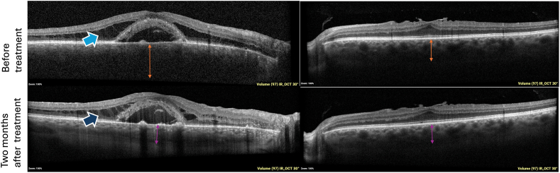
Observations: A 66-year-old male presented with unilateral macular retinoschisis and choroidal thickening in the right eye had an unrevealing systemic work-up for inflammatory or neoplastic processes. The patient eventually developed mild vitritis and a diagnostic vitrectomy was performed. Flow cytometry of the vitreous showed clonal expansion consistent with a mature B cell lymphoma of the uveal tract. Repeat systemic work-up including bone marrow biopsy, however, revealed two additional systemic conditions: Waldenstrom macroglobulinemia (WM) and monoclonal B cell lymphocytosis. Based on B cell gene rearrangement testing, the choroidal lymphoma was found to be distinct from the systemic lymphoma. In total, the patient had three lymphoproliferative disorders. Soon after the diagnosis, the left eye also developed choroidal thickening. The patient received orbital external beam radiation and zanubrutinib to treat the systemic WM and the choroidal lymphoma with improvement of macular retinoschisis and choroidal thickening.
Conclusions: We present a case of macular retinoschisis in a patient with choroidal lymphoma and multiple systemic lymphoproliferative disorders. Both the systemic disease and the uveal lymphoma contributed to the unique ocular findings in this patient. Our case highlights the challenges of uveal lymphoma diagnosis and the importance of tissue biopsy, systemic work-up, and close monitoring.
© 2025 The Authors. Published by Elsevier Inc.
Conflict of interest statement
The authors declare that they have no known competing financial interests or personal relationships that could have appeared to influence the work reported in this paper.
Figures


Similar articles
-
Anti-vascular endothelial growth factor combined with intravitreal steroids for diabetic macular oedema.Cochrane Database Syst Rev. 2018 Apr 18;4(4):CD011599. doi: 10.1002/14651858.CD011599.pub2. Cochrane Database Syst Rev. 2018. PMID: 29669176 Free PMC article.
-
Systemic pharmacological treatments for chronic plaque psoriasis: a network meta-analysis.Cochrane Database Syst Rev. 2017 Dec 22;12(12):CD011535. doi: 10.1002/14651858.CD011535.pub2. Cochrane Database Syst Rev. 2017. Update in: Cochrane Database Syst Rev. 2020 Jan 9;1:CD011535. doi: 10.1002/14651858.CD011535.pub3. PMID: 29271481 Free PMC article. Updated.
-
Systemic pharmacological treatments for chronic plaque psoriasis: a network meta-analysis.Cochrane Database Syst Rev. 2021 Apr 19;4(4):CD011535. doi: 10.1002/14651858.CD011535.pub4. Cochrane Database Syst Rev. 2021. Update in: Cochrane Database Syst Rev. 2022 May 23;5:CD011535. doi: 10.1002/14651858.CD011535.pub5. PMID: 33871055 Free PMC article. Updated.
-
Laser therapy for retinopathy in sickle cell disease.Cochrane Database Syst Rev. 2022 Dec 12;12(12):CD010790. doi: 10.1002/14651858.CD010790.pub3. Cochrane Database Syst Rev. 2022. PMID: 36508693 Free PMC article.
-
Blue-light filtering intraocular lenses (IOLs) for protecting macular health.Cochrane Database Syst Rev. 2018 May 22;5(5):CD011977. doi: 10.1002/14651858.CD011977.pub2. Cochrane Database Syst Rev. 2018. PMID: 29786830 Free PMC article.
References
-
- Mashayekhi A., Shukla S.Y., Shields J.A., Shields C.L. Choroidal lymphoma: clinical features and association with systemic lymphoma. Ophthalmology. 2014;121(1):342–351. - PubMed
-
- Aronow M.E., Portell C.A., Sweetenham J.W., Singh A.D. Uveal lymphoma: clinical features, diagnostic studies, treatment selection, and outcomes. Ophthalmology. 2014;121(1):334–341. - PubMed
-
- Sharara N., Holden J.T., Wojno T.H., et al. Ocular adnexal lymphoid proliferations: clinical, histologic, flow cytometric, and molecular analysis of forty-three cases. Ophthalmology. 2003;110:1245–1254. - PubMed
-
- Menke M.N., Feke G.T., McMeel J.W., Branagan A., Hunter Z., Treon S.P. Hyperviscosity-related retinopathy in waldenström macroglobulinemia. Arch Ophthalmol. 2006;124(11):1601–1606. - PubMed
Publication types
LinkOut - more resources
Full Text Sources
