

Dynamic change in maternal cardiac function during pregnancy
- PMID: 40547503
- PMCID: PMC12179202
- DOI: 10.3389/fcvm.2025.1577213
Dynamic change in maternal cardiac function during pregnancy
Abstract
Background: Pregnant women experience various physiological changes that, if uncompensated, may result in varying degrees of cardiac dysfunction, and adverse pregnancy outcomes. Left ventricular (LV) global longitudinal strain (GLS) and P-wave to A' duration on tissue Doppler imaging (PA-TDI) have been shown to be able to detect subtle cardiac dysfunction.
Methods: The present study was a prospective cross-sectional study. A total of 506 healthy pregnant women were enrolled, including 149 during early pregnancy (before 13 weeks' gestation, T1 group), 99 during mid-pregnancy (14-27 weeks' gestation, T2 group), and 258 during late pregnancy (after 28 weeks' gestation, T3 group), while 172 age- and baseline weight-matched healthy nonpregnant women served as the control group (NPC group). Clinical and echocardiographic data of the subjects were collected. The difference in cardiac structure and function among the 4 groups were analyzed. Multivariate regression analysis was conducted to identify the independent factors influencing change in cardiac function.
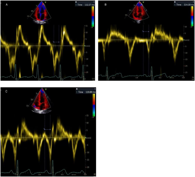
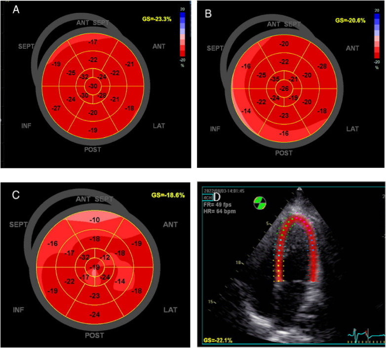
Results: The median age of the 4 groups were comparable [T1 group, 31.0 (28.5,34.0) years; T2 group, 31.0 (29.0,34.0) years; T3 group, 31.0 (29.0,34.0) years; the NPC group, 31.0 (28.0,34.0) years, P = 0.905). Left ventricular ejection fraction (LVEF) during late pregnancy was lower than that during early pregnancy and the control group, but remained within normal range. With the increase of gestational age, the absolute value of LV-GLS decreased gradually [T1 group, -19.00 (-21.40, -16.70); T2 group, -17.40 (-20.10, -15.30); T3 group, -16.35 (-17.93, -13.97); P < 0.001]. PA-TDI during the third trimester was longer than that in the first [117.65 (108.45,128.03) ms vs. 114.19 (105.61,121.11) ms, P = 0.012] or the second trimester [111.32 (107.27,121.11) ms, P = 0.010]]. Multivariate regression analysis showed that gestational age was independently associated with LV-GLS (b = 0.096, t = 2.212, P = 0.027) and PA-TDI (b = 0.158, t = 2.449, P = 0.014).
Conclusion: Pregnant women show a trend toward decreased left ventricular systolic and diastolic function. PA-TDI and LV-GLS can be used to evaluate subtle change in left cardiac function in pregnant women.
Keywords: cardiac function; pregnancy; remodeling; speckle-tracking echocardiography; tissue doppler imaging.
© 2025 Wang, Chen, Hong, Kong, Xiang, Fu, Li and Liu.
Conflict of interest statement
The authors declare that the research was conducted in the absence of any commercial or financial relationships that could be construed as a potential conflict of interest.
Figures
References
LinkOut - more resources
Full Text Sources
Miscellaneous