. 2025 Jun 3;10(23):24587-24600.
doi: 10.1021/acsomega.5c01334. eCollection 2025 Jun 17.

Identification of a Natural Small Molecule Dauricine for Glioma Therapy through Targeting p53 and VEGFA Pathways

Affiliations

Identification of a Natural Small Molecule Dauricine for Glioma Therapy through Targeting p53 and VEGFA Pathways

Hanjing Ding et al. ACS Omega. .

Abstract

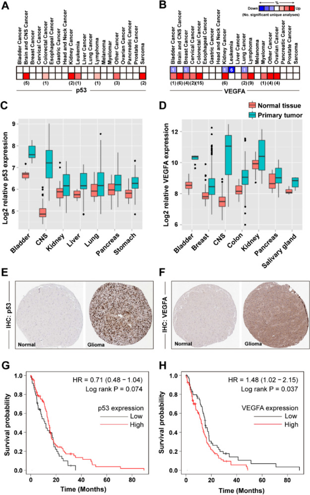
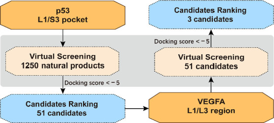
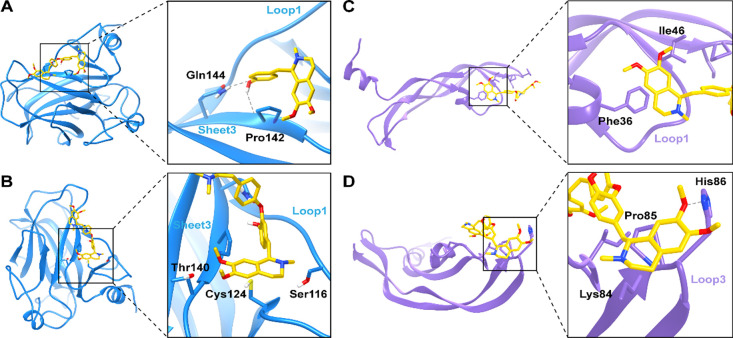
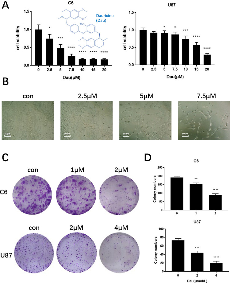
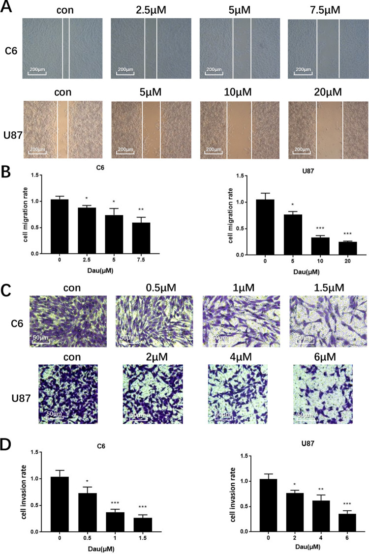
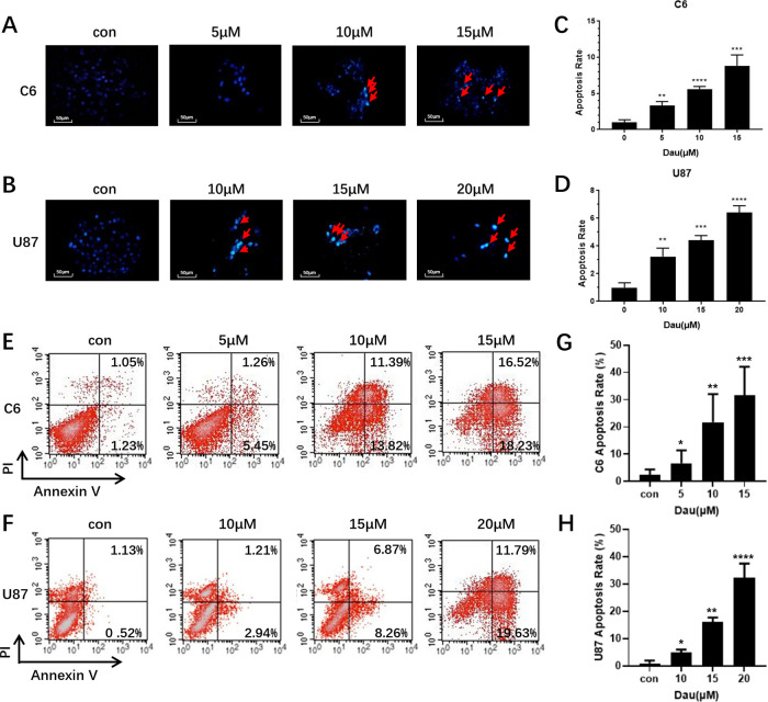
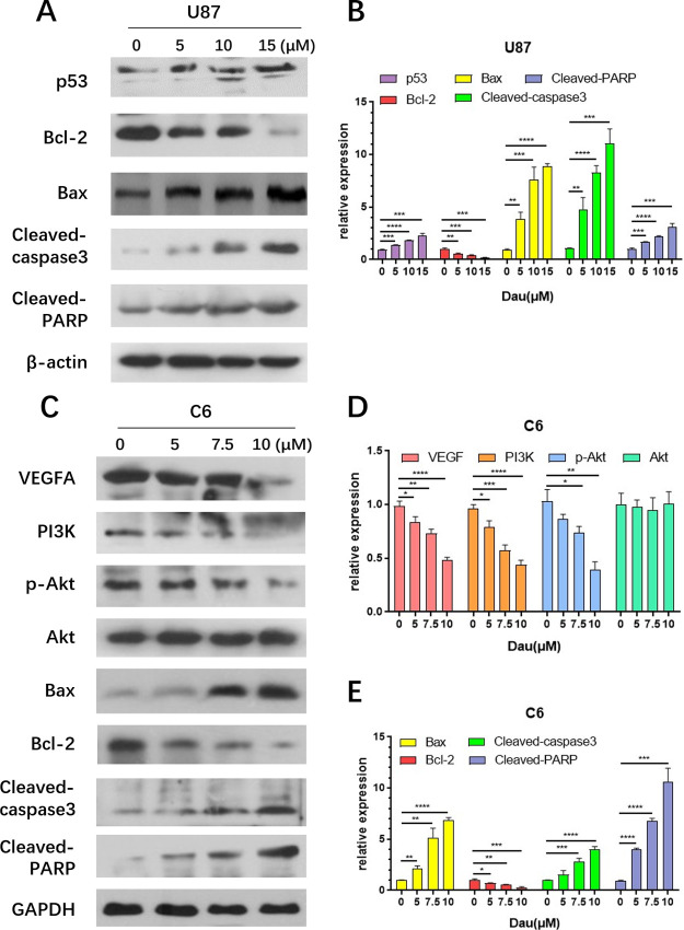
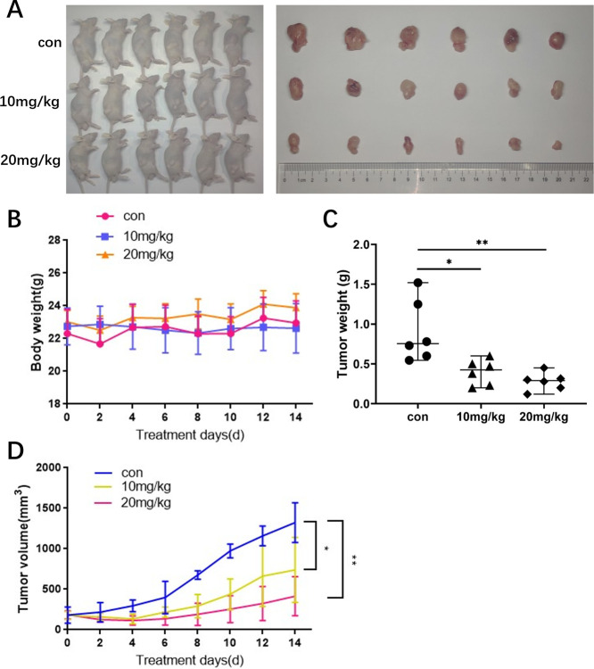
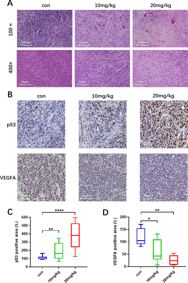
Glioma is a highly malignant primary brain tumor with a poor prognosis. Current treatments for intracranial glioma are often ineffective, necessitating the development of novel therapeutic agents. In this work, molecular docking-based screening analyses were conducted to identify dauricine as a dual-targeted agent of p53 and VEGFA by targeting the Loop1/Sheet3 (L1/S3) pocket of p53 and the Loop1/Loop3 (L1/L3) regions of VEGFA. The anticancer activity of dauricine was further evaluated using MTT, colony formation, wound-healing, transwell invasion, Western blot, and apoptosis assays, including Hoechst staining and flow cytometry on U87 and C6 glioma cell lines. A C6 xenograft model in BALB/c-nu mice was used to assess tumor growth inhibition. The in vivo anticancer effect of dauricine was detected by HE staining, and the levels of p53 and VEGFA were tested by IHC staining. The results showed that dauricine significantly inhibited the proliferation, migration, and invasion of glioma cells, induced apoptosis, and reduced tumor growth both in vitro and in vivo. Western blot analyses showed that dauricine upregulated p53 and downregulated VEGFA pathways. In vivo experiments demonstrated that dauricine inhibited the growth of subcutaneous tumors in nude mice by targeting p53 and VEGFA. Overall, these results demonstrated that dauricine suppresses glioma growth by targeting the p53 and VEGFA pathways, making it a promising anticancer agent for glioma treatment.


Figures

1
1
Upregulation of p53 and VEGFA in human cancers. (A,B) Comparison of p53 (n = 443) or VEGFA (n = 452) expression in different types of cancer analyzed by Oncomine. (C,D) p53 and VEGFA expression in different primary tumors and corresponding normal tissues analyzed by Metabolic gEne RApid Visualizer. (E,F) p53 and VEGFA expression in normal brain tissue and corresponding glioblastoma tissue as revealed by IHC from Human Protein Atlas. (G,H) Survival probability analyses of p53 and VEGFA expression by Kaplan–Meier plotter (n = 153).
1
1. Work Flow of the Virtual Screening
2
2
Molecular docking of dauricine and the p53 core domain as well as VEGFA. (A) Dauricine binds to the L1/S3 pocket of p53, forming two hydrogen bonds with Gln144 and Pro142 from Sheet3. P53 is shown in blue, and dauricine in yellow. The hydrogen bonds are shown with gray dashed lines. The rest part of the dauricine is hidden for the clarity of view. (B) Dauricine interacts with Ser116, Cys124, and Thr140 in the L1/S3 pocket of p53. (C) Dauricine binds to the L1/L3 region of VEGFA, interacting with Phe36 and Ile46 in the Loop1 (L1) region. (D) Dauricine binds to the Loop3 (L3) region of VEGFA, forming a hydrogen bond with His86 and interacting with Lys84 as well as Pro85.
3
3
Dauricine (Dau) inhibits the viability and proliferation of glioma cells. (A) Inhibitory effects of dauricine on C6 and U87 cell proliferation at 24 h using MTT analyses. The cell viability of the control group was designated as 1, the viability values for all treatment groups were normalized to the control. (B) Changes in cell morphology and growth after dauricine treatment on U87 proliferation at 24 h. (C) Clone formation assay of C6 and U87 cells after 14 days of dauricine treatment with indicated concentrations, cells were fixed and stained with crystal violet. (D) Statistical analyses of colony numbers (*P < 0.05, **P < 0.01, ***P < 0.001, ****P < 0.0001 vs control, n = 3).
4
4
Dauricine inhibits the migration and invasion of glioma. (A) Wound healing of cells at 48 h after dauricine treatment. The scratch was made to each group in the plate with a 200 μL pipet tip before treatment. The white lines indicate the front edges of cell growth after treatment. (B) Statistical analyses for the scratch assay. Cell migration rates were calculated for C6 and U87 cells after dauricine treatment. (C) Dauricine inhibited the invasion of glioma cells in the transwell assay. (D) Statistical analyses for the transwell assay (*P < 0.05, **P < 0.01, ***P < 0.001 vs control, n = 3).
5
5
Dauricine induces apoptosis in glioma cells. (A,B) C6 and U87 cells undergoing apoptosis analyzed by Hoechst staining after 24 h of dauricine treatment. (C,D) Statistical analyses of the apoptosis rates from Hoechst staining (**P < 0.01, ***P < 0.001, ****P < 0.0001, n = 3). (E,F) Apoptosis of C6 and U87 cells after dauricine treatment analyzed by flow cytometry. (G,H) Statistical analyses of the apoptosis rates from flow cytometry (*P < 0.05, **P < 0.01, ***P < 0.001, ****P < 0.0001, n = 3).
6
6
Dauricine upregulates p53 expression, downregulates VEGFA expression, and induces apoptosis in glioma cells. (A) Western blot analyses of p53 and its downstream genes. (B) Gray scale statistics of protein expression in U87 cells. (C) Western blot analyses of VEGFA and its downstream genes. (D, E) Gray scale statistics of protein expression in C6 cells (*P < 0.05, **P < 0.01, ***P < 0.001, ****P < 0.0001 vs control, n = 3).
7
7
Dauricine inhibits the growth of glioma xenograft. (A) Xenograft tumors of the nude mice 14 days after inoculation. (B) Minimal changes in body weight of nude mice showing the safety of dauricine at the tested doses. (C) Statistical analyses of subcutaneous tumor weight after sacrificing nude mice. (D) Growth curve of subcutaneous tumor volume from day 0 to day 14 (*P < 0.05, **P < 0.01 vs control, n = 6).
8
8
Pathological analyses of C6 glioma xenograft tissue. (A) HE staining was performed on three groups of C6 tumor tissue sections. Photos were taken under the microscope at 100× and 400×. Blue-violet staining indicates tumor cell nuclei. (B) IHC analyses of the tumor sections. p53 and VEGFA were detected in the subcutaneous xenograft tumor sections of nude mice. Brown staining indicates positive signals. (C,D) Statistical analyses of p53 and VEGFA expression in IHC staining of the xenograft (*P < 0.05, **P < 0.01, ****P < 0.0001 vs Control, n = 6).

Similar articles

References

    1. Bi J., Chowdhry S., Wu S., Zhang W., Masui K., Mischel P. S.. Altered cellular metabolism in gliomas - an emerging landscape of actionable co-dependency targets. Nat. Rev. Cancer. 2020;20(1):57–70. doi: 10.1038/s41568-019-0226-5. - DOI - PubMed
    1. Tan A. C., Ashley D. M., Lopez G. Y., Malinzak M., Friedman H. S., Khasraw M.. Management of glioblastoma: State of the art and future directions. CA Cancer J. Clin. 2020;70(4):299–312. doi: 10.3322/caac.21613. - DOI - PubMed
    1. Jackson C. M., Choi J., Lim M.. Mechanisms of immunotherapy resistance: lessons from glioblastoma. Nat. Immunol. 2019;20(9):1100–1109. doi: 10.1038/s41590-019-0433-y. - DOI - PubMed
    1. Bush N. A. O., Chang S. M., Berger M. S.. Current and future strategies for treatment of glioma. Neurosurg. Rev. 2017;40(1):1–14. doi: 10.1007/s10143-016-0709-8. - DOI - PubMed
    1. Zhong L., Li Y., Xiong L., Wang W., Wu M., Yuan T., Yang W., Tian C., Miao Z., Wang T., Yang S.. Small molecules in targeted cancer therapy: advances, challenges, and future perspectives. Signal. Transduction Targeted Ther. 2021;6(1):201. doi: 10.1038/s41392-021-00572-w. - DOI - PMC - PubMed

LinkOut - more resources