

Enhancing Osteogenesis Differentiation and In Vitro Degradation in Polymer Scaffolds with Spike-like Strontium Carbonate Microrods
- PMID: 40547639
- PMCID: PMC12177776
- DOI: 10.1021/acsomega.4c09683
Enhancing Osteogenesis Differentiation and In Vitro Degradation in Polymer Scaffolds with Spike-like Strontium Carbonate Microrods
Abstract
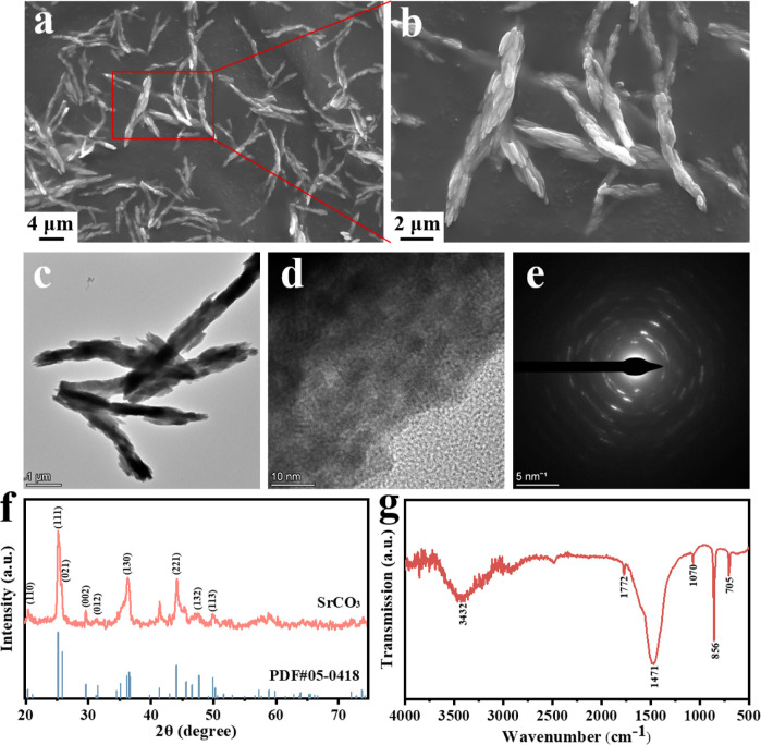
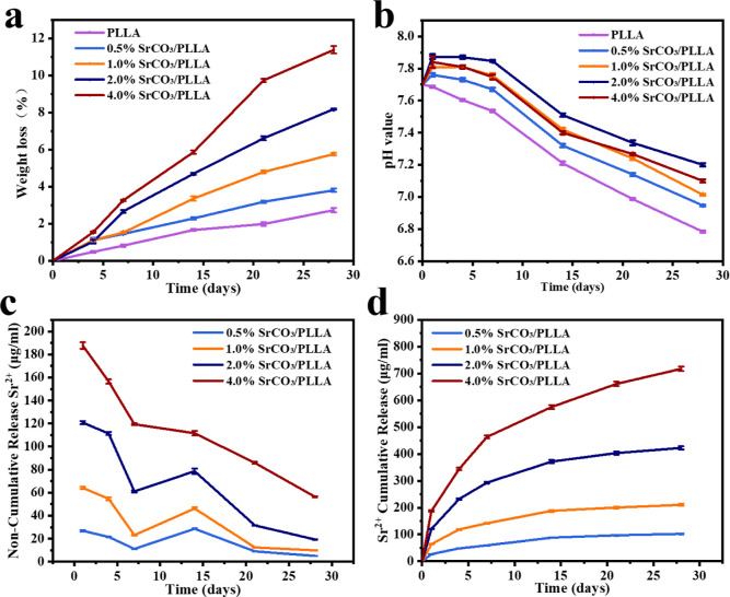
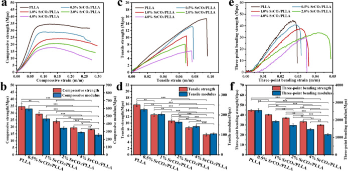
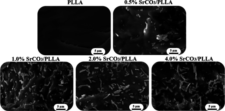
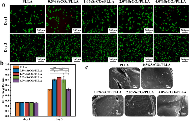
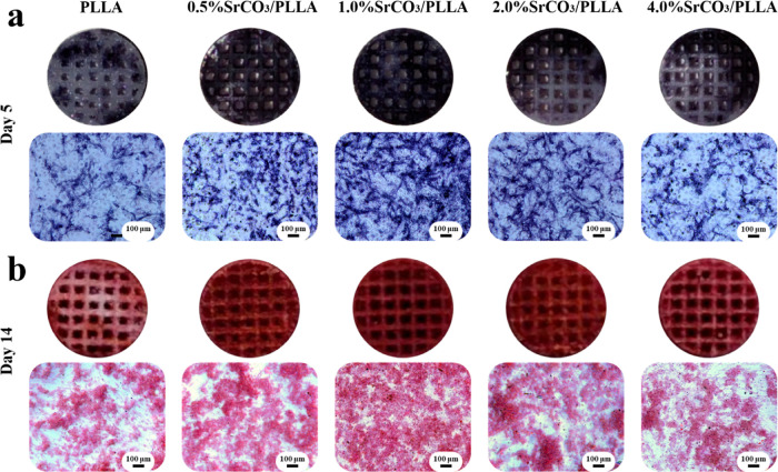
Poly-(L-lactic-acid) (PLLA) has shown significant promise in the fields of orthopedics and dentistry. However, its lack of inherent osteogenic activity limits its potential for further application in bone repair and fixation. In this study, we synthesized strontium carbonate (SrCO3) nanoparticles and incorporated them into PLLA to prepare bioactive composite scaffolds using selective laser sintering technology. The synthesized SrCO3 particles exhibited a unique microrod morphology with spike-like structures. In vitro degradation experiments demonstrated that SrCO3 effectively neutralized acidic byproducts and promoted the degradation of PLLA in a dose-dependent manner. Furthermore, in vitro cell culture experiments revealed that composite scaffolds containing 1-2 wt % SrCO3 significantly enhanced the adhesion, proliferation, and osteogenic differentiation of mouse bone marrow mesenchymal stem cells compared to the PLLA scaffolds. This can be attributed to the sustained release of Sr ions from the composite scaffolds. Overall, our study elucidates the positive effects of incorporating SrCO3 into the PLLA matrix, highlighting its potential as an alternative to the currently used PLLA implants for promoting degradation and facilitating osteogenesis.
© 2025 The Authors. Published by American Chemical Society.
Figures
Similar articles
-
Mineralized osteoblast-derived exosomes and 3D-printed ceramic-based scaffolds for enhanced bone healing: A preclinical exploration.Acta Biomater. 2025 Jun 15;200:686-702. doi: 10.1016/j.actbio.2025.05.051. Epub 2025 May 21. Acta Biomater. 2025. PMID: 40409510
-
Survivor, family and professional experiences of psychosocial interventions for sexual abuse and violence: a qualitative evidence synthesis.Cochrane Database Syst Rev. 2022 Oct 4;10(10):CD013648. doi: 10.1002/14651858.CD013648.pub2. Cochrane Database Syst Rev. 2022. PMID: 36194890 Free PMC article.
-
The clinical effectiveness and cost-effectiveness of strontium ranelate for the prevention of osteoporotic fragility fractures in postmenopausal women.Health Technol Assess. 2007 Feb;11(4):1-134. doi: 10.3310/hta11040. Health Technol Assess. 2007. PMID: 17280622
-
Direct composite resin fillings versus amalgam fillings for permanent posterior teeth.Cochrane Database Syst Rev. 2021 Aug 13;8(8):CD005620. doi: 10.1002/14651858.CD005620.pub3. Cochrane Database Syst Rev. 2021. PMID: 34387873 Free PMC article.
-
pH-Responsive nanoplatform synergistic gas/photothermal therapy to eliminate biofilms in poly(L-lactic acid) scaffolds.J Mater Chem B. 2024 Jan 31;12(5):1379-1392. doi: 10.1039/d3tb02600k. J Mater Chem B. 2024. PMID: 38247429
References
-
- Khouri N. G., Bahú J. O., Blanco-Llamero C.. et al. Polylactic Acid (PLA): Properties, Synthesis, and Biomedical Applications-A Review of the Literature. J. Mol. Struct. 2024;1309:138243. doi: 10.1016/j.molstruc.2024.138243. - DOI
-
- Lopes M. S., Jardini A. L., Filho R. M.. Poly (lactic acid) production for tissue engineering applications. Procedia Eng. 2012;42:1402–1413. doi: 10.1016/j.proeng.2012.07.534. - DOI
-
- Paula E. L., Mano V., Duek E. A. R., Pereira F. V.. Hydrolytic degradation behavior of PLLA nanocomposites reinforced with modified cellulose nanocrystals. Química Nova. 2015;38:1014–1020. doi: 10.5935/0100-4042.20150108. - DOI
LinkOut - more resources
Full Text Sources
Research Materials