

Giant spindle cell lipoma of the left inguinal region: A rare case with diagnostic challenges on MRI
- PMID: 40547940
- PMCID: PMC12182281
- DOI: 10.1016/j.radcr.2025.05.034
Giant spindle cell lipoma of the left inguinal region: A rare case with diagnostic challenges on MRI
Abstract
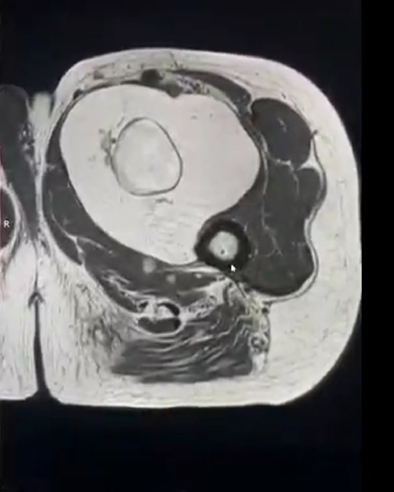
Spindle cell lipomas (SCLs) are rare benign adipocytic tumors, predominantly occurring in elderly men's posterior neck, upper back, and shoulders. Accounting for 1.5% of lipomas, SCLs in atypical locations like the inguinal region are exceptionally rare and pose diagnostic challenges due to similarities with malignant tumors. We present a 64-year-old male with a 6-month history of a painless, enlarging left inguinal mass. MRI revealed a 20 × 13 × 10 cm well-defined lipomatous lesion displacing femoral vessels, showing high T1/T2 signals, fat suppression, and septations. Core needle biopsy confirmed SCL, featuring mature adipocytes and bland spindle cells in a collagenous matrix, though histopathological images were unavailable. Definitive surgery was deferred due to the patient's inability to return amid the Syrian conflict. Inguinal SCLs are seldom reported and often mimic well-differentiated liposarcoma. This case, among the largest documented in the groin, underscores the importance of integrating clinical, radiological, and histopathological data for accurate diagnosis. MRI findings aligned with classic SCL features, aiding differentiation from malignancies. While surgical resection remains standard, recognizing benign SCLs in unusual sites is critical to avoid overtreatment. Preoperative diagnosis through imaging and biopsy is vital, particularly when management is delayed or lost to follow-up. This case highlights challenges in resource-limited settings and emphasizes multidisciplinary evaluation for deep-seated lipomatous tumors.
Keywords: Adipocytic tumor; Case report; Inguinal region; MRI; Soft tissue mass; Spindle cell lipoma.
© 2025 The Authors. Published by Elsevier Inc. on behalf of University of Washington.
Figures


References
-
- Takeda T., Ando R., Unno R., Iida K., Iwatsuki S., Umemoto Y., Kawai N., Tozawa K., Yasui T. Hinyokika kiyo. Acta Urologica Japonica. 2016;62(4):205–208. - PubMed
Publication types
LinkOut - more resources
Full Text Sources
Research Materials
Miscellaneous
