

Imaging Epilepsy: Past, Passing, and to Come
- PMID: 40548097
- PMCID: PMC12176802
- DOI: 10.1177/15357597251332191
Imaging Epilepsy: Past, Passing, and to Come
Abstract
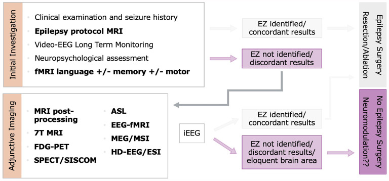
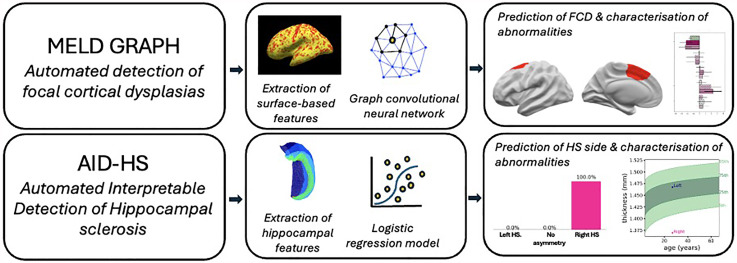
New imaging techniques appearing over the last few decades have replaced procedures that were uncomfortable, of low specificity, and prone to adverse events. While computed tomography remains useful for imaging patients with seizures in acute settings, structural magnetic resonance imaging (MRI) has become the most important imaging modality for epilepsy evaluation, with adjunctive functional imaging also increasingly well established in presurgical evaluation, including positron emission tomography (PET), single photon ictal-interictal subtraction computed tomography co-registered to MRI and functional MRI for preoperative cognitive mapping. Neuroimaging in inherited metabolic epilepsies is integral to diagnosis, monitoring, and assessment of treatment response. Neurotransmitter receptor PET and magnetic resonance spectroscopy can help delineate the pathophysiology of these disorders. Machine learning and artificial intelligence analyses based on large MRI datasets composed of healthy volunteers and people with epilepsy have been initiated to detect lesions that are not found visually, particularly focal cortical dysplasia. These methods, not yet approved for patient care, depend on careful clinical correlation and training sets that fully sample broad populations.
Keywords: artificial intelligence; genetic epilepsy syndromes; image processing; imaging.
© The Author(s) 2025.
Conflict of interest statement
The authors declared no potential conflicts of interest with respect to the research, authorship, and/or publication of this article.
Figures
References
-
- Charash LI, Dunning HS. An appraisal of pneumoencephalography in mental retardation and epilepsy. Pediatrics. 1956;18(5):716–720. PMID: 13370236. - PubMed
-
- Harden CL, Huff JS, Schwartz TH, et al. Reassessment: neuroimaging in the emergency patient presenting with seizure (an evidence-based review): report of the Therapeutics and Technology Assessment Subcommittee of the American Academy of Neurology. Neurology. 2007;69:1772–1780. doi: 10.1212/01.wnl.0000285083.25882.0e. PMID: 17967993. - DOI - PubMed
Publication types
LinkOut - more resources
Full Text Sources
Miscellaneous
