

Grape seed proanthocyanidins improve lymphatic drainage and blood perfusion in secondary lymphedema models
- PMID: 40548114
- PMCID: PMC12179177
- DOI: 10.3389/fonc.2025.1553090
Grape seed proanthocyanidins improve lymphatic drainage and blood perfusion in secondary lymphedema models
Abstract
Introduction: Secondary lymphedema (SLE) is a chronic and debilitating condition that frequently arises following cancer treatments, particularly in breast cancer patients. Despite its increasing global prevalence and impact on patients' quality of life, there remains no effective pharmacological treatment for SLE. Grape seed proanthocyanidin extract (GSPE), a compound known for treating venous insufficiency, is hypothesized to enhance lymphatic function and may offer therapeutic value for managing SLE. This study aimed to evaluate the efficacy of GSPE in a rat model of secondary lymphedema.
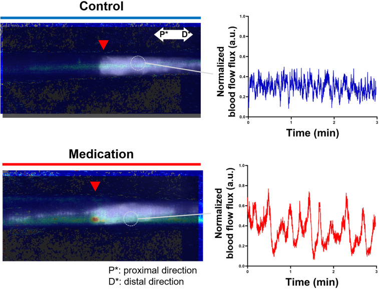
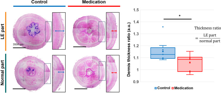
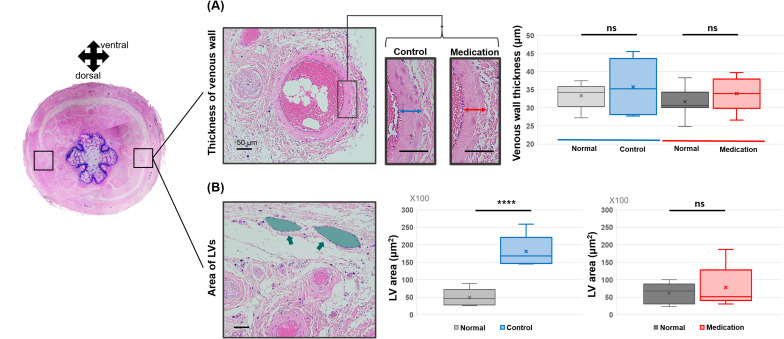
Methods: Fifteen nine-week-old Sprague-Dawley rats (weighing 250-300 g) were used in this study. Tail lymphedema was surgically induced in 12 rats to model SLE, while 3 rats served as normal controls. The lymphedema-induced rats were randomly assigned to either a treatment group (n=6) or a control group (n=6). The treatment group received intraperitoneal injections of GSPE powder dissolved in saline, whereas the control group received saline alone. Tail volume was measured periodically to monitor edema progression. Lymphatic and blood flow were assessed using near-infrared fluorescence indocyanine green lymphangiography (NIRF-ICGL) and laser Doppler flowmetry imaging (LDFI), respectively. Histological analysis was conducted using hematoxylin and eosin (H&E) staining.
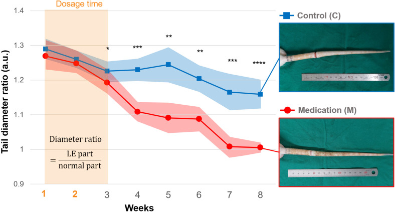
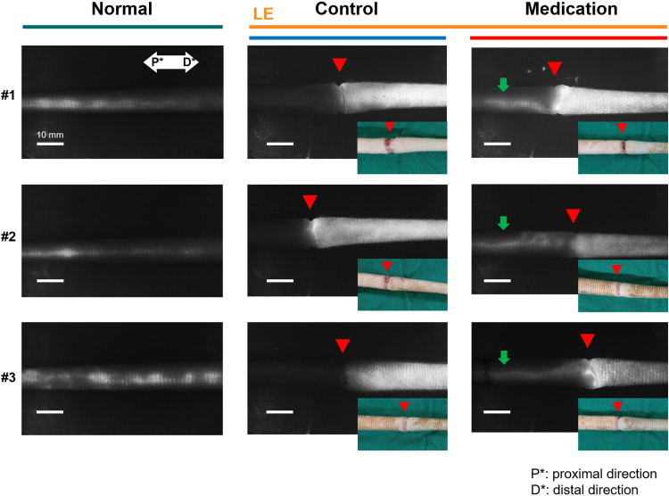
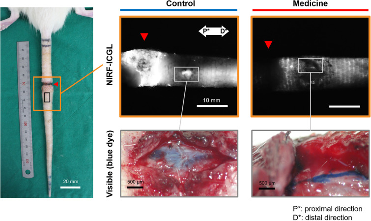
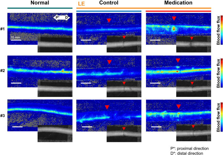
Results: The treatment group demonstrated a significant reduction of edema in the tail compared to the control group. NIRF-ICGL revealed improved lymphatic drainage, while LDFI analysis indicated enhanced blood perfusion in GSPE-treated animals. Histopathological examination showed reduced extracellular matrix deposition and fewer lymphatic abnormalities in the treatment group, suggesting mitigation of tissue fibrosis and lymphatic dysfunction.
Discussion: These findings highlight the therapeutic potential of GSPE in treating secondary lymphedema. The observed improvements in lymphatic drainage, tissue perfusion, and histological features suggest that GSPE may exert beneficial effects beyond its established role in venous insufficiency. Considering the current lack of effective pharmacologic therapies for SLE, GSPE represents a promising candidate for future clinical applications. Further studies are warranted to validate its efficacy and safety in human subjects.
Keywords: Doppler flowmetry; grape seed proanthocyanidin extract (GSPE); lymphangiography; lymphedema; pharmacological treatment; preclinical (in vivo) studies.
Copyright © 2025 Cheon, Kim and Jeon.
Conflict of interest statement
The authors declare that the research was conducted in the absence of any commercial or financial relationships that could be construed as a potential conflict of interest.
Figures
Similar articles
-
Inhaled mannitol for cystic fibrosis.Cochrane Database Syst Rev. 2018 Feb 9;2(2):CD008649. doi: 10.1002/14651858.CD008649.pub3. Cochrane Database Syst Rev. 2018. Update in: Cochrane Database Syst Rev. 2020 May 1;5:CD008649. doi: 10.1002/14651858.CD008649.pub4. PMID: 29424930 Free PMC article. Updated.
-
Systemic pharmacological treatments for chronic plaque psoriasis: a network meta-analysis.Cochrane Database Syst Rev. 2021 Apr 19;4(4):CD011535. doi: 10.1002/14651858.CD011535.pub4. Cochrane Database Syst Rev. 2021. Update in: Cochrane Database Syst Rev. 2022 May 23;5:CD011535. doi: 10.1002/14651858.CD011535.pub5. PMID: 33871055 Free PMC article. Updated.
-
Saline irrigation for allergic rhinitis.Cochrane Database Syst Rev. 2018 Jun 22;6(6):CD012597. doi: 10.1002/14651858.CD012597.pub2. Cochrane Database Syst Rev. 2018. PMID: 29932206 Free PMC article.
-
Ear drops for the removal of ear wax.Cochrane Database Syst Rev. 2018 Jul 25;7(7):CD012171. doi: 10.1002/14651858.CD012171.pub2. Cochrane Database Syst Rev. 2018. PMID: 30043448 Free PMC article.
-
Cost-effectiveness of using prognostic information to select women with breast cancer for adjuvant systemic therapy.Health Technol Assess. 2006 Sep;10(34):iii-iv, ix-xi, 1-204. doi: 10.3310/hta10340. Health Technol Assess. 2006. PMID: 16959170
References
LinkOut - more resources
Full Text Sources