Review
. 2025 Jun 6:15:1499283.
doi: 10.3389/fonc.2025.1499283. eCollection 2025.

Nanomaterials targeting cancer stem cells to overcome drug resistance and tumor recurrence

Affiliations
Review

Nanomaterials targeting cancer stem cells to overcome drug resistance and tumor recurrence

Divya Vishambhar Kumbhakar et al. Front Oncol. .

Abstract

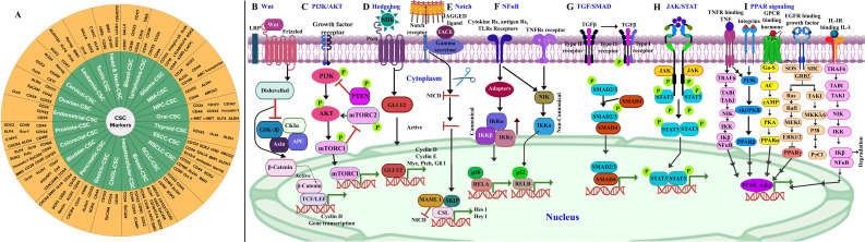
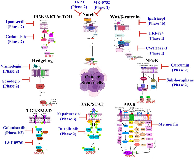
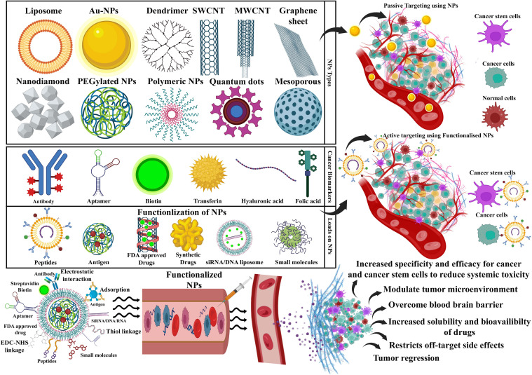
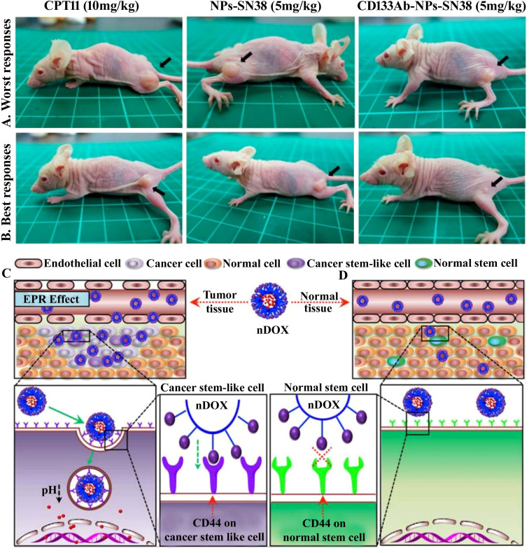
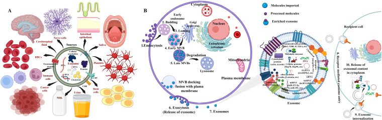
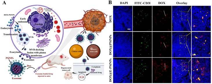
A cancer stem cell (CSC) is an immortal cell that is capable of self-renewal, continuous proliferation, differentiation into various cancer cell lineages, metastatic dissemination, tumorigenesis, maintaining tumor heterogeneity, and resistance to conventional treatments. Targeted therapies have made huge advances in the past few years, but resistance is still a major roadblock to their success, in addition to their life-threatening side effects. Progressive treatments are now available, including immunotherapies, CRISPR-Cas 9, sonodynamic therapy, chemodynamic therapy, antibody-drug nanoconjugates, cell-based therapies, gene therapy, and ferroptosis-based therapy, which have replaced surgery, chemotherapy, and radiotherapy for cancer treatment. The challenge is to develop targeted treatment strategies that are effective in eradicating CSCs, as they are resistant to anticancer drugs, causing treatment failure, relapse, and recurrence of cancer. An overview of the fundamental characteristics of CSCs, drug resistance, tumor recurrence, and signaling pathways as well as biomarkers associated with their metastatic potential of CSC is elucidated in this review. The regulatory frameworks for manufacturing and conducting clinical trials on cancer therapy are explicated. Furthermore, we summarize a variety of promising nanocarriers (NCs) that have been used directly and/or synergistic therapies coupled with the therapeutic drug of choice for the detection, targeting, and imaging of CSCs to surmount therapeutic resistance and stemness-related signaling pathways and eradicate CSCs, hence alleviating the limitation of conventional therapies. Nanoparticle-mediated ablation therapies (NMATs) are also being argued as a method for burning or freezing cancer cells without undergoing open surgery. Additionally, we discuss the recent clinical trials testing exosomes, CRISPR/Cas9, and nanodrugs, which have already received approval for several new technologies, while others are still in the early stages of testing. The objective of this review is to elucidate the advantages of nanocarriers in conquering cancer drug resistance and to discuss the most recent developments in this field.

Keywords: biomarkers; cancer stem cells; exosomes; nanocarriers; nanoparticle-mediated ablation therapies; signaling pathways; targeted therapy.


Conflict of interest statement

The authors declare that the research was conducted in the absence of any commercial or financial relationships that could be construed as a potential conflict of interest.

Figures

Figure 1
Figure 1
(A) Cancer stem cell markers in different types of cancer (, –110). A schematic illustration of signaling pathways involved in cancer stem cells. (B) Wnt/β-catenin: targets include Wnt/Frizzled complexes, β-catenin/TCF, and CK1α. (C) PI3K/AKT: targets include PI3K complex, AKT1/2/3, and mTORC1/2. (D) Hedgehog: targets include SHh-Ptch interaction, SMO, and GLI. (E) Notch: targets include Notch and γ-secretase. (F) NF-κB: targets include IKKα/β/γ and NF-κB-inducing kinase (NIK). (G) TGF/SMAD: targets include TGF-β1/β2/β3, TβRI/II, and Smad3/4/5. (H) JAK/STAT: targets include JAK1/2/3 and STAT1/2/3/4/5. (I) PPAR: targets include PPARα/γ/δ signaling pathways. Adapted and modified using BioRender for illustrative purposes with permission from Chu et al. (2) (Copyright 2024).
Figure 2
Figure 2
An overview of a few cancer stem cell (CSC)-targeted therapeutic agents that have been approved or are undergoing clinical trials. Adapted and modified with permission from PS Kharkar (Copyright 2020, American Chemical Society) (, –123).
Figure 3
Figure 3
A schematic diagram showing that an array of nanocarriers modified with targeting ligands can, however, specifically attach to tumor cells’ receptors, permitting localized drug delivery or endocytosis. An illustration of active and passive targeting in antitumor nano-delivery systems. Passive targeting is achieved by delivering nanocarriers into tumor tissues via leaky tumor blood vessels, where they accumulate due to enhanced permeability and retention (EPR) effects. Illustration showing the ability of targeted cancer cells to absorb nanocarriers and their accumulation in tumors that exhibit tumor suppression. Image created with BioRender.com.
Figure 4
Figure 4
(A, B) CD133-positive (CD133+) cell was targeted with topoisomerase inhibitor SN-38-loaded nanoparticles conjugated with anti-CD133 antibody to resolve chemotherapy failure. HCT116 overexpress CD133 glycoprotein, which was efficiently bound by anti-CD133 antibody-conjugated SN-38-loaded nanoparticles (CD133Ab-NP-SN-38) demonstrated by the in vivo study. The tumor size depiction in mice treated with CPT11 (irinotecan, DNA topoisomerase I inhibitor) as control group, SN-38 nanoparticles (NPs), and CD133Ab-SN38 NPs in HCT116 xenograft model. This CD133Ab-NP-SN-38 combination thwarted tumor growth and hindered recurrence in xenograft model. Reprinted with permission from Ning et al. (381) (Copyright 2016, American Chemical Society). (C) CD44-overexpressing breast cancer stem cells (CSCs) were eradicated via chitosan-modified poly(ethylene glycol) (PEG)–poly(propylene glycol) (PPG)–PEG micelle crosslinking loaded with doxorubicin (DOX) in comparison with the free DOX application. DOX-loaded micelles facilitated increased DOX cytotoxicity on cancer stem cell (CSC)-expressing MCF7 breast tumor mouse model exhibiting CD44+ overexpression by six times compared to (D) normal tissue. Enhanced permeability and retention (EPR) effect of conjugated nanoparticles caused them to accumulate in tumor tissues more than they did in normal tissues. In addition, no noticeable systemic side effects were observed. Reused for illustrative purposes with permission from Rao et al. (382) (© 2015, American Chemical Society).
Figure 5
Figure 5
(A) Schematic depiction for sources of exosome: stem cells, cancer cells, immune cells, nerve cells, saliva, platelets, red blood cells (RBCs), breast milk, cerebrospinal fluid, intestinal epithelium, and urine, among others. (B) The generalized structure of exosomes and overview describing the biogenesis of exosomal stem cells and cargos and consequent intake of exosomes by recipient cells. (1) Fusion with target recipient cell, (2) endocytosis, and (3) interaction with specific receptors on cell surface and internalization. Source: Vakil et al. (442). Image was modified and recreated using BioRender.com.
Figure 6
Figure 6
(A) The exosome-encapsulated porous silicon nanoparticles (E-PSiNPs) and DOX@E-PSiNP preparation as antitumor drug carriers are shown in schematic diagram. After incubation, DOX@E-PSiNPs are endocytosed into cancer cells, localized to multivesicular bodies (MVBs), and form autophagosomes. Exocytosis of DOX@E-PSiNPs occurs upon fusion of MVBs with cell membranes. A systemic injection of DOX@E-PSiNPs in tumor-bearing mice resulted in strong anticancer activity displaying accumulation in both cancer cells and cancer stem cells (CSCs) and penetrating deeply into tumor tissues. (B) Florescent images showing the localization of both DOX and CD31-labeled tumor blood vessels in tumors isolated from H22 tumor-bearing mice at 24 h after intravenous infusion of DOX alone, DOX@PSiNPs, and functional exosome-encapsulated DOX-PSiNPs (DOX@E-PSiNPs) at DOX dosage of 0.5 mg/kg; scale bar, 200 µm. A wide distribution of DOX@E-PSiNPs was evident after treatment; conversely, DOX and DOX@PSiNPs accumulated mostly around blood vessels as evidenced by stronger co-localization with FITC-CD31-labeled endothelial cells. A white line outlines the gap between the DOX distribution in blood vessels and the tumor parenchyma. Reused for illustrative purposes with permission from Yong et al. (467) (Copyright 2019, source: Tumor exosome-based nanoparticles are efficient drug carriers for chemotherapyNature Communications).
Figure 7
Figure 7
(A) A comparison of microscopy descriptions of gastric cancer stem cell (GCSC) spheroid colonies treated with GNS-PEG-CD44v6 and GNS-PEG irradiated for 24 h with near-infrared (NIR) laser (790 nm, 1.5 W/cm2, 5 min) showing damaged spheroid colonies in NIR irradiated GNS-PEG-CD44v6. (B) Photoacoustic (PA) images attained before and after injection (0, 2, 4, and 24 h) with GNS-PEG-CD44, GNS-PEG-CD44v6 (the first and third row depict a subtumor, while the second and fourth display orthotopic tumor taken as test group), and GNS-PEG (the fifth row denotes a subtumor taken as control). Observation of (C) deionized water and (D) GNS in a tube exposed to NIR radiation (790 nm, 0.3 W/cm2, 3 min) using infrared microscopic imaging. On NIR laser irradiation, subcutaneous GC tumors are shown (E) without and (F) with injections of GNS-PEG-CD44v6. An injection of GNS-PEG-CD44v6 was administered to a nude mouse of GC subcutaneous xenograft (G) with and (H) without laser exposure (790 nm, 0.3 W/cm2, 3 min). (I) Growth curves of GC tumors exposed to NIR laser treatment (790 nm, 0.8 W/cm2, 5 min), which were additionally treated with GNS-PEG-CD44v6, GNS-PEG, and PBS, along with the untreated control group. (J) Treatment-induced survivability rate (%) was assessed for GC tumor-bearing mice after 8 weeks in comparison to controls. Reused for illustrative purposes with permission from Liang et al. (499) (Copyright 2015, Ivyspring International Publisher, source: https://www.ncbi.nlm.nih.gov/pmc/articles/PMC4493535/).
Figure 8
Figure 8
(A) A tumor xenograft model was established in mice on day 8 with the inoculation of HT-29 control cells and transfected TRPV2. The treatments PBS, PCHN, and TRPV2–PCNH were administered intraperitoneally (i.p.) on day 0 and administered every other day until day 16. Near-infrared (NIR) laser of 1,064 nm (1 W to 50 mW mm−2) was applied for 5 min to the right side of tumor on days 2, 6, 9, 13, and 16. (B) A thermographic infrared camera was used to monitor the surface temperature of the body during laser irradiation. (C) A laser-induced increase in temperature was observed in mice with HT-29 or HT-29–TRPV2 tumors after nanocomplex injection and measured at 24 (h) Tumor volume measured in (D) HT-29 and (E) HT-29-TRPV2 in different treatment groups with/without NIR laser exposure. A significant reduction in tumor volumes was observed in nude mice bearing HT-29–TRPV2 following TRPV2–PCNH+laser treatment. (F) Tumor-bearing nude mice with HT-29 and HT-29–TRPV2 photographed on day 16 (black arrows indicating irradiated tumors). Xenograft models overexpressing TRPV2 and TRPV2–PCNH+laser inhibit tumor reinitiation. Immunohistochemical analysis showed that expressions of (G) Ki-67 and (H) CD133 expressions were documented to reduce with TRPV2–PCNH+laser in primary tumor sections of xenografts overexpressing TRPV2. (I) The RT-qPCR analysis of tumors with TRPV2 overexpression from mice treated with TRPV2–PCNH and laser irradiation exhibited declines in mRNA of stemness-associated markers (Nanog, ALDH1, CD133, CD44, and CD9). (J) Methodology followed that of Yu et al. (517) for tumorigenesis experiments involving resection, isolation, re-implantation, and tumor initiation investigation. (K) The cells isolated from xenograft tumors showed reduction in tumor proliferation following TRPV2–PCNH+laser treatment in comparison with PBS and TRPV2–PCNH tested groups. (L) TRPV2–PCNH+laser irradiation in osteosarcoma cell line (U2OS) demonstrated reduced β-catenin expression as demonstrated by immunostaining images (Hoechst specifies nuclei, mCherry specifies TRPV2, and Alexa488 specifies β-catenin). (M) Western blotting illustrated downregulated non-phosphorylated and total β-catenin expression and upregulated PKCα expression in MCF7–TRPV2 cells with TRPV2–PCNH+laser treatment. β-Actin was used as a control for normalization. Reused for illustrative purposes with permission from Yu et al. (517) (Copyright 2020, Nature Communications, source: Photothermogenetic inhibition of cancer stemness by near-infrared-light-activatable nanocomplexes - PubMed (nih.gov)).
Figure 9
Figure 9
MXene-mediated photothermal ablation of cancer stem cells (CSCs). (A) An overview of the synthesis and use of “nano-lymphatic” (M/CdS/HA) for increased tumor penetration, photothermal (NIR-II), and hydrodynamic therapies with the decrease in tumor interstitial pressure (TIP) via pyroelectric catalysis. (B) Pyroelectric effect with temperature variation depicted schematically. (C) The infrared thermal image (ITI) demonstrated the excellent photothermal effect of M/CdS under laser irradiation at 3 min and could confirm further usage for both PTT and pyroelectric catalysis (here, 0 min was considered as control). (D) Photothermal consequence of CdS, MXene, and MXene/CdS. (E) The curves of O2 generation due to CdS, MXene, and M/CdS under laser irradiation (1,064 nm, 1.0 W/cm2) in comparison to control. (F) The illustration of multicellular spheroid (MCS) formation and the penetration mechanism showing increased diffusion with rise in temperature (T↑), reduction in TIP, and transcytosis in MXene+laser, MXene/CdS, MXene/CdS+laser, and MXene/CdS+laser+genistein (Gen) treatments. (G) Confocal fluorescence imaging of MCSs on exposure with MXene/CdS-HA/RB with NIR-II laser irradiation (1,064 nm) from day 0 to 6 (scale bar = 100 μm). (H) Representation of pyroelectric catalysis for water splitting and induced reactive oxygen species (ROS) production in case of treatment. (I) Enhanced tumor penetration with improved blood perfusion due to TIP reduction and induced ROS generation due to lactic acid (LA) in the tumor. (J) MXene/CdS-HA treatment of tumor blood vessels with/without 1,064-nm NIR-II laser exposure at various time intervals (0, 15, and 30 min) was visualized using photoacoustic illustration. (K) Concentration of oxygen in tumor blood vessels treated with saline as a control and MXene/CdS-HA (with/without 2 min of 1,064-nm irradiation exposure) at various time intervals of 0, 15, and 30 min. Reused for illustrative purposes with permission from He et al. (502) (Copyright 2021, American Chemical Society, source: Pyroelectric Catalysis-Based “Nano-Lymphatic” Reduces Tumor Interstitial Pressure for Enhanced Penetration and Hydrodynamic TherapyACS Nano).

Similar articles

References

    1. Siegel RL, Kratzer TB, Giaquinto AN, Sung H, Jemal A. Cancer statistics 2025. CA Cancer J Clin. (2025) 75:10–45. doi: 10.3322/caac.21871 - DOI - PMC - PubMed
    1. Chu X, Tian W, Ning J, Xiao G, Zhou Y, Wang Z, et al. Cancer stem cells: advances in knowledge and implications for cancer therapy. Signal Transduct Target Ther. (2024) 9:170. doi: 10.1038/s41392-024-01851-y - DOI - PMC - PubMed
    1. Dakal TC, Bhushan R, Xu C, Gadi BR, Cameotra SS, Yadav V, et al. Intricate relationship between cancer stemness, metastasis, and drug resistance. MedComm (Beijing). (2024) 5:e710. doi: 10.1002/mco2.710 - DOI - PMC - PubMed
    1. Kapoor-Narula U, Lenka N. Cancer stem cells and tumor heterogeneity: Deciphering the role in tumor progression and metastasis. Cytokine. (2022) 157:155968. doi: 10.1016/j.cyto.2022.155968 - DOI - PubMed
    1. Abbas Z, Rehman S. An overview of cancer treatment modalities. In: Neoplasm. London, United Kingdom: InTech; (2018), 254. doi: 10.5772/intechopen.76558 - DOI

LinkOut - more resources