. 2025 Jun 15;16(6):102727.
doi: 10.4239/wjd.v16.i6.102727.

Mechanism of trypsin-mediated differentiation of pancreatic progenitor cells into functional islet-like clusters

Affiliations

Mechanism of trypsin-mediated differentiation of pancreatic progenitor cells into functional islet-like clusters

Ling Gao et al. World J Diabetes. .

Abstract

Background: Endogenous regeneration of pancreatic islet β-cells is a path to cure both type 1 and advanced type 2 diabetes. Pancreatic cancer cell line-1 (PANC-1), a human pancreatic islet progenitor cell line, can be induced by trypsin to differentiate into insulin-secreting islet-like aggregates (ILAs). However, the underlying mechanism has not been explored.

Aim: To explore the mechanism and signaling pathway of trypsin-induced differentiation of islet progenitor cells into insulin-secreting cells.

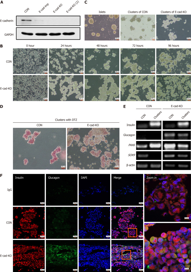
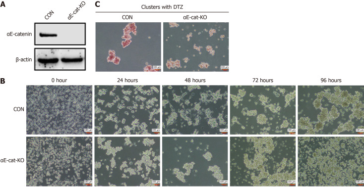
Methods: PANC-1 cells were induced by trypsin to form ILAs and differentiate into insulin-secreting cells. Clustered regularly interspaced short palindromic repeats (CRISPR)-associated protein 9 knockout and small interfering RNA knockdown techniques were used to investigate membrane proteins and downstream signaling pathways involved in the process.

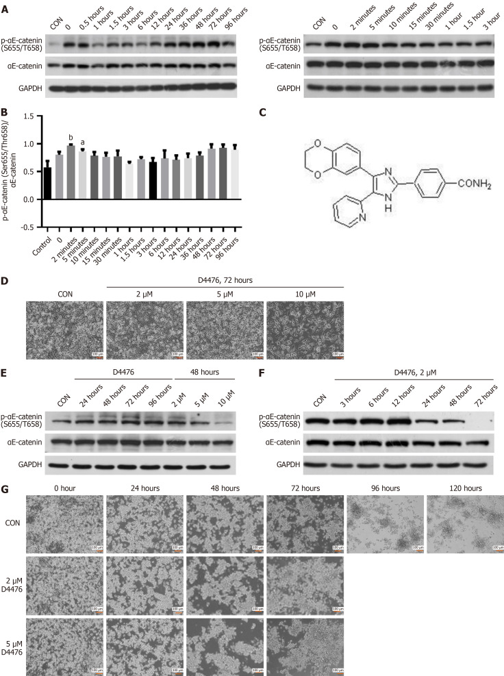
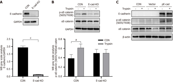
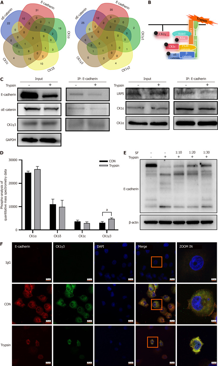
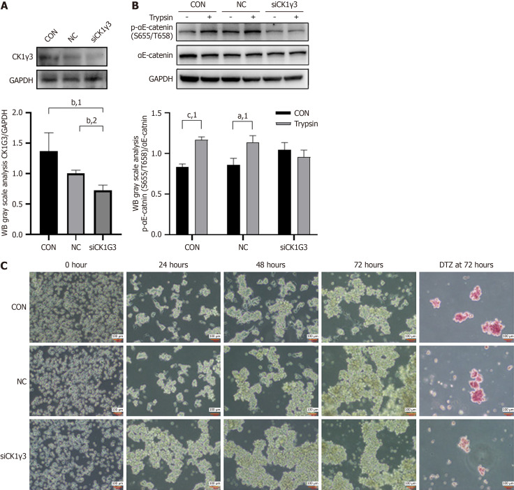
Results: The extracellular domain of membrane receptor E-cadherin hydrolyzed by trypsin induced the aggregation of PANC-1 cells and stimulated E-cadherin-recruited casein kinase-1γ3, which specifically phosphorylated the Ser655/Thr658 site of α-catenin in the cadherin-catenin complex, participating in the process of PANC-1 differentiation and affecting the maturation of differentiated ILAs.

Conclusion: The current study reveals the mechanism by which trypsin promotes PANC-1 cell differentiation into islet-like cells, providing a novel approach for endogenous islet β-cell regeneration.

Keywords: Alpha-catenin; E-cadherin; Endogenous islet β-cell regeneration; Pancreatic cancer cell line-1; Pancreatic islet progenitor cell; Trypsin.


Conflict of interest statement

Conflict-of-interest statement: The authors have no conflicts of interest to declare.

Figures

Figure 1
Figure 1
E-cadherin contributes to aggregation of human pancreatic cancer cell line-1 into islet-like clusters induced by trypsin. A: Expression of E-cadherin protein was detected by western blotting, reflecting knockout (KO) efficiency of E-cadherin 1 gene (CDH1), E-cad-imp group: Stable transgenic cells with CDH1 gene KO by clustered regularly interspaced short palindromic repeats (CRISPR)- -associated 9 (Cas9) system impure, E-cad-KO and E-cad-KO (2): Monoclonal cells screening from stable KO cells; B: Representative images of KO cells at five time points after digestion with trypsin before and after CDH1 KO, magnification 10 ×; C: Morphology of islet-like cell clusters differentiated in vitro was compared with that of natural islets; D: Representative image of diphenylterazine (DTZ) staining of cell clusters formed at 96 hours before and after CDH1 KO, magnification 10 ×; E: Polymerase chain reaction detection reflected the changes in expression of the transcripts for mature islet-related genes insulin, glucagon, paired box 6 (PAX6), SRY-box transcription factor 9 (SOX9) before and after CDH1 KO; F: Representative immunofluorescence images of insulin, glucagon, and 4’,6-diamidino-2-phenylindole (DAPI) signal in differentiated cell clusters before and after CDH1 KO, magnification 20 ×, increased to 63 ×. CON: Control; GAPDH: Glyceraldehyde-3-phosphate dehydrogenase.
Figure 2
Figure 2
αE-catenin is related to trypsin-mediated human pancreatic cancer cell line-1 aggregation and differentiation. A: Expression of αE-catenin protein was detected by western blotting, reflecting knockout (KO) efficiency of catenin alpha 1 (CTNNA1); B: Representative images of KO cells at five time points after digestion with trypsin before and after CTNNA1 KO, magnification 10 ×; C: Representative images of diphenylterazine (DTZ) staining of cell clusters formed at 96 hours before and after KO of CTNNA1, magnification 10 ×. CON: Control.
Figure 3
Figure 3
Phosphorylation of αE-catenin at Ser655/Thr658 during human pancreatic cancer cell line-1 differentiation and effects of casein kinase-1 inhibitor D4476 on human pancreatic cancer cell line-1. A: Representative western blot images reflecting protein expression of phospho-αE-catenin (p-αE-catenin) (Ser655/Thr658) at different time points during human pancreatic cancer cell line-1 (PANC-1) differentiation; B: Statistical analyses of the gray value of western blot images using ImageJ and GraphPad Prism, reflecting the differences in expression of p-αE-catenin (Ser655/Thr658); C: Chemical structure of D4476, inhibitor of casein kinase-1; D: Representative images of PANC-1 cells treated with 2, 5, or 10 μM D4476 for 72 hours, magnification 10 ×; E: Western blot images showed expression of p-αE-catenin (Ser655/Thr658) of PANC-1 cells treated with 2 μM D4476; F: Western blot images showed expression of p-αE-catenin (Ser655/Thr658) of PANC-1 cells treated with 2 μM D4476 at different times; G: Representative images of PANC-1 after 72 hours treatment with 2 μM D4476 at different time points after digestion with trypsin, magnification 10 ×. aP < 0.05. bP < 0.01. P calculated vs control (CON). GAPDH: Glyceraldehyde-3-phosphate dehydrogenase.
Figure 4
Figure 4
E-cadherin effects on phosphorylation of αE-catenin (Ser655/T658) mediated by trypsin. A: Representative western blot images reflecting protein expression of E-cadherin of cells with knockout (KO) of E-cadherin gene (CDH1); B: Representative western blot images reflecting protein expression of phospho-αE-catenin (p-αE-catenin) (Ser655/Thr658) in CDH1 KO cells and difference in trypsin-mediated αE-catenin (Ser655/Thr658) phosphorylation before and after KO; C: Representative western blot images reflecting protein expression of p-αE-catenin (Ser655/Thr658) of restoration of E-cadherin in CDH1 KO cells and difference in trypsin-mediated αE-catenin (Ser655/Thr658) phosphorylation before and after restoration (n = 2). aP < 0.05. cP < 0.001. P calculated vs control (CON). GAPDH: Glyceraldehyde-3-phosphate dehydrogenase.
Figure 5
Figure 5
Trypsin promotes binding of casein kinase-1γ3 to the E-cadherin complex. A: Wayne diagram, showing intersection of E-cadherin, αE-catenin, and casein kinase-1 (CK1) subtypes interacting proteins; B: Hypothetical model of the interaction among CK1, E-cadherin, and αE-catenin; C: Immunoprecipitation (IP) reflecting the protein composition of the E-cadherin complex; D: Quantitative mass spectrometric data analysis of phosphorylated protein, control group and trypsin representing the samples before and after trypsin treatment, and the ordinate reflects the level of phosphorylation; E: Western blot images reflect the hydrolysis of E-cadherin by trypsin and the blocking effect of different concentrations of trypsin inhibitor soybean flour (SF); F: Representative confocal images show colocalization changes of E-cadherin and CK1γ3 before and after trypsin treatment, Cy3 labeling E-cadherin (red), fluorescein isothiocyanate isomer labeling CK1γ3 (green), 4’,6-diamidino-2-phenylindole (DAPI) labeling nucleus (blue), magnification 20 ×, increased to 63 ×. aP < 0.05. P calculated vs control (CON). GAPDH: Glyceraldehyde-3-phosphate dehydrogenase.
Figure 6
Figure 6
Casein kinase-1γ3 participated in the αE-catenin (Ser655/Thr658) phosphorylation mediated by trypsin. A: Representative western blot (WB) images reflecting the efficiency of small interfering RNA knockdown of casein kinase-1γ3 (CK1γ3); B: Representative WB images reflecting protein expression of phospho-αE-catenin (Ser655/Thr658) in CK1γ3 knockdown cells and differences in trypsin-mediated αE-catenin (Ser655/Thr658) phosphorylation before and after knockdown; C: Representative images of knockout (KO) cells at five time points after digestion with trypsin and diphenylterazine (DTZ) staining of cell clusters formed at 72 hours before and after CK1γ3 knockdown, magnification 10 ×. aP < 0.05. bP < 0.01. cP < 0.001. 1P calculated vs control. 2P calculated vs negative control (NC). GAPDH: Glyceraldehyde-3-phosphate dehydrogenase; CON: Control.

Similar articles

References

    1. Sun H, Saeedi P, Karuranga S, Pinkepank M, Ogurtsova K, Duncan BB, Stein C, Basit A, Chan JCN, Mbanya JC, Pavkov ME, Ramachandaran A, Wild SH, James S, Herman WH, Zhang P, Bommer C, Kuo S, Boyko EJ, Magliano DJ. IDF Diabetes Atlas: Global, regional and country-level diabetes prevalence estimates for 2021 and projections for 2045. Diabetes Res Clin Pract. 2022;183:109119. - PMC - PubMed
    1. Lorenzati B, Zucco C, Miglietta S, Lamberti F, Bruno G. Oral Hypoglycemic Drugs: Pathophysiological Basis of Their Mechanism of ActionOral Hypoglycemic Drugs: Pathophysiological Basis of Their Mechanism of Action. Pharmaceuticals (Basel) 2010;3:3005–3020. - PMC - PubMed
    1. Kim HS, Lee MK. β-Cell regeneration through the transdifferentiation of pancreatic cells: Pancreatic progenitor cells in the pancreas. J Diabetes Investig. 2016;7:286–296. - PMC - PubMed
    1. Zhang J, Liu F. The De-, Re-, and trans-differentiation of β-cells: Regulation and function. Semin Cell Dev Biol. 2020;103:68–75. - PubMed
    1. Zhang S, Huang F, Tian W, Lai J, Qian L, Hong W, Chen H, Li LC. Andrographolide promotes pancreatic duct cells differentiation into insulin-producing cells by targeting PDX-1. Biochem Pharmacol. 2020;174:113785. - PubMed

LinkOut - more resources