. 2025 May 28:14:117-128.
doi: 10.1016/j.ncrna.2025.05.014. eCollection 2025 Oct.

LncRNA MEG3/CTCF-CXCR4 axis functions in the regulation of breast cancer cell migration

Affiliations

LncRNA MEG3/CTCF-CXCR4 axis functions in the regulation of breast cancer cell migration

Gusai Elhassan et al. Noncoding RNA Res. .

Abstract

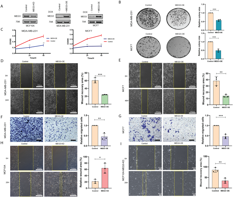
Loss or decreased expression of lncRNA MEG3 is a frequent event in the progression of many different malignancies. Overexpression of MEG3 in breast cancer cell lines MCF7 or MDA-MB-231 prevented cell migration, whereas depletion of MEG3 in human mammary epithelial cell line MCF10A strikingly promoted cell migration. As RNA-protein interactions are vital for RNA to function, RNP assembled on MEG3 in vivo was purified using affinity purification followed by mass spectrometry, which revealed ∼600 proteins with the potential to interact with MEG3. Bioinformatic analysis on RNA-seq data from MCF7 with MEG3 overexpression and MCF10A with MEG3 depletion led to the identification of CXCR4 as the major downstream mediator negatively regulated by MEG3 that facilitated breast cancer cell migration. In addition, the chromatin regulator CTCF emerged as the MEG3-binding protein that might regulate CXCR4 expression after comparison of proteins presenting in MEG3 lncRNP to ChIP-seq data and GPSAdb data of CXCR4. Further evidence was provided to show CTCF upregulated the expression of CXCR4 at transcriptional level, whereas co-expression of MEG3 with CTCF abolished transcriptional activation of CXCR4. Overall, our study pinpoints the importance of MEG3/CTCF-CXCR4 axis in regulating migration of breast cancer cells and provides novel insight into the mechanism of lncRNA MEG3 in cancer development.

Keywords: Breast cancer; CTCF; CXCR4; Cell migration; lncRNA MEG3.


Conflict of interest statement

The authors declare that they have no known competing financial interests or personal relationships that could have appeared to influence the work reported in this paper.

Figures

Fig. 1
Fig. 1
Decreased expression of nuclear lncRNA MEG3 in breast cancer. (A) Detection of MEG3 in normal, tumor, and metastatic breast cancer in TNMplot database. (B) Kaplan-Meier recurrence-free survival analysis of MEG3 using breast cancer data in Kaplan-Meier Plotter database. (C) RT-PCR of endogenous lncRNA MEG3 expression in various breast cancer cell lines. (D) Schematic of the MEG3 construct, β-globulin intron showed as dotted line, numbers represent the length of MEG3 transcript, cDNA showed as box, pA (poly A signal), probe showed as red hybridized sequence. (E) RNA-FISH to visualize the localization of MEG3 transcript post-transfection in HeLa, MCF10A, MCF7 and MDA-MB-231. DAPI staining was utilized to identify the nucleus. (F) Silver stain for lncRNPs assembled on the sense strand of MEG3 with the antisense strand used as a control, separated on SDS-PAGE. (G) Western blot analysis for the selected proteins for MEG3 mass spectrometry data verification.
Fig. 2
Fig. 2
MEG3 is crucial for breast cancer cell proliferation and migration. (A) RT-PCR analysis of MEG3 expression in MEG3-KD cell line (MCF10A), MEG3-OE cell lines (MDA-MB-231, and MCF7), respectively. KD: knockdown, OE: overexpression. (B) Colony formation assay to assess the impact of MEG3 overexpression in MDA-MB-231 and MCF7 cell lines (n = 3, ∗∗∗p < 0.001). (C) MTT analysis of cell viability in MEG3-OE cell lines MDA-MB-231 and MCF7 (n = 3, ∗∗p < 0.01). (D–E) Wound healing assay to demonstrate the migratory capacity of MDA-MB-231 and MCF7 cell lines following MEG3 overexpression (the scale bar represents 500 μm) and further evaluated using a Transwell assay (the scale bar represents 200 μm) (F–G) (n = 3, ∗∗p < 0.01, ∗∗∗p < 0.001). (H) Wound healing assay in MCF10A-MEG3-KD cell line (n = 3, ∗p < 0.05) (the scale bar represents 500 μm). (I) Wound healing assay in MCF10A-MEG3-KD cell line following re-expression of MEG3 (the scale bar represents 50 μm) (n = 3, ∗∗p < 0.01).
Fig. 3
Fig. 3
CXCR4 is the major downstream mediator regulated by MEG3. (A) Venn diagram illustrating the overlap of differentially expressed genes upon MEG3 knockdown and overexpression. (B) Heatmap representation of the differentially expressed genes with a log2 fold change more than −1.5 and 1.5 and a significance level of padj<0.05. (C) Proteins association network analysis using String database. (D–E) RT-PCR analysis showing the expression of CXCR4 at RNA level upon MEG3 knockdown or overexpression (n = 4, ∗∗p < 0.01, ∗∗∗p < 0.001). (F–G) Western blot analysis showing the expression of CXCR4 at protein level upon MEG3 knockdown or overexpression (n = 4, ∗∗p < 0.01, ∗∗∗p < 0.001).
Fig. 4
Fig. 4
MEG3 regulates cell migration via CXCR4. (A) Expression of CXCR4 in normal, tumor, and metastatic breast cancer from TNMplot database. (B) Kaplan-Meier recurrence-free survival analysis for CXCR4 using breast cancer data in Kaplan-Meier Plotter database. (C) Spearman correlation analysis between MEG3 and CXCR4 in breast cancer data from TNMplot database. (D) Western blot analysis of CXCR4 knockdown using two siRNAs. (E) Wound healing assay for MCF7 following CXCR4 knockdown using two siRNAs (n = 3, ∗p < 0.05, ∗∗p < 0.01) (the scale bar represents 500 μm). (F–G) Wound healing and Transwell assay for MEG3 re-expression in MCF7 after depletion of CXCR4 (n = 3, ∗p < 0.05, ∗∗p < 0.01, ∗∗∗p < 0.001, ns: no significance) (the scale bar represents 500 μm for Wound healing assay and 200 μm for Transwell assay).
Fig. 5
Fig. 5
MEG3 interacts with CTCF to restrain the transcriptional activation on CXCR4. (A) Venn diagram illustrates the proteins detected in our MEG3 mass spectrometry data, predicted MEG3 binding proteins from the AnnoLnc2 database, CXCR4-ChIP-seq data from the Cistrome toolkit database, and data from genetic perturbation similarity analysis for CXCR4 upstream regulators from the GPSAdb (Duplicated gene names were removed). (B) RT-PCR was performed to analyze MEG3 lncRNA precipitated from RIP experiment with a CTCF antibody, IgG antibody was used as a control. (C) RNA-FISH was used to demonstrate the co-localization of the full-length MEG3 transcript and the CTCF-EGFP protein. (D) Western blot analysis was conducted on MDA-MB-231 cells following the knockdown of CTCF using two siRNAs to assess the impact on CXCR4 expression (n = 3, ∗∗p < 0.01, ∗∗∗p < 0.001). (E) Wound healing assay to evaluate the migratory capacity of MDA-MB-231 cells after the downregulation of CXCR4 caused by CTCF knockdown (n = 3, ∗∗p < 0.01, ∗∗∗p < 0.001) (the scale bar represents 500 μm). (F) Transwell assay for MDA-MB-231 after CTCF depletion (n = 3, ∗∗∗p < 0.001) (the scale bar represents 200 μm). (G&H) Wound healing assay and Transwell assay for MEG3 overexpression and CTCF depletion dual treatment (n = 3, ∗∗p < 0.01, ∗∗∗p < 0.001, ns: no significance) (the scale bar represents 500 μm for Wound healing assay and 200 μm for Transwell assay). (I) Western blot analysis showing the effect of MEG3 overexpression in combination with CTCF depletion on CXCR4 gene expression (n = 3, ∗∗∗p < 0.001, ns: no significance). (J) Western blot analysis showing the effect of dual overexpression of MEG3 and CTCF on CXCR4 gene expression (n = 3, ∗p < 0.05, ∗∗p < 0.01, ns: no significance).
Fig. 6
Fig. 6
MEG3/CTCF-CXCR4 axis regulates breast cancer cell migration. Study model: The expression of lncRNA MEG3 in the nucleus of normal cells allows interaction of CTCF protein to MEG3, which restrains CTCF from activating CXCR4 transcription. Consequently, CXCR4 is expressed at a low level that cannot promote cell migration. In contrast, since the expression of MEG3 is lost or significantly reduced in breast cancer cells, CTCF acts freely to activate the expression of CXCR4. Breast cancer cells with a high level of CXCR4 are subject to enhanced cell migration. Created with BioRender.com.
Supplementary Fig. 1
GO analysis for MEG3 mass spectrometry and RNA seq data (A) Gene ontology analysis (Molecular function) based on Enrichr database for our MEG3 mass spectrometry data overlapped with the predicted MEG3-binding proteins using Annolnc2. (B) Gene ontology analysis (Biological process) for the differentially expressed genes with a log2 fold change more than −1.5 and 1.5 and a significance level of padj < 0.05 using String database. (B) Pathway analysis for the differentially expressed genes using Enrich database.
Supplementary Fig. 2
RNA seq data analysis for TCGA breast cancer using UCSC Xena Database (A) Heatmap of MEG3 and CXCR4 expression in TCGA breast cancer data (n = 1218) according to sample type from UCSC Xena database. (B) Violin view of MEG3 expression in TCGA breast cancer data. (C) Violin view of CXCR4 expression in TCGA breast cancer data.

Similar articles

References

    1. Wylie A.A. Novel imprinted DLK1/GTL2 domain on human chromosome 14 contains motifs that mimic those implicated in IGF2/H19 regulation. Genome Res. 2000;10:1711–1718. doi: 10.1101/gr.161600. - DOI - PMC - PubMed
    1. Azam S., Hou S., Zhu B., Wang W., Hao T., Bu X., Khan M., Lei H. Nuclear retention element recruits U1 snRNP components to restrain spliced lncRNAs in the nucleus. RNA Biol. 2019;16:1001–1009. doi: 10.1080/15476286.2019.1620061. - DOI - PMC - PubMed
    1. Cabili M.N., Dunagin M.C., McClanahan P.D., Biaesch A., Padovan-Merhar O., Regev A., Rinn J.L., Raj A. Localization and abundance analysis of human lncRNAs at single-cell and single-molecule resolution. Genome Biol. 2015;16:20. doi: 10.1186/s13059-015-0586-4. - DOI - PMC - PubMed
    1. Wang Y., Li R.-W., Li X.-T., Xin Y. Decreased long non-coding RNA MEG3 expression is associated with survival outcome and lymph node metastasis: a meta-analysis. Int. J. Clin. Exp. Med. 2017;10(4):5913–5921.
    1. Zhou Y., Zhang X., Klibanski A. MEG3 noncoding RNA: a tumor suppressor. J. Mol. Endocrinol. 2012;48:R45–R53. doi: 10.1530/JME-12-0008. - DOI - PMC - PubMed

LinkOut - more resources