

Imaging diagnosis of paranasal sinus mucocele in a Yorkshire Terrier dog
- PMID: 40548342
- PMCID: PMC12178644
- DOI: 10.17221/207/2020-VETMED
Imaging diagnosis of paranasal sinus mucocele in a Yorkshire Terrier dog
Abstract
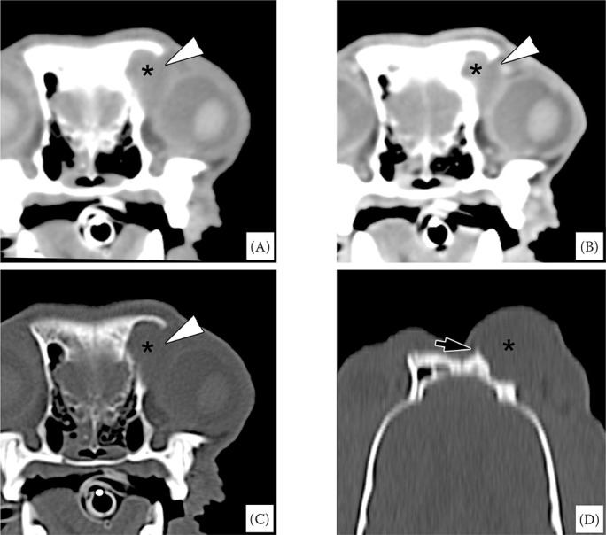
An 8-year-old, neutered male Yorkshire Terrier dog presented with left ventromedial canthus swelling over a one-month period, refractory to pharmacological therapy. There was no history of trauma. On ultrasonography, the lesion had a cystic character filled with anechoic fluid and hyperechoic sedimenting debris. The computed tomography (CT) and CT-dacryocystography showed a cystic lesion protruding from the lacrimal sac fossa and occupying a defect in the orbital plate and an ethmoidal ectoturbinate surrounded by a bony structure with an intact nasolacrimal system. The dog underwent the surgical resection of the cyst and its fluid content was aspirated. Ethmoid mucocele was diagnosed based on the CT, cytologic examination, bacterial culture and histopathologic findings. This case describes the imaging characteristics of an ethmoid mucocele and highlights the importance of CT and CT-dacryocystography in dogs with ventromedial canthus swelling that had poor response to medical treatment.
Keywords: canine; computed tomography; dacryocystography; ethmoid mucocele.
Copyright: © 2021 Noh et al.
Conflict of interest statement
The authors declare no conflict of interest.
Figures


References
-
- Adamo PF. Intracranial epidural mucocele in a cat. J Am Anim Hosp Assoc. 2005 Jan-Feb;41(1):74-7. - PubMed
-
- Bleier B, Palmer JN, Woodworth BA. Recurrent mucoceles. In: Kountakis SE, Jacobs JB, Gosepath J, editors. Revision sinus surgery. Berlin, Germany: Springer; 2018. p. 185-92.
-
- Eggesbo HB. Radiological imaging of inflammatory lesions in the nasal cavity and paranasal sinuses. Eur Radiol. 2006 Apr;16(4):872-88. - PubMed
Publication types
LinkOut - more resources
Full Text Sources