. 2025 Jun 23:13:RP104475.
doi: 10.7554/eLife.104475.

Cognitive control of behavior and hippocampal information processing without medial prefrontal cortex

Affiliations

Cognitive control of behavior and hippocampal information processing without medial prefrontal cortex

Eun Hye Park et al. Elife. .

Abstract

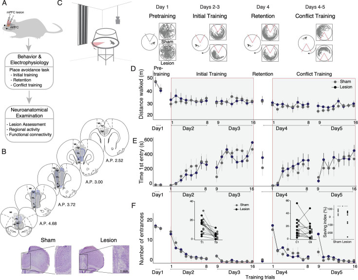
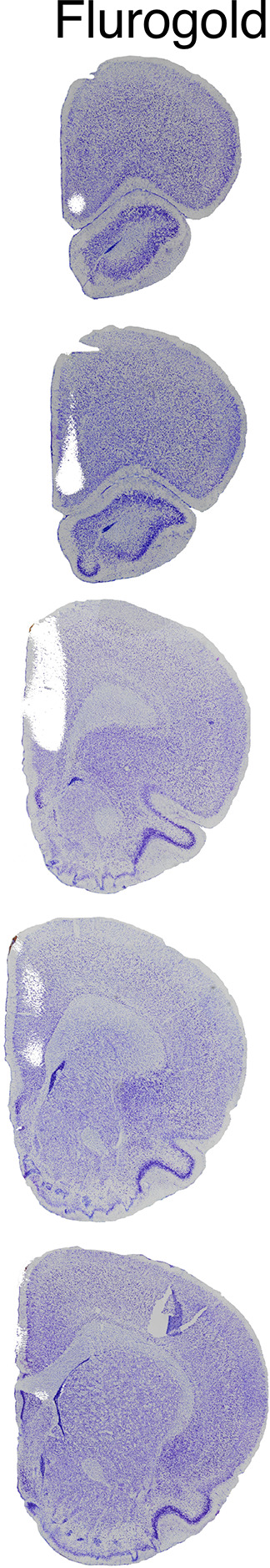
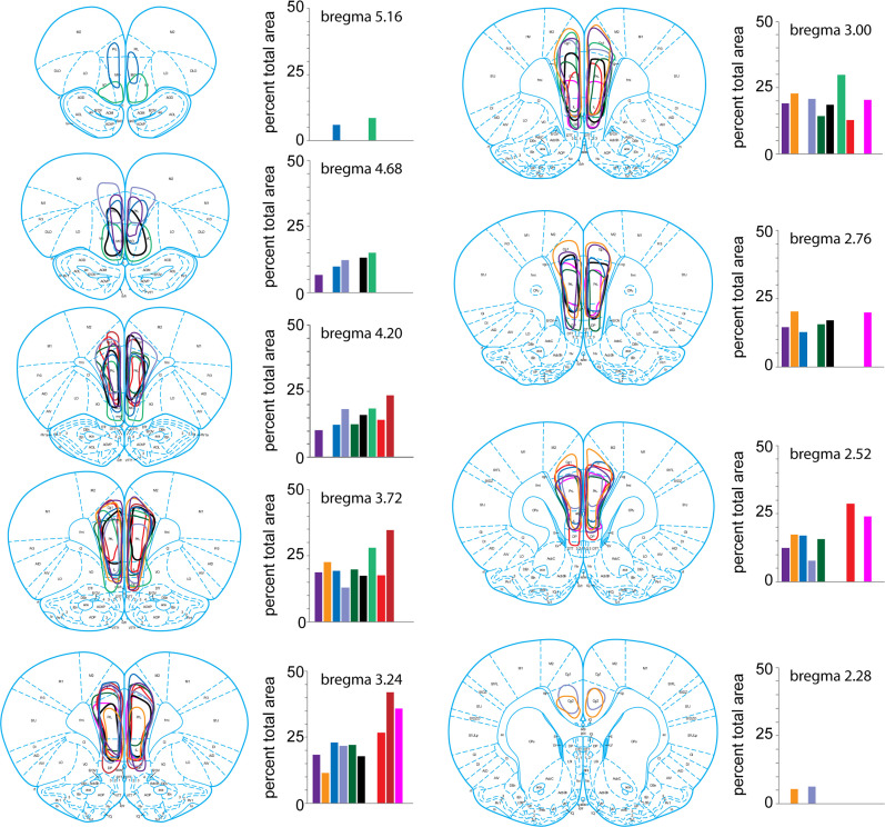
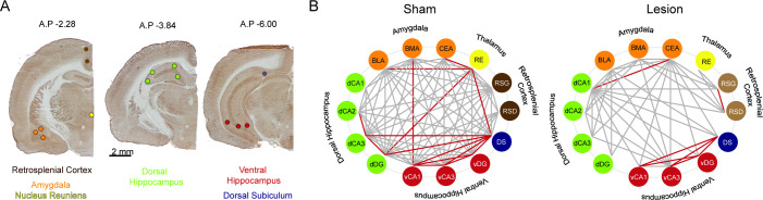
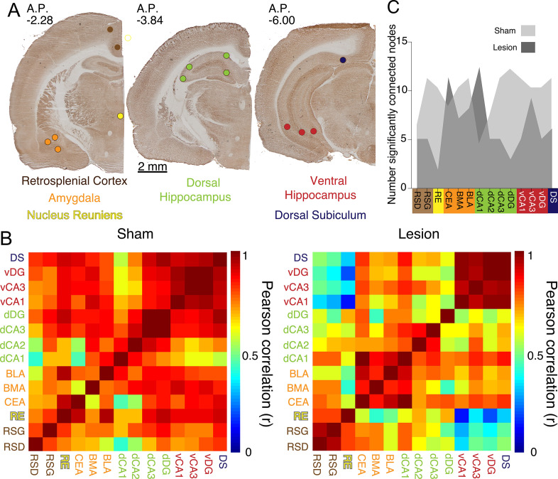
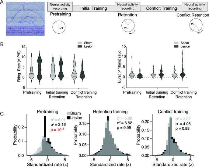
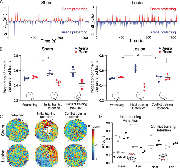
Cognitive control tasks require using one class of information while ignoring competing classes of information. The central role of the medial prefrontal cortex (mPFC) in cognitive control is well established in the primate literature and largely accepted in the rodent literature because mPFC damage causes deficits in tasks that may require cognitive control, as inferred, typically from the task design. In prior work, we used an active place avoidance task where a rat or mouse on a rotating arena is required to avoid the stationary task-relevant locations of a mild shock and ignore the rotating task-irrelevant locations of those shocks. The task is impaired by hippocampal manipulations, and the discharge of hippocampal place cell populations judiciously alternates between representing stationary locations near the shock zone and rotating locations far from the shock zone, demonstrating cognitive control concurrently in behavior and the hippocampal representation of spatial information. Here, we test whether rat mPFC lesion impairs the active place avoidance task to evaluate two competing hypotheses, a 'central-computation' hypothesis that the mPFC is essential for the computations required for cognitive control and an alternative 'local-computation' hypothesis that other brain areas can perform the computations required for cognitive control, independent of mPFC. Ibotenic acid lesion of the mPFC was effective, damaging the cingulate, prelimbic, and infralimbic cortices. The lesion also altered the normal coordination of metabolic activity across remaining structures. The lesion did not impair learning to avoid the initial location of shock or long-term place avoidance memory, but impaired avoidance after the shock was relocated. The lesion also did not impair the alternation between task-relevant and task-irrelevant hippocampal representations of place information. These findings support the local-computation hypothesis that computations required for cognitive control can occur locally in brain networks independently of the mPFC.

Keywords: decision; executive control; hippocampus; learning; memory; neural coordination; neuroscience; rat.


Conflict of interest statement

EP, KO, GG, DT, KN, AA, NR, NK, SS, AF No competing interests declared

Figures

Figure 1.
Figure 1.. Medial prefrontal cortex (mPFC) lesion does not impair initial active place avoidance learning, but impairs cognitive flexibility in the conflict task variant.
(A) Workflow to assess the impact of mPFC or sham lesions on spatial cognitive control. (B) Assessment and impact of the mPFC lesion in three representative subjects (light gray [intermediate with dorsal anterior lesion], dark gray [intermediate with ventral anterior lesion – includes medial orbital cortex], and purple [largest lesion]). The smallest lesion spanned A-P 3.24–2.52. PrL (prelimbic), IL (infralimbic), Cg (cingulate cortex), M2 (secondary motor cortex). Representative mPFC from a sham and a lesion rat (bottom). (C) Tracked room-frame positions from two example rats across active place avoidance training. The shock zone is indicated as a 60° sector, gray (shock off) and red (shock on), and arena rotation by the curved arrow. Day 1– pretraining: free exploration with shock off; days 2 and 3 – initial training: eight daily trials to avoid the shock zone. Day 4 – retention: one trial with shock on. Days 4 and 5 – conflict training: eight daily trials to avoid the shock zone relocated 180° from the initial location. Sham and mPFC lesion rats did not differ in (D) locomotor activity, (E) avoidance memory, or (F) place learning. (F) The left inset compares the number of entrances during the first 5 min of the first (D2T1) and second days of initial training (D3T9). The right inset compares the first 5 min of the first (D4C1) and second (D5C9) days of conflict trials. The percent difference in number of entrances between D5C9 and D4C1 of conflict training was computed as a savings index. Savings was not related to lesion size r=0.009, p=0.98. *p<0.05. Sham: n=8; lesion: n=10.
Figure 1—figure supplement 1.
Figure 1—figure supplement 1.. Targeting the medial prefrontal cortex (mPFC) injections.
To determine the appropriate coordinates for injection of ibotenic acid, we first injected fluorogold and immediately examined the extent of fluorescent labeling, followed by Nissl counterstaining. Fluorogold labeling was confirmed in the cingulate, prelimbic, and infralimbic cortices.
Figure 1—figure supplement 2.
Figure 1—figure supplement 2.. Medial prefrontal cortex (mPFC) lesions.
The extent of each rat’s lesion was traced and quantified as the percentage of the total mPFC at each of nine coronal planes through the A-P extent of the mPFC.
Figure 2.
Figure 2.. Cytochrome oxidase (CO) analysis demonstrates widespread metabolic consequences of medial prefrontal cortex (mPFC) lesion.
(A) Representative CO staining and locations of the optical density readings. CO activity was measured in the dysgranular and granular retrosplenial cortices (RSD and RSG, respectively), the nucleus reuniens (RE), the central nucleus of the amygdala (CEA), basomedial and basolateral amygdala (BMA and BLA, respectively), the dorsal hippocampus CA1, CA2, CA3 and dentate gyrus areas (dCA1, dCA2, dCA3, dDG, respectively) the ventral hippocampus CA1, CA3 and dentate gyrus areas (vCA1, vCA3, vDG, respectively), and the dorsal subiculum (DS) marked as colored circle areas. (B) Interarea covariations of CO activity by graph theory analysis. Each line indicates a significant correlation (p<0.05) between the two brain regions (‘nodes’) it connects; only the red lines survived false discovery rate (FDR <0.01) correction. Sham: n=8; lesion: n=8.
Figure 2—figure supplement 1.
Figure 2—figure supplement 1.. Medial prefrontal cortex (mPFC) lesions change covarying resting-state metabolic activity between the dorsal and ventral hippocampus.
(A) Representative cytochrome oxidase staining and locations of the optical density readings. Cytochrome oxidase activity was measured in the dysgranular and granular retrosplenial cortices (RSD and RSG, respectively), the nucleus reuniens (RE), the central nucleus of the amygdala (CEA), basomedial and basolateral amygdala (BMA and BLA, respectively), the dorsal hippocampus CA1, CA2, CA3, and dentate gyrus areas (dCA1, dCA2, dCA3, dDG, respectively), the ventral hippocampus CA1, CA2, and CA3 areas (vCA1, vCA3, vDG, respectively), and the dorsal subiculum (DS) marked as colored circles. (B) The matrix of interregional cytochrome oxidase activity correlations indicates that mPFC lesions reduce Pearson correlations in the lesion group. Univariate correlations among hippocampus, nucleus reunions, amygdala, and DS decreased after mPFC lesion, but these decreases did not survive the p<0.0005 Bonferroni correction for the 91 comparisons (see Supplementary file 1 for abbreviations: BLA-RE: z=1.91, p=0.05, dCA1-CEA: z=3.12, p=0.001, dDG-dCA3: z=2.79, p=0.005, vCA1-RE: z=1.94, p=0.05, DS-RE: z=1.92, p=0.05, DS-dDG: z=1.9, p=0.05). (C) Number of significantly connected nodes at each site before FDR correction. Sham: n=8; lesion: n=8.
Figure 3.
Figure 3.. Medial prefrontal cortex (mPFC) lesion does not change basic discharge properties, but decreases hippocampus place cell overdispersion only in the absence of the cognitive control challenge.
(A) Left: representative histology with overlaid recording traces from the Neuropixel probe. Right: recording schedule workflow during the cognitive control task. (B) There is no difference between sham and mPFC lesion rats in firing rate (sham: 3.29±0.27, lesion: 3.68±0.33, t184=0.91, p=0.36) and burst ratio in hippocampal neurons (sham: 1.53±0.25, lesion: 2.02±0.28, t174=1.29, p=0.19). (C) Distribution of standardized place cell discharge (z scores) computed during every 5 s episode in which the rat passed through place cell firing fields in the data set. Different numbers of passes qualified for evaluation during the pretraining (sham = 2777, lesion = 3398), retention (sham = 4039, lesion = 3366), and conflict (sham = 1748, lesion = 2336) recordings. The variance of the histograms characterizes overdispersion, statistically evaluated by their ratio as an F-test (pretraining: F2776,3397 = 2.19, p=5.8 × 10⁻⁴). Sham: n=3; lesion: n=3 rats.
Figure 4.
Figure 4.. Sham and medial prefrontal cortex (mPFC) lesion rats do not differ in expressing cognitive control of spatial frame-specific representations of location.
(A) Individual ensemble examples during the day 4 retention session and (B) group statistics of spatial frame ensemble preference (SFEP), demonstrating cognitive control in both groups. During retention, SFEP is biased to the arena frame in both the sham (t4=4.16, #p=0.01) and lesion rats (t4=6.62, #p=0.003). *p<0.05 post-hoc differences. (C) Group spatial probability distribution of SFEP for room frame preference during the pretraining, initial, and conflict retention sessions with no shock. (D) Summary of average probability of room-preferring SFEP discharge in half the arena near and far from the shock zone during the initial and conflict retention sessions. Two-way group × location ANOVA (sham: n=3, lesion: n=3) during retention of the initial shock zone location: Group: F1,4 = 0.17, p=0.68, η2=10–3; location: F1,4 = 54.69, p=0.001, η2=0.84; group × location F1,4 = 2.04, p=0.2, η2=10–2; during retention of the conflict shock zone location: group: F1,4 = 0.71, p=0.45, η2=0.05; location: F1,4 = 0.42, p=0.55, η2=0.056; group × location F1,4 = 0.67, p=0.45, η2=0.08. Sham: n=3; lesion: n=3 rats.

Update of

  • doi: 10.1101/2019.12.20.884262
  • doi: 10.7554/eLife.104475.1
  • doi: 10.7554/eLife.104475.2

Similar articles

References

    1. Adhikari A, Topiwala MA, Gordon JA. Synchronized activity between the ventral hippocampus and the medial prefrontal cortex during anxiety. Neuron. 2010;65:257–269. doi: 10.1016/j.neuron.2009.12.002. - DOI - PMC - PubMed
    1. Badre D, Bhandari A, Keglovits H, Kikumoto A. The dimensionality of neural representations for control. Current Opinion in Behavioral Sciences. 2021;38:20–28. doi: 10.1016/j.cobeha.2020.07.002. - DOI - PMC - PubMed
    1. Badre D. Cognitive Control. Annual Review of Psychology. 2024;76:167–195. doi: 10.1146/annurev-psych-022024-103901. - DOI - PubMed
    1. Birrell JM, Brown VJ. Medial frontal cortex mediates perceptual attentional set shifting in the rat. The Journal of Neuroscience. 2000;20:4320–4324. doi: 10.1523/JNEUROSCI.20-11-04320.2000. - DOI - PMC - PubMed
    1. Botvinick MM, Braver TS, Barch DM, Carter CS, Cohen JD. Conflict monitoring and cognitive control. Psychological Review. 2001;108:624–652. doi: 10.1037/0033-295x.108.3.624. - DOI - PubMed

LinkOut - more resources