. 2025 Aug 5;13(8):e0342324.
doi: 10.1128/spectrum.03423-24. Epub 2025 Jun 23.

African swine fever virus MGF505-3R facilitates ferroptosis to restrict TBK1-IRF3 pathway

Affiliations

African swine fever virus MGF505-3R facilitates ferroptosis to restrict TBK1-IRF3 pathway

Sai Niu et al. Microbiol Spectr. .

Abstract

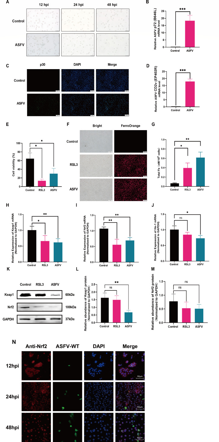
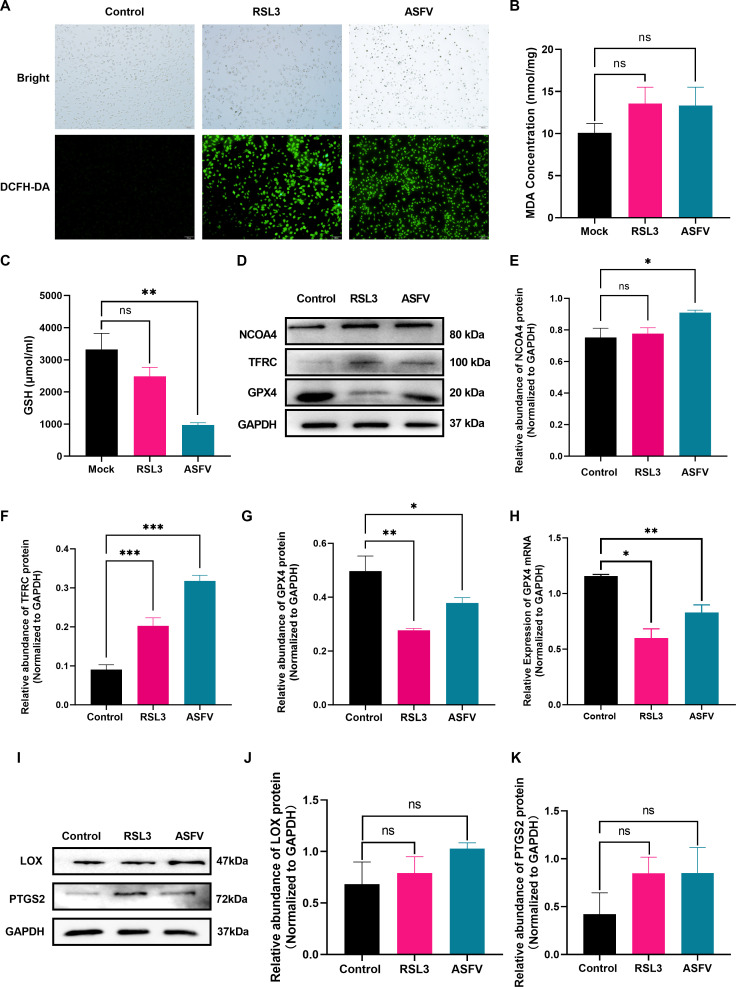
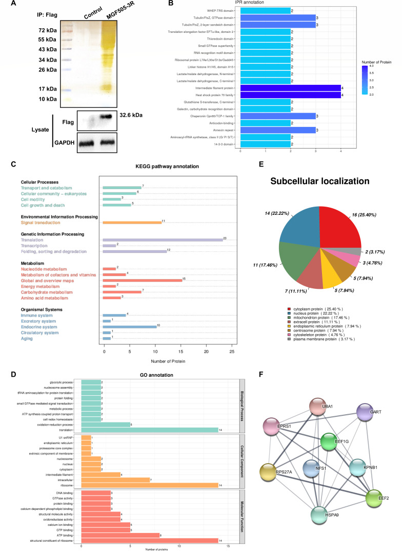
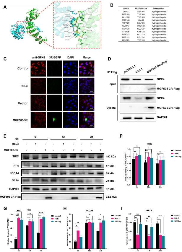
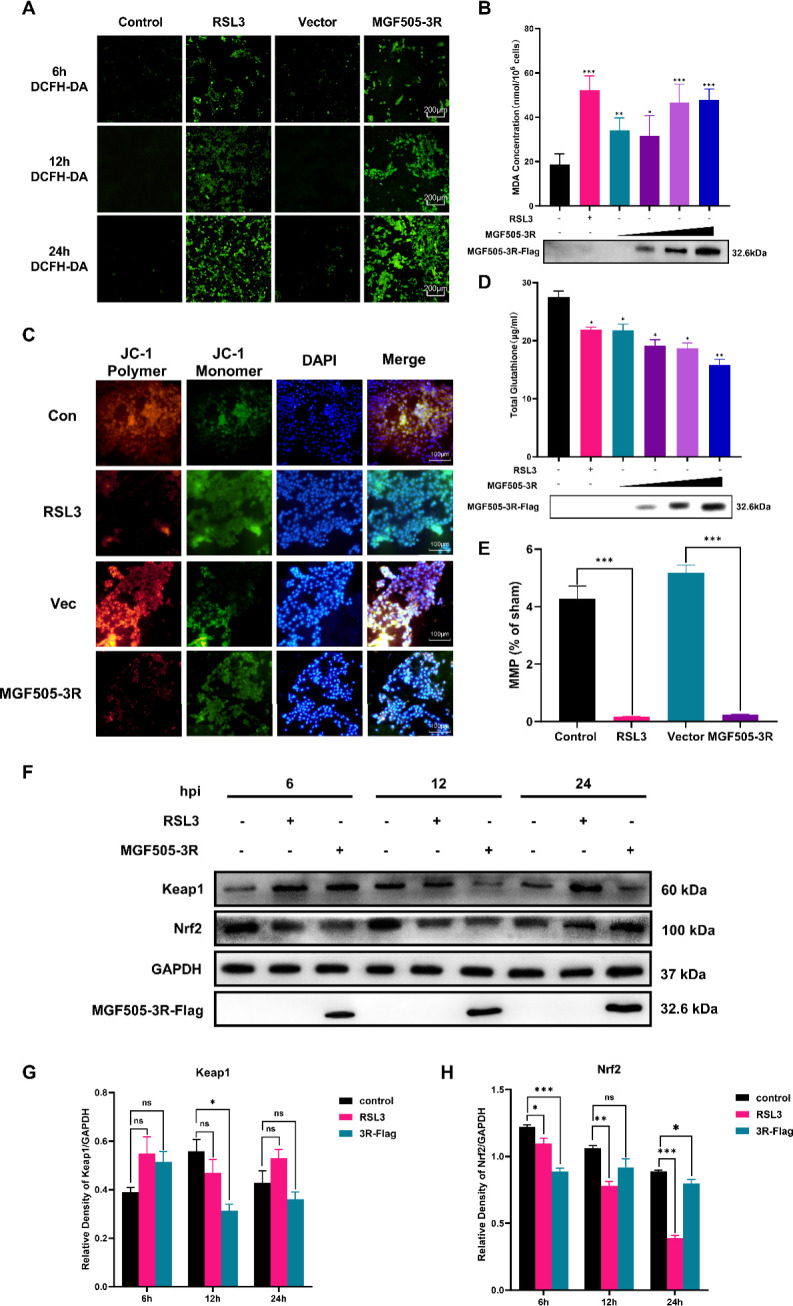
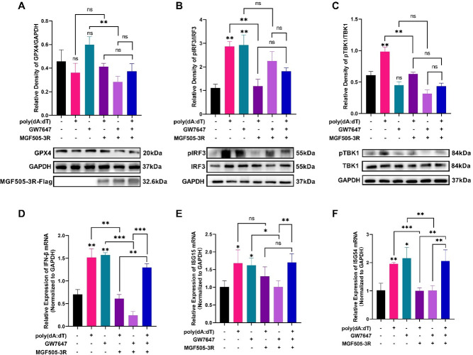
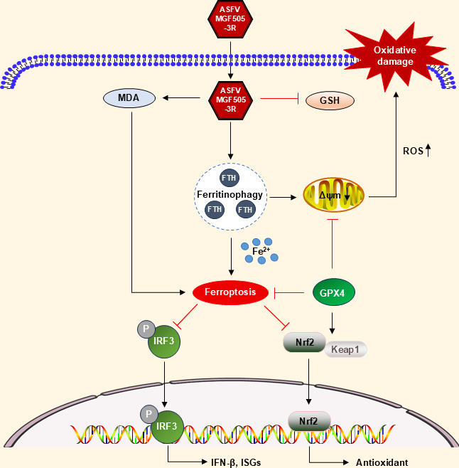
African swine fever virus (ASFV) causes hemorrhagic, severe infectious diseases and serious economic losses to the pig industry. ASFV multigene family 505 can antagonize the host's innate immunity through multiple signaling pathways and is considered an important target for vaccine development. However, the mechanism by which it induces host cell damage remains unclear. In this study, we observed that ASFV infection, similar to RSL3, can induce ferroptosis with the accumulation of reactive oxygen species (ROS) and iron, decrease glutathione peroxidase 4 (GPX4) expression, and restrain the Kelch-like ECH-associated protein 1-nuclear factor E2-related factor (Keap1-Nrf2) pathway. Moreover, the expression of ferroptosis biomarkers (LOX and PTGS2) has been moderately upregulated. Some proteins related to ASFV replication, invasion, and infection were evaluated for evidence of ferroptosis. MGF505-3R interacts with GPX4 to undergo ferroptosis, resulting in ROS accumulation, mitochondrial membrane potential destruction, and NCOA4-mediated ferritinophagy elevation. In addition, MGF505-3R suppressed the Keap1-Nrf2 pathway, while GPX4 activation counteracted its stimulatory effect on TANK-binding kinase 1 (TBK1)-IRF3 phosphorylation. Importantly, the transcription levels of interferon beta (IFN-β), ISG15, and ISG54 were elevated after GPX4 activation, suggesting that ferroptosis resistance could reverse the inhibition of the TBK1-IRF3 pathway and IFN-β levels induced by MGF505-3R. These findings provide new ideas and directions for elucidating the mechanism of ASFV-induced oxidative damage and lay a significant foundation for revealing the pathogenic mechanism of the virus by targeting ferroptosis.

Importance: We revealed that ASFV infection and MGF505-3R transfection induced the accumulation of iron and ROS, resulting in NCOA4-mediated ferritinophagy and ferroptosis, as well as restricted GPX4 expression and the Keap1-Nrf2 pathway. GPX4 activation promotes the TBK1-IRF3-IFN-β pathway and exerts antiviral activity. These findings indicate that ASFV facilitates ferroptosis, providing a proof of principle that may be applicable to oxidative damage and lipid peroxidation manipulation-based therapy for ASFV infection. Given the GPX4 downregulation in ASFV infection, GPX4 activation and ferroptosis resistance highlight its potential as a therapeutic target for viral infection.

Keywords: African swine fever virus; GPX4; IRF3; MGF505-3R; ferroptosis.


Conflict of interest statement

The authors declare no conflict of interest.

Figures

Fig 1
Fig 1
ASFV infection elevates the intracellular iron levels and restricts Keap1-Nrf2 signaling in the PAM cells. PAM cells were treated with ASFV and RSL3. The cells and RNA were harvested and subjected to microscopy, qPCR, or immunofluorescence. (A) The observations of the PAM cells under microscope after ASFV infection (multiplicity of infection of 1.0) at 12, 24, and 48 h. The phenomenon of cell death and shedding was obviously observed after treatment for 24 h. The subsequent assays were carried out at 24 h after ASFV treatment. (B) p72 mRNA expression. (C) p30 protein expression. (D) CD2v mRNA expression. (E) Cell activity. (F and G) Intracellular iron levels. (H) Keap1 mRNA expression. (I) Nrf2 mRNA expression. (J) HO-1 mRNA expression. (K and L) Keap1 expression. (K and M) Nrf2 expression. (N) The dynamic changes of Nrf2 localization. ASFV-WT indirect immunofluorescence of p30 protein. *P < 0.05, **P < 0.01, ***P < 0.001. ns, not significant.
Fig 2
Fig 2
ASFV infection induces ferroptosis in host cells. The PAM cells were treated with ASFV and RSL3 for 24 h. The cells, protein, and RNA were harvested. The samples were subjected to ROS level assay, Western blot, and qPCR. (A) ROS accumulation. (B) MDA concentration. (C) GSH levels. (D and E) NCOA4 protein expression. (D and F) TFRC protein expression. (D and G) GPX4 protein expression. (H) GPX4 mRNA expression. (I and J) LOX protein expression. (I and K) PTGS2 protein expression. *P < 0.05, **P < 0.01, ***P < 0.001. ns, not significant.
Fig 3
Fig 3
Identification of the proteins interacting with MGF505-3R in PK-15 cells. (A) Western blotting and silver staining were used to detect the expression of exogenous MGF505-3R and the enrichment of MGF505-3R interacting proteins in PK-15 cells. Empty Flag vector or Flag-MGF505-3R was transfected into PK-15 cells. Pull down was used with anti-Flag monoclonal antibody 24 h after transfection. MGF505-3R interacting host proteins were eluted from protein A + G agarose gels and analyzed on SDS-PAGE followed by silver staining. GAPDH was used as an internal control. (B) Gene ontology (GO) analysis. (C) Subcellular localization analysis of interacting proteins. (D) InterPro (IPR) database analysis. (E) Kyoto Encyclopedia of Genes and Genomes (KEGG) pathway database analysis. (F) Drawn MGF505-3R interacting protein grid with Cytoscape and STRING software.
Fig 4
Fig 4
MGF505-3R interacts with endogenous GPX4 to facilitate ferroptosis in the PK-15 cells. Molecular docking was used to analyze the interactions of MGF505-3R and GPX4. The cells were transfected with pcDNA3.1-MGF505-3R-Flag plasmid or treated with RSL3. The cells and proteins were collected. Immunoprecipitation and Western blot were carried out with the indicated antibodies. (A) The interaction structure between GPX4 and MGF505-3R was predicted by AlphaFold3 and visualized by Pymol. (B) Hydrogen bonding sites were predicted based on the structure by Pymol. (C) The colocalization of MGF505-3R and endogenous GPX4. (D) The interaction of MGF505-3R and GPX4. (E and F) TFRC protein expression. (E and G) FTH protein expression. (E and H) NCOA4 protein expression. (E and I) GPX4 protein expression. *P < 0.05, **P < 0.01,***P < 0.001. ns, not significant.
Fig 5
Fig 5
MGF505-3R impairs mitochondrial function and restricts the Keap1-Nrf2 signaling pathway to facilitate oxidative damage. PK-15 cells were transfected with pcDNA3.1-MGF505-3R-Flag plasmid and treated as indicated. Cells and proteins were extracted for analyzing the oxidative damage biomarker and kKeap1-Nrf2 pathway. (A) ROS detection by using fluorescent dye DCFH-DA. (B) MDA levels. (C) GSH levels. (D and E) MMP detection by using JC-1 staining. (F and G) Keap1 expression. (F and H) Nrf2 expression. *P < 0.05, **P < 0.01, ***P < 0.001. ns, not significant.
Fig 6
Fig 6
MGF505-3R blocks the TBK1-IRF3 phosphorylation induced by GPX4 activation to resist IFN-β expression. PK-15 cells were transfected with pcDNA3.1-MGF505-3R-Flag plasmid and treated with GW7647 or poly(dA:dT). Protein and RNA were extracted for analyzing the TBK1-IRF3 pathway and IFN-β mRNA expression. (A) GPX4 expression. (B) pIRF3 and IRF3 expression. (C) pTBK1 and TBK1 expression. (D) IFN-β mRNA expression. (E) ISG15 mRNA expression. (F) ISG54 mRNA expression. *P < 0.05, **P < 0.01, ***P < 0.001. ns, not significant.
Fig 7
Fig 7
A working model of MGF505-3R promoting ferroptosis to restrict type I IFN production. ASFV infection and MGF505-3R transfection facilitate iron and ROS accumulation, resulting in NCOA4-mediated ferritinophagy and ferroptosis, while the GPX4 and Keap1-Nrf2 pathway is restricted. GPX4 activation promotes the TBK1-IRF3-IFN-β pathway and exerts antivirus capacity.

Similar articles

References

    1. Almendral JM, Almazán F, Blasco R, Viñuela E. 1990. Multigene families in African swine fever virus: family 110. J Virol 64:2064–2072. doi: 10.1128/JVI.64.5.2064-2072.1990 - DOI - PMC - PubMed
    1. González A, Calvo V, Almazán F, Almendral JM, Ramírez JC, de la Vega I, Blasco R, Viñuela E. 1990. Multigene families in African swine fever virus: family 360. J Virol 64:2073–2081. doi: 10.1128/JVI.64.5.2073-2081.1990 - DOI - PMC - PubMed
    1. Rodriguez F, Alcaraz C, Eiras A, Yáñez RJ, Rodriguez JM, Alonso C, Rodriguez JF, Escribano JM. 1994. Characterization and molecular basis of heterogeneity of the African swine fever virus envelope protein p54. J Virol 68:7244–7252. doi: 10.1128/JVI.68.11.7244-7252.1994 - DOI - PMC - PubMed
    1. Yozawa T, Kutish GF, Afonso CL, Lu Z, Rock DL. 1994. Two novel multigene families, 530 and 300, in the terminal variable regions of African swine fever virus genome. Virology (Auckl) 202:997–1002. doi: 10.1006/viro.1994.1426 - DOI - PubMed
    1. Burrage TG, Lu Z, Neilan JG, Rock DL, Zsak L. 2004. African swine fever virus multigene family 360 genes affect virus replication and generalization of infection in Ornithodoros porcinus ticks. J Virol 78:2445–2453. doi: 10.1128/jvi.78.5.2445-2453.2004 - DOI - PMC - PubMed

MeSH terms

Substances

LinkOut - more resources