. 2025 Aug 6;69(8):e0170024.
doi: 10.1128/aac.01700-24. Epub 2025 Jun 23.

Synthetic host defense peptide inhibits SARS-CoV-2 replication in vitro

Affiliations

Synthetic host defense peptide inhibits SARS-CoV-2 replication in vitro

Rhodri Harfoot et al. Antimicrob Agents Chemother. .

Abstract

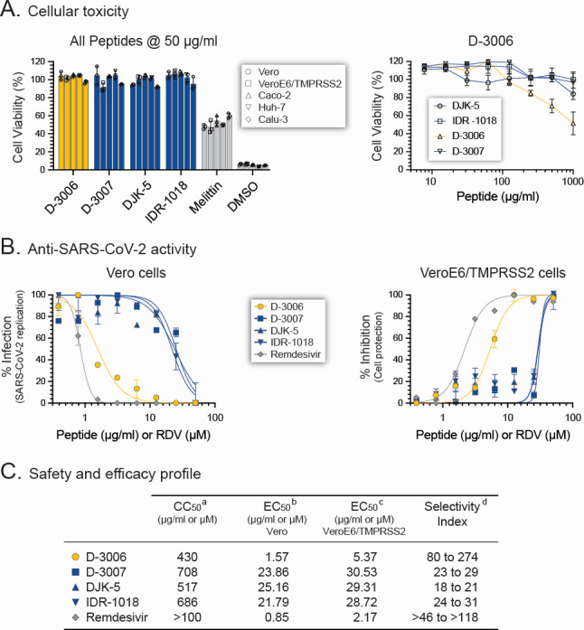
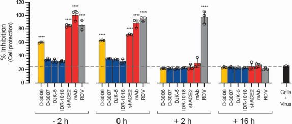
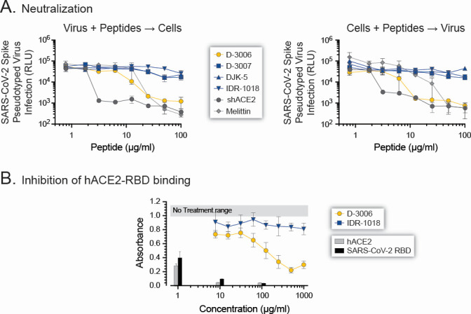
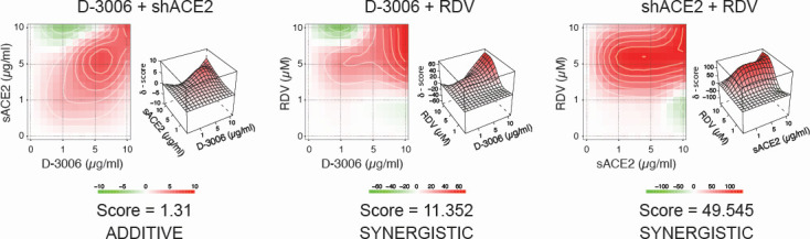
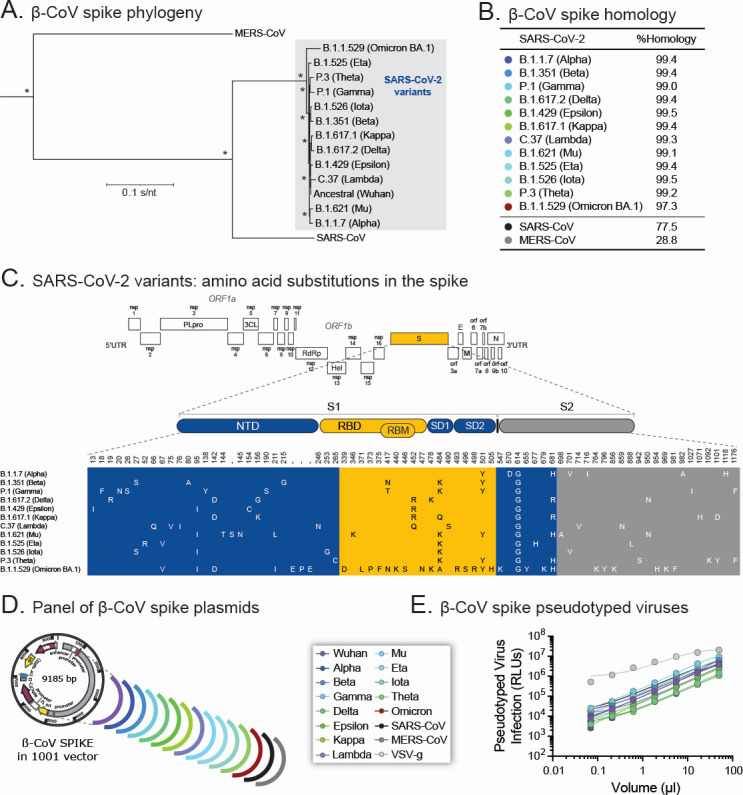
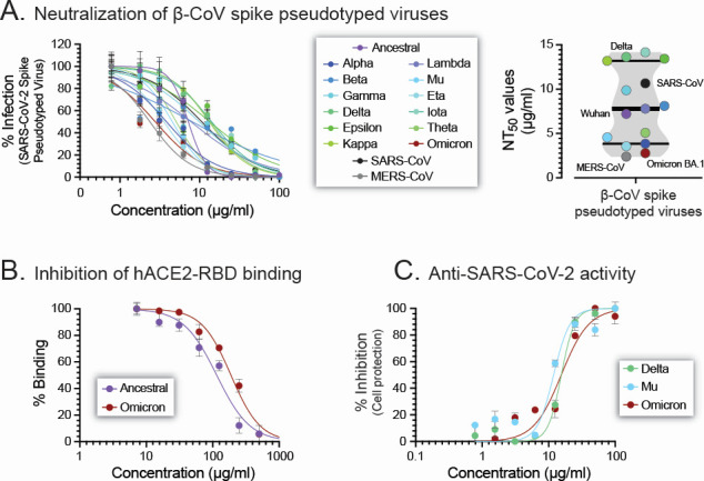
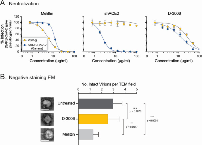
Although myriads of potential antiviral agents have been tested against SARS-CoV-2, only a handful have proven to be effective in clinical trials. During the COVID-19 pandemic, many known or novel peptides were evaluated for their ability to inhibit SARS-CoV-2 replication; however, testing of D-enantiomers that resist body and viral proteases has been limited. Here, we characterized the ability of D-3006, a D-enantiomeric synthetic host defense peptide, to inhibit SARS-CoV-2 replication in vitro. A battery of authentic SARS-CoV-2 variants (ancestral, Mu, Delta, and Omicron BA.1) and a comprehensive panel of β-coronavirus spike pseudotyped lentiviruses were used to demonstrate that D-3006 safely (CC50value = 430 µg/mL) blocked spike-mediated entry (EC50 values ranging from 1.57 to 5.37 µg/mL) and also had synergistic anti-SARS-CoV-2 activity in vitro when combined with the viral polymerase inhibitor remdesivir. We also showed that D-3006 inhibited influenza A virus (H1N1) replication in vitro, suggesting that this synthetic host defense peptide could have potential broad antiviral activity against multiple enveloped viruses. These data, together with negative-stain transmission electron microscopy analysis, suggest that the mechanism of action of D-3006 is associated with non-specific binding to the viral membrane, most likely causing virus aggregation and interfering with virus attachment and entry. The potential broad-spectrum antiviral activity of D-3006, its innate resistance to host proteases, as well as the possibility of being used in combination with other antiviral drugs suggest that this host synthetic peptide could be developed as a candidate for the treatment of SARS-CoV-2 and/or other respiratory viral infections.

Keywords: COVID-19; SARS-CoV-2; antiviral; host defense peptides; influenza virus.


Conflict of interest statement

The authors declare no conflict of interest.

Figures

Fig 1
Fig 1
Cellular toxicity and anti-SARS-CoV-2 activity of synthetic host defense peptides. (A) The viability of Vero, VeroE6/TMPRSS2, Caco-2, Huh-7, and Calu-3 cells exposed to 50 µg/mL of D-3006, D-3007, DJK-5, or IDR-1016, as well as melittin or DMSO as controls, was determined in triplicate (left panel). The dose-dependent cytotoxic effect was calculated for D-3006 in VeroE6/TMPRSS2 cells in triplicate (right panel), with the cell viability percentage fitted by a four-parameter log-logistic model to determine half-maximum (50%) cytotoxic concentration (CC50) values. (B) Susceptibility of the authentic SARS-CoV-2 hCoV-19/New Zealand/NZ1_patient/2020 isolate (56) to the four synthetic host defense peptides (D-3006, D-3007, DJK-5, or IDR-1018) and remdesivir in Vero or VeroE6/TMPRSS2 cells. SARS-CoV-2 replication was quantified 72 h post-infection by CPE, RT-qPCR assay (62), or a cell protection assay based on the Pierce BCA protein assay kit (Thermo Fisher Scientific). EC50 values, determined using the non-linear regression model (log inhibitor vs. normalized response-variable slope), are depicted in µg/mL for the synthetic peptides or µM for remdesivir. (C) In vitro safety and efficacy (CC50 and EC50 values, respectively) profiles, calculated in Vero and VeroE6/TMRPSS2 cells, were used to determine the selectivity index (CC50/EC50) for each synthetic host defense peptide and remdesivir.
Fig 2
Fig 2
Time-of-drug addition analysis of synthetic host defense peptides. VeroE6/TMPRSS2 cells were infected with authentic SARS-CoV-2 hCoV-19/New Zealand/NZ1_patient/2020 in the presence and absence of the four synthetic peptides (D-3006, D-3007, DJK-5, or IDR-1018) or the control agents (shACE2, RDV, or a SARS-CoV-2 spike neutralizing antibody mAb) added 2 h prior (−2 h), during (0 h), or 2 h (+2 h) or 16 h (+16 h) after infection at a concentration equal to 10-fold of their respective EC50 values (as described in Fig. 1). Inhibition of SARS-CoV-2 replication was quantified 72 h post-infection using a cell protection assay based on the Pierce BCA protein assay kit (Thermo Fisher Scientific) (56). Cells + virus, SARS-CoV-2 replication in the absence of inhibitory agent. Depicted values represent medians ± standard deviations (SD) from three replicates. Wilcoxon-Mann-Whitney test was used to compare the % inhibition of SARS-CoV-2 replication between the different conditions and the Cells + virus control. **** P < 0.0001.
Fig 3
Fig 3
D-3006 neutralized SARS-CoV-2 spike-pseudotyped viruses. (A) Synthetic host defense peptides (D-3006, D-3007, DJK-5, or IDR-1018) or control agents (shACE2 or melittin) were incubated with the SARS-CoV-2 (Wuhan) spike-pseudotyped virus (left panel) or with HEK293T-hACE2 cells (right panel) for 1 h prior to adding the cells or pseudotyped virus, respectively. SARS-CoV-2 spike-mediated entry was quantified 72 h post-infection, and neutralizing titers (50% neutralizing titer, NT50) were determined. (B) Dose-dependent inhibition of hACE2-RBD binding by the synthetic peptides D-3006 and IDR-1018, as well as soluble hACE2 and SARS-CoV-2 RBD as controls, using a competitive ELISA as described in Fig. S2. Absorbance was determined at 650 nm. Depicted values represent medians ± standard deviations (SD) from three replicates. The horizontal gray bar represents the no treatment control, ranging from 1 to 1.18.
Fig 4
Fig 4
Combination treatment of D-3006 with shACE2 or remdesivir. VeroE6/TRMPSS2 cells were infected with authentic SARS-CoV-2 (hCoV-19/New Zealand/NZ1_patient/2020 strain) in the presence and absence of each agent alone and in combination at the following concentrations: 0, 1, 5, and 10 µg/mL (D-3006 and shACE2) or µM (remdesivir), and the degree of synergistic, additivity, or antagonistic effect quantified using SynergyFinder (67). Scores for drug combinations were visualized as heatmaps and 3D synergy maps and interpreted as the average excess due to drug interactions. Scores < −10, in the range of −10 to 10, or >10 suggest that interaction between the two agents is likely to be antagonistic, additive, or synergistic, respectively (67).
Fig 5
Fig 5
Generation of a comprehensive panel of betacoronavirus (β-CoV) spike-pseudotyped viruses. (A) Spike protein sequences from 13 SARS-CoV-2 variants from the ancestral Wuhan to Omicron BA.1, together with SARS-CoV and MERS-CoV, were aligned, and a maximum likelihood phylogenetic tree was constructed. Bootstrap resampling (1,000 data sets) of the multiple amino acid alignment tested the statistical robustness of the tree, with percentage values above 75% indicated by an asterisk. SARS-CoV-2 variants are enclosed in a gray box. S/nt, substitutions per nucleotide. (B) A pairwise comparison analysis of spike protein sequence identities, quantified as % homology, was used to determine the relatedness of the 14 betacoronavirus spike protein sequences with that of the SARS-CoV-2 Wuhan strain. (C) Schema of the SARS-CoV-2 Wuhan genome listing the amino acid substitutions of the 12 SARS-CoV-2 variants of concern (Alpha to Omicron BA.1) in the spike (S) gene compared with the reference Wuhan strain. S1 and S2 spike regions; NTD, N-terminal domain; RBD, receptor-binding domain; RBM, receptor-binding motif; SD1 and SD2, spike subdomains 1 and 2, respectively. (D) Schema of the 16 viral proteins, 15 betacoronavirus spikes, and one VSV-g, cloned into the 1001 vector (a modified pcDNA3.1 plasmid) to generate pseudotyped lentiviruses. (E) Viral titers (TCID50 values) were determined using serial dilutions of the betacoronavirus spike or VSV-g pseudotyped lentiviruses to infect HEK293T-hACE2 cells (for all SARS-CoV-2 variants, SARS-CoV, and VSV-g) or HEK293-hDPP4 cells (for MERS-CoV). RLUs, relative light units.
Fig 6
Fig 6
Broad neutralization activity of D-3006. (A) The synthetic host defense peptide neutralized lentiviruses pseudotyped with spike proteins from 13 SARS-CoV-2 strains (ancestral/Wuhan to Omicron BA.1), SARS-CoV, and MERS-CoV. See Fig. 4 and 5 legends, as well as Materials and Methods, for details. (B) Dose-dependent inhibition of hACE2-RBD binding by D-3006 using a competitive ELISA based on the RBD protein sequence from the ancestral (Wuhan) and Omicron (BA.1). (C) Susceptibility of the authentic SARS-CoV-2 Delta, Mu, and Omicron BA.1 strains to D-3006 in VeroE6/TMPRSS2 cells as described in Fig. S2. Depicted values represent medians ± standard deviations (SD) from three replicates. NT50, 50% neutralizing titer.
Fig 7
Fig 7
D-3006 did not show a virucidal effect. (A) Lentiviruses pseudotyped with the SARS-CoV-2 Gamma spike of VSV-g proteins were exposed to melittin, shACE2, or D-3006 for 1 h before adding them to HEK293T-hACE2 cells. Dose-dependent viral entry inhibition was quantified 72 h post-infection, and neutralizing titers (NT50) were determined. (B) Aliquots of SARS-CoV-2 hCoV-19/New Zealand/NZ1_patient/2020 strain were incubated with 100 µg/mL of D-3006, melittin, or 1× DMEM (Untreated) for 1 h at 37°C and 5% CO2, fixed with 2% formaldehyde, purified (20% sucrose cushion), and prepared for negative staining transmission electron microscopy analysis. Viral particles were evaluated and quantified in 15 random fields per condition. Single SARS-CoV-2 particles, per condition, are shown; however, additional electron microscopy images - as well as raw data - are available upon request. Depicted values represent medians ± standard deviations (SD). Wilcoxon-Mann-Whitney test was used to compare the number of intact virions per field between the untreated, D-3006, and melittin conditions. n.s., not significant.

Similar articles

References

    1. Zhu N, Zhang D, Wang W, Li X, Yang B, Song J, Zhao X, Huang B, Shi W, Lu R, Niu P, Zhan F, Ma X, Wang D, Xu W, Wu G, Gao GF, Tan W, China Novel Coronavirus Investigating and Research Team . 2020. A novel coronavirus from patients with pneumonia in China, 2019. N Engl J Med 382:727–733. doi: 10.1056/NEJMoa2001017 - DOI - PMC - PubMed
    1. Baker MG, Wilson N, Anglemyer A. 2020. Successful elimination of covid-19 transmission in New Zealand. N Engl J Med 383:e56. doi: 10.1056/NEJMc2025203 - DOI - PMC - PubMed
    1. Al-Jighefee HT, Najjar H, Ahmed MN, Qush A, Awwad S, Kamareddine L. 2021. COVID-19 vaccine platforms: challenges and safety contemplations. Vaccines (Basel) 9:1196. doi: 10.3390/vaccines9101196 - DOI - PMC - PubMed
    1. Worobey M, Pekar J, Larsen BB, Nelson MI, Hill V, Joy JB, Rambaut A, Suchard MA, Wertheim JO, Lemey P. 2020. The emergence of SARS-CoV-2 in Europe and North America. Science 370:564–570. doi: 10.1126/science.abc8169 - DOI - PMC - PubMed
    1. Wu N, Joyal-Desmarais K, Ribeiro PAB, Vieira AM, Stojanovic J, Sanuade C, Yip D, Bacon SL. 2023. Long-term effectiveness of COVID-19 vaccines against infections, hospitalisations, and mortality in adults: findings from a rapid living systematic evidence synthesis and meta-analysis up to December, 2022. Lancet Respir Med 11:439–452. doi: 10.1016/S2213-2600(23)00015-2 - DOI - PMC - PubMed

MeSH terms

Supplementary concepts

LinkOut - more resources