. 2025 Aug 13;16(8):e0130325.
doi: 10.1128/mbio.01303-25. Epub 2025 Jun 23.

Evolutionary dynamics of heparan sulfate utilization by SARS-CoV-2

Affiliations

Evolutionary dynamics of heparan sulfate utilization by SARS-CoV-2

Shuhei Higuchi et al. mBio. .

Abstract

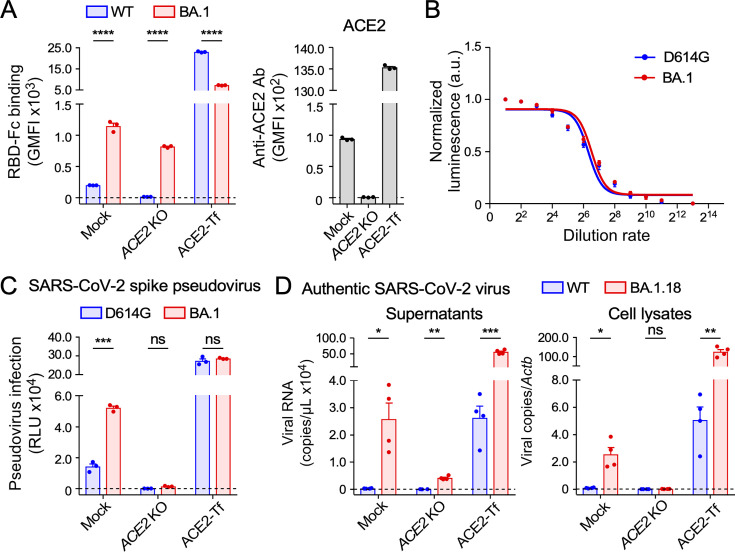
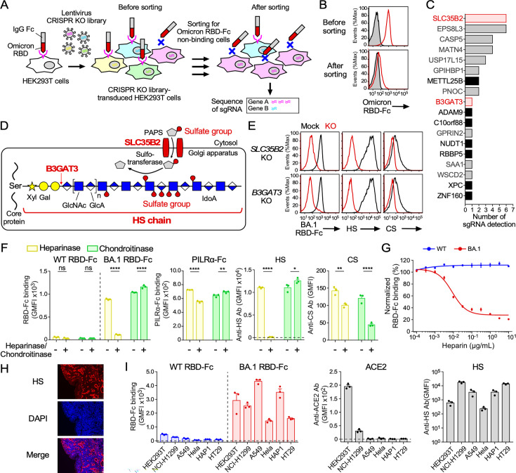
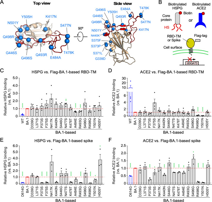
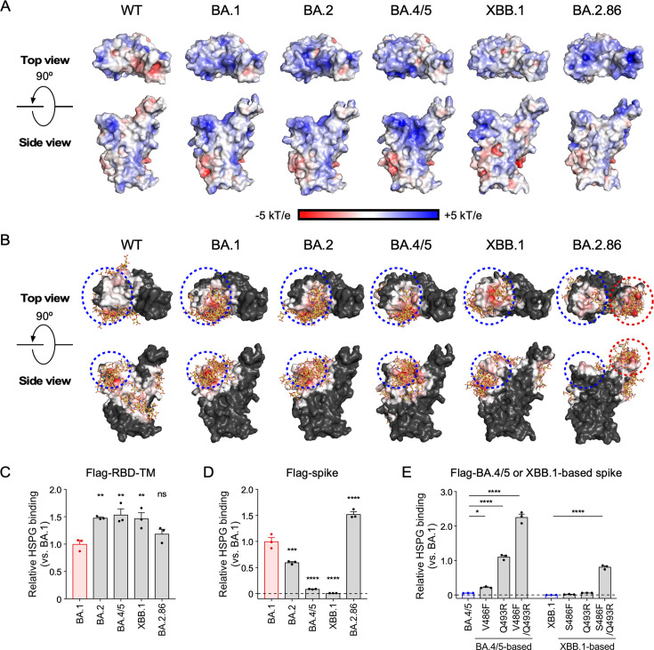
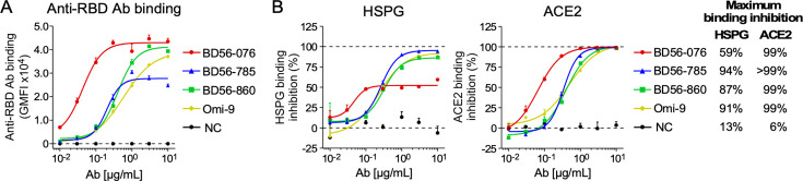
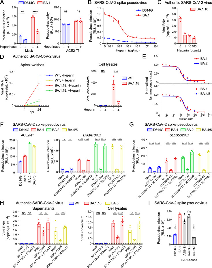
Severe acute respiratory syndrome coronavirus 2 (SARS-CoV-2) Omicron variants have acquired enhanced infectivity compared to earlier variants. To elucidate the underlying molecular mechanisms, we conducted CRISPR library screening to identify cell surface molecules that interact with the Omicron spike protein. Our findings revealed a significantly higher affinity between the Omicron spike and cell surface heparan sulfate compared to the wild-type spike. This increased binding affinity enables Omicron variants to infect cells expressing low levels of ACE2, which are minimally infected by the wild-type virus. Mutational analysis of heparan sulfate binding sites on the Omicron spike protein, coupled with electrostatic potential mapping, suggested that the accumulation of positively charged mutations has contributed to the enhanced heparan sulfate binding. Comparative analysis of heparan sulfate binding among Omicron subvariants-including BA.1, BA.2, BA.4, BA.5, XBB.1, and BA.2.86-revealed that most are likely to bind efficiently to heparan sulfate, but potential heparan sulfate binding sites of the spike protein have shifted from the early Omicron variants to more recent ones. Furthermore, we discovered that cell surface heparan sulfate proteoglycans are cleaved by TMPRSS2, a protease essential for wild-type SARS-CoV-2 infection. These findings suggest that SARS-CoV-2 is evolving to enhance its infectivity by optimizing its interaction with cell surface heparan sulfate.IMPORTANCEThe Omicron variant has evolved to become highly infectious by acquiring numerous mutations. Understanding the impact of these mutations can provide valuable insights into the drivers of viral evolution and aid in the development of improved viral surveillance and vaccines. Our study demonstrates that the Omicron variants contain mutations that enhance their ability to bind to heparan sulfate. Highly infectious human viruses often utilize heparan sulfate for infection, suggesting that heparan sulfate likely plays a crucial role in viral adaptation to human hosts. Furthermore, we found that cell surface heparan sulfate proteoglycans are sensitive to TMPRSS2, while most other cell surface proteins are resistant to TMPRSS2. Given that TMPRSS2 is known to enhance the infectivity of earlier severe acute respiratory syndrome coronavirus 2 variants but cleaves heparan sulfate proteoglycans, it is probable that the high heparan sulfate binding acquired by the Omicron variant contributes to its decreased infectivity against TMPRSS2-expressing cells compared to earlier variants.

Keywords: Omicron; SARS-CoV-2; TMPRSS2; heparan sulfate.


Conflict of interest statement

The authors declare no conflict of interest.

Figures

Fig 1
Fig 1
The Omicron variant infects HEK293T cells expressing low levels of endogenous ACE2. (A) Binding of WT (B lineage) or Omicron (BA.1) RBD fused to human IgG Fc (RBD-Fc), or anti-ACE2 Ab to mock, ACE2 KO, or ACE2-transfected (Tf) HEK293T cells. GMFI, geometric mean fluorescence intensity. (B) Titration of pseudoviruses bearing the SARS-CoV-2 D614G or BA.1 spike using VSV-G-Tf HEK293T cells. (C) Infection of mock, ACE2 KO, or ACE2-Tf HEK293T cells with D614G or BA.1 pseudovirus. RLU, relative luminescence units. (D) Infection of mock, ACE2 KO, or ACE2-Tf HEK293T cells with authentic SARS-CoV-2 WT or Omicron (BA.1.18) variant. Viral RNA in supernatants or cell lysates at 24 hours post-inoculation (hpi) is shown. Lysate RNA was normalized to Actb. Data are mean ± SEM of three to four technical replicates. Statistical analysis was performed using two-way analysis of variance (ANOVA) with Sidak’s multiple comparison tests in panels A and C and unpaired two-tailed Welch’s t-tests between WT and BA.1.18 in panel D; *P < 0.05, **P < 0.01, ***P < 0.001, and ****P < 0.0001; ns, not significant. Data are representative of two to three independent experiments.
Fig 2
Fig 2
ACE2-independent binding of the Omicron RBD to cell surface HS. (A) CRISPR KO library screening scheme to identify Omicron RBD binding molecules expressed on HEK293T cells. (B) Binding of CRISPR KO library-transduced HEK293T cells with (red line) or without (shaded gray) Omicron RBD-Fc, before and after sorting. (C) Number of sgRNA sequences identified in Omicron RBD-Fc non-binding cells after sorting. Red: GAG-synthesis-related genes; black: other expressed genes; gray: non-expressed genes in HEK293T cells. (D) HS biosynthetic pathway highlighting SLC35B2 and B3GAT3. PAPS, 3'-phosphoadenosine-5'-phosphosulfate; Xyl, xylose; Gal, galactose; GlcNAc, N-acetylglucosamine; GlcA, glucuronic acid; IdoA, iduronic acid. (E) Binding of BA.1 RBD-Fc, anti-HS Ab, or anti-CS Ab to mock (black line), or SLC35B2 or B3GAT3 KO (red line) HEK293T cells. (F) Binding of WT or BA.1 RBD-Fc, PILRα-Fc, anti-HS Ab, or anti-CS Ab to ACE2 KO HEK293T cells pretreated with (+) or without (–) heparinase or chondroitinase. (G) Binding of WT or BA.1 RBD-Fc to HEK293T cells at different heparin concentrations. RBD-Fc binding was normalized to binding in the absence of heparin. (H) Immunofluorescence of human nasal tissue with anti-HS Ab and 4', 6-diamidino-2-phenylindole (DAPI). Scale bar, 200 µm. (I) Binding of WT or BA.1 RBD-Fc, anti-ACE2 Ab, or anti-HS Ab to cell lines. Data are mean ± SEM of three to four technical replicates. Statistical analysis was performed using two-way ANOVA with Sidak’s multiple comparison tests in panel F; *P < 0.05, **P < 0.01, and ****P < 0.0001; ns, not significant. Data are representative of two to three independent experiments.
Fig 3
Fig 3
Enhanced binding of RBD to cell surface HS by the mutations acquired by the Omicron variant. (A) Structure of BA.1 RBD (PDB: 7WBP) with mutation sites compared to WT (blue spheres) and ACE2-binding sites (receptor-binding motif; red). (B) Schematic of the binding assay using biotinylated HSPG or ACE2 with B3GAT3 KO HEK293T cells expressing Flag-tagged RBD fused to a transmembrane domain (Flag-RBD-TM) or Flag-tagged whole spike protein (Flag-spike). (C and D) The binding of biotinylated HSPG (C) or ACE2 (D) to Flag-RBD-TM transfectants of BA.1-based revertants is shown. Each revertant contains a single mutation reverted to the WT sequence. The expression levels of Flag-RBD-TM were adjusted by anti-Flag Ab staining. Data were normalized to the binding of BA.1. The dashed horizontal red lines indicate the value of parental BA.1. (E and F) The binding of biotinylated HSPG (E) or ACE2 (F) to Flag-spike transfectants of BA.1-based revertants is shown. The expression levels of Flag-spike were adjusted by anti-Flag Ab staining. Data are mean ± SEM of three to four biological replicates. Each dot represents one independent experiment. Statistical analysis was performed using unpaired two-tailed Student’s t-tests between parental BA.1 and each revertant; *P < 0.05, **P < 0.01, ***P < 0.001, and ****P < 0.0001.
Fig 4
Fig 4
The binding manner of spike protein to HS among the Omicron subvariants. (A) Electrostatic potential maps for WT (PDB: 6M0J), BA.1 (7WBP), BA.2 (7XB0), BA.4/5 (7XWA), XBB.1 (8IOV), and BA.2.86 (8QSQ) RBDs are shown. Blue and red colors indicate electropositive and electronegative surfaces, respectively. (B) Contact maps showing interactions with 10 predicted heparin poses (yellow sticks) docked to the RBDs of WT, BA.1, BA.2, BA.4/5, XBB.1, and BA.2.86. Red indicates regions with frequent contacts, white indicates fewer contacts, and black indicates no contacts. Blue and red dotted circles indicate putative heparin-binding sites I and II, respectively. (C and D) The binding of biotinylated HSPG to B3GAT3 KO HEK293T cells expressing Flag-RBD-TM (C) or Flag-spike (D) from BA.1, BA.2, BA.4/5, XBB.1, or BA.2.86. (E) The binding of biotinylated HSPG to B3GAT3 KO HEK293T cells expressing Flag-spike of BA.4/5- or XBB.1-based revertants. Each revertant contains a single or double mutation reverted to the BA.1 sequence. The expression levels of Flag-RBD-TM or Flag-spike were adjusted by anti-Flag Ab staining. Data were normalized to BA.1 binding. Data are mean ± SEM of three technical replicates. Statistical analysis was performed using two-way ANOVA with Dunnett’s multiple comparison test compared to BA.1 in panels C and D and two-way ANOVA with Dunnett’s multiple comparison test compared to parental BA.4/5 or XBB.1 in panel E; *P < 0.05, **P < 0.01, and ****P < 0.0001; ns, not significant. Data are representative of three independent experiments.
Fig 5
Fig 5
Inhibition of HS binding to Omicron BA.1 spike by anti-RBD neutralizing Abs from BA.1-infected patients. (A) Binding of anti-RBD Abs from BA.1-infected patients to BA.1 spike. B3GAT3 KO HEK293T cells lacking HS were transfected with BA.1 spike and were used for Ab binding. (B) Inhibition of HSPG or ACE2 binding to BA.1 spike by anti-RBD Abs. HSPG or ACE2 binding to B3GAT3 KO HEK293T cells transfected with BA.1 spike was analyzed in the presence or absence of anti-RBD Abs. The maximum binding inhibition of Abs for HSPG or ACE2 binding to BA.1 spike is shown. Data are mean ± SEM of three technical replicates. Data are representative of two independent experiments.
Fig 6
Fig 6
HS-dependent infection of the Omicron variants to low-level ACE2-expressing cells. (A) Infection of D614G or BA.1 pseudovirus to mock or ACE2-Tf HEK293T cells pretreated with (+) or without (–) heparinase. (B) Infection of D614G or BA.1 pseudovirus to HEK293T cells at different heparin concentrations. (C) Infection of authentic SARS-CoV-2 BA.1.18 variant to HEK293T cells at different heparin concentrations. Viral RNA in the supernatants at 24 hours post-inoculation (hpi) is shown. (D) Infection of authentic SARS-CoV-2 WT or BA.1.18 variant to primary human nasal epithelial cells in the presence (+) or absence (–) of 100 µg/mL heparin. Viral RNA in apical washes at 1 and 24 hpi or in cell lysates at 24 hpi is shown. Lysate RNA was normalized to Actb. (E) Titration of BA.2 or BA.4/5 pseudovirus using VSV-G-Tf HEK293T cells. (F) Infection of D614G, BA.1, BA.2, or BA.4/5 pseudovirus to ACE2-Tf HEK293T cells or to mock, B3GAT3 KO, or B3GAT3-Tf B3GAT3 KO HEK293T cells. (G) Infection of D614G, BA.1, BA.2, or BA.4/5 pseudovirus to mock, SLC35B2 KO, or SLC35B2-Tf SLC35B2 KO HEK293T cells. (H) Infection of authentic SARS-CoV-2 WT, BA.1.18 or BA.5 variant to mock, B3GAT3 KO, or B3GAT3-Tf B3GAT3 KO HEK293T cells. Viral RNA in supernatants or cell lysates at 24 hpi is shown. Lysate RNA was normalized to Actb. (I) Infection of D614G, BA.1, or BA.1-based revertant (A484E, R493Q, and R498Q) pseudovirus to HEK293T cells. Data are mean ± SEM of three to four technical replicates. Statistical analysis was performed using two-way ANOVA with Sidak’s multiple comparison tests in panels A and D, two-way ANOVA with Dunnett’s multiple comparison test compared to B3GAT3 KO HEK293T cells in panels F and H, two-way ANOVA with Dunnett’s multiple comparison test compared to SLC35B2 KO HEK293T cells in panel G, and unpaired two-tailed Student’s t-tests between parental BA.1 and each revertant in panel I; *P < 0.05, **P < 0.01, ***P < 0.001, and ****P < 0.0001; ns, not significant. Data are representative of two to three independent experiments.
Fig 7
Fig 7
TMPRSS2 cleaves cell surface HS proteoglycans. (A) Binding of WT or BA.1 RBD-Fc, anti-syndecan-1 (SDC1) Ab, anti-glypican-4 (GPC4) Ab, anti-HS Ab, anti-ACE2 Ab, anti-HLA class I Ab, anti-integrin αV (CD51) Ab, anti-CD46 Ab, or anti-CD59 Ab to HEK293T cells pretreated with (red line) or without (black line) trypsin. (B) Representative gating strategy for TMPRSS2-negative (GFPneg, black), low-expressing (GFPlow, blue), or high-expressing (GFPhigh, red) cells in TMPRSS2/GFP-Tf HEK293T cells. (C) Top: representative histograms showing the binding of WT or BA.1 RBD-Fc, anti-SDC1 Ab, anti-GPC4 Ab, anti-HS Ab, anti-ACE2 Ab, anti-HLA class I Ab, anti-CD51 Ab, anti-CD46 Ab, or anti-CD59 Ab to TMPRSS2-negative (black line, GFPneg), low-expressing (blue line, GFPlow) or high-expressing (red line, GFPhigh) cells. Bottom: quantification of binding is shown as GMFI. (D) Infection of D614G or BA.1 pseudovirus to mock or ACE2-Tf HEK293T cells with (+) or without (–) TMPRSS2 expression. (E) Infection of authentic SARS-CoV-2 WT or BA.1.18 variant to mock or ACE2-Tf HEK293T cells with (+) or without (–) TMPRSS2 expression. Viral RNA in cell lysates at 24 hours post-inoculation is shown. Lysate RNA was normalized to Actb. Data are mean ± SEM of three to four technical replicates. Statistical analysis was performed using two-way ANOVA with Sidak’s multiple comparison tests in panel D and unpaired two-tailed Welch’s t-tests between WT and BA.1.18 in panel E; *P < 0.05, **P < 0.01, and ****P < 0.0001; ns, not significant. Data are representative of two to three independent experiments.

Similar articles

References

    1. Viana R, Moyo S, Amoako DG, Tegally H, Scheepers C, Althaus CL, Anyaneji UJ, Bester PA, Boni MF, Chand M, et al. 2022. Rapid epidemic expansion of the SARS-CoV-2 Omicron variant in southern Africa. Nature 603:679–686. doi: 10.1038/s41586-022-04411-y - DOI - PMC - PubMed
    1. Zhou P, Yang XL, Wang XG, Hu B, Zhang L, Zhang W, Si HR, Zhu Y, Li B, Huang CL, et al. 2020. A pneumonia outbreak associated with a new coronavirus of probable bat origin. Nature 579:270–273. doi: 10.1038/s41586-020-2012-7 - DOI - PMC - PubMed
    1. Lan J, Ge J, Yu J, Shan S, Zhou H, Fan S, Zhang Q, Shi X, Wang Q, Zhang L, Wang X. 2020. Structure of the SARS-CoV-2 spike receptor-binding domain bound to the ACE2 receptor. Nature 581:215–220. doi: 10.1038/s41586-020-2180-5 - DOI - PubMed
    1. Daly JL, Simonetti B, Klein K, Chen KE, Williamson MK, Antón-Plágaro C, Shoemark DK, Simón-Gracia L, Bauer M, Hollandi R, Greber UF, Horvath P, Sessions RB, Helenius A, Hiscox JA, Teesalu T, Matthews DA, Davidson AD, Collins BM, Cullen PJ, Yamauchi Y. 2020. Neuropilin-1 is a host factor for SARS-CoV-2 infection. Science 370:861–865. doi: 10.1126/science.abd3072 - DOI - PMC - PubMed
    1. Cantuti-Castelvetri L, Ojha R, Pedro LD, Djannatian M, Franz J, Kuivanen S, van der Meer F, Kallio K, Kaya T, Anastasina M, et al. 2020. Neuropilin-1 facilitates SARS-CoV-2 cell entry and infectivity. Science 370:856–860. doi: 10.1126/science.abd2985 - DOI - PMC - PubMed

LinkOut - more resources