. 2025 Jul 22;99(7):e0009825.
doi: 10.1128/jvi.00098-25. Epub 2025 Jun 23.

METTL3 regulates PRRSV replication by suppressing interferon beta through autophagy-mediated IKKε degradation

Affiliations

METTL3 regulates PRRSV replication by suppressing interferon beta through autophagy-mediated IKKε degradation

Yunyun Zhai et al. J Virol. .

Abstract

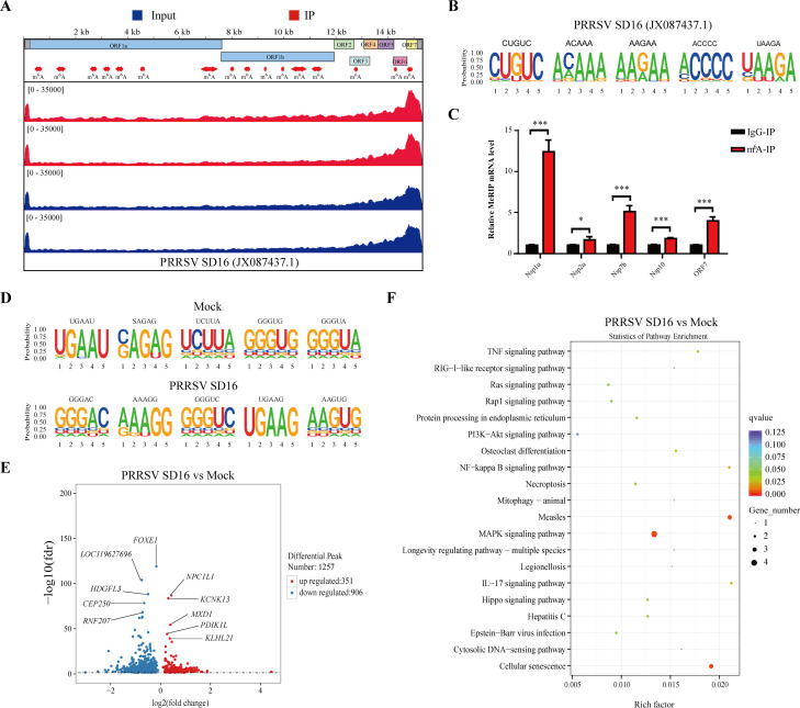
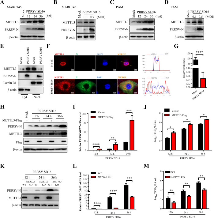
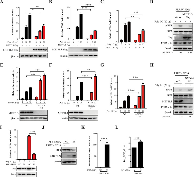
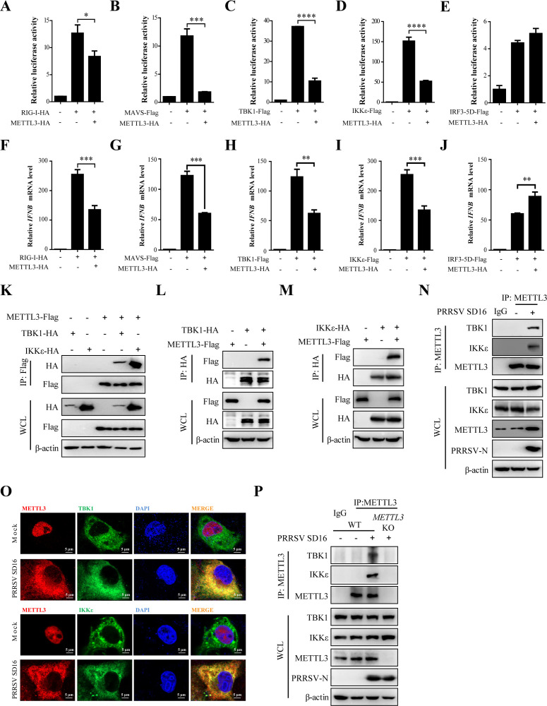
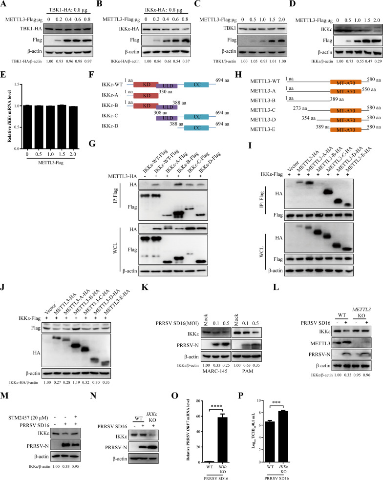
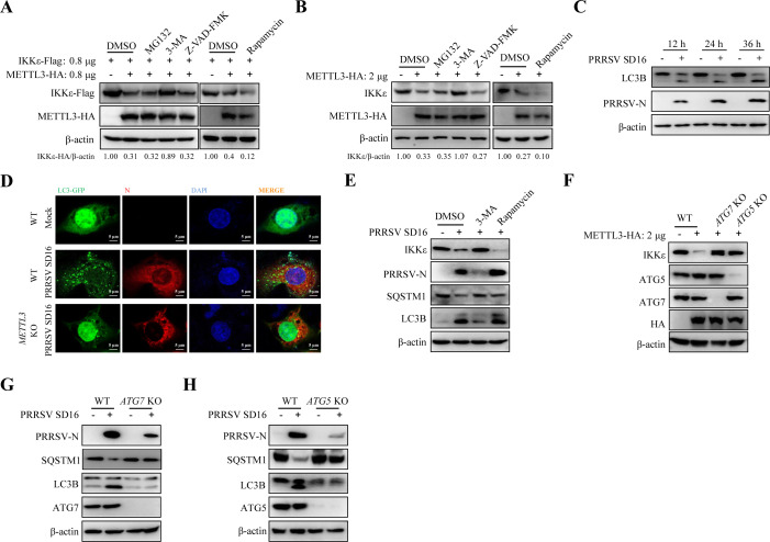
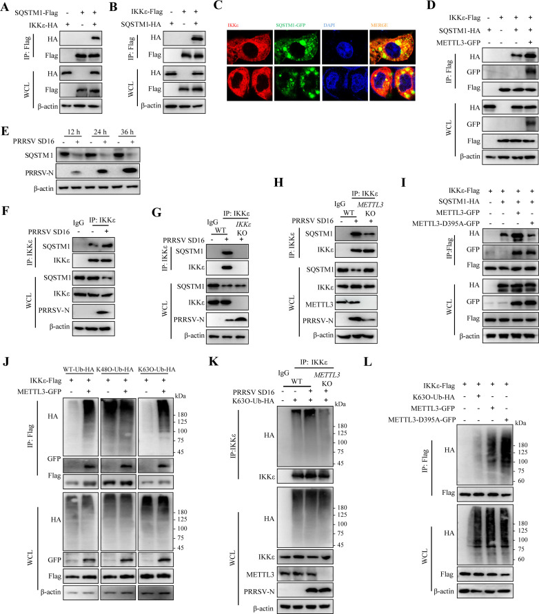
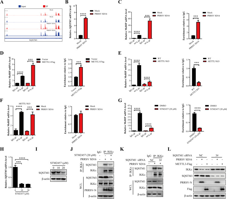
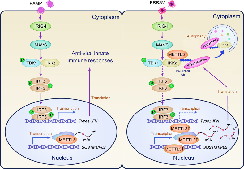
Methyltransferase-like-3 (METTL3)-mediated N6-methyladenosine (m6A) modification of messenger RNAs plays a pivotal role in regulating innate immune responses, either promoting or combating virus replication. However, the biological function of METTL3 during porcine reproductive and respiratory syndrome virus (PRRSV) infection remains unclear. In this study, we found that PRRSV infection reprograms m6A modifications in cellular transcripts, enhances METTL3 expression, and alters its subcellular distribution. Intriguingly, METTL3 overexpression facilitates PRRSV replication, whereas its deficiency suppresses it, primarily through the negative regulation of type I interferon (IFN-I) production. Further investigation revealed that METTL3 interacts with and promotes the degradation of IκB kinase-ε (IKKε) during PRRSV infection. Mechanistically, METTL3-mediated m6A modification of SQSTM1 (sequestosome 1) enhances SQSTM1 messenger RNA (mRNA) expression, increasing autophagy levels. Moreover, METTL3 facilitates the formation of K63-linked ubiquitin chains on IKKε, targeting it for degradation via SQSTM1-dependent selective autophagy. Collectively, our findings unveil a novel mechanism whereby METTL3 facilitates PRRSV replication by suppressing antiviral innate immunity, thereby offering potential targets for antiviral therapy.IMPORTANCEPorcine reproductive and respiratory syndrome (PRRS), induced by the porcine reproductive and respiratory syndrome virus (PRRSV), poses a highly contagious threat to the global swine industry, leading to substantial economic losses. The genetic variability and immune evasion capabilities of PRRSV complicate the development of effective vaccines and control strategies. Thus, a comprehensive understanding of PRRSV's immune evasion mechanisms is imperative. In this study, we reveal that METTL3 plays a pivotal role in PRRSV's evasion of interferon (IFN) immunity. Specifically, METTL3 targets IKKε, inducing its autophagy degradation and subsequently inhibiting the expression of interferon beta 1 (IFNB1). Furthermore, PRRSV infection alters the N6-methyladenosine (m6A) modification of various host genes, with notable changes observed in the m6A modification and transcriptional levels of SQSTM1, which are regulated by METTL3. This regulation is crucial for SQSTM1-mediated autophagy degradation of IKKε. Our findings offer novel insights into the mechanisms underlying host protein involvement in PRRSV's immune evasion.

Keywords: IKKε; METTL3; N6-methyladenosine (m6A); PRRSV; SQSTM1/p62; type I interferon.


Conflict of interest statement

The authors declare no conflict of interest.

Figures

Fig 1
Fig 1
PRRSV RNA is m6A modified, and viral infection reprograms host m6A epitranscriptome. (A) Genome-wide distribution of m6A peaks across the PRRSV SD16 genome visualized using Integrative Genomics Viewer (IGV). (B) Consensus m6A methylation motifs identified in PRRSV genomic RNA during in vitro infection. (C) Validation of representative m6A peaks in nsp1a, nsp2a, nsp7b, nsp10, and ORF7 by MeRIP-qPCR. (D) Comparison of enriched m6A motifs between PRRSV-infected and uninfected MARC-145 cells. (E) Volcano plot depicting peaks with significantly altered m6A modification levels in PRRSV-infected versus untreated MARC-145 cells. (F) Heat map showing significantly enriched pathways in PRRSV-infected cells, sorted by log2-transformed P-values.
Fig 2
Fig 2
METTL3 positively regulates PRRSV infection. (A) Western blot analysis of METTL3 and PRRSV-N expression in MARC-145 cells infected with PRRSV at an MOI of 0.1 or 0.5, collected at 36 h. (B) Time-course analysis of METTL3 and PRRSV-N protein expression in MARC-145 cells infected with PRRSV at an MOI of 0.5, collected at 12, 24, and 36 h. (C) Time-course analysis of METTL3 and PRRSV-N protein expression in PAMs infected with PRRSV at an MOI of 0.5, collected at 12, 24, and 36 h. (D) Western blot analysis of METTL3 and PRRSV-N protein expression in PAMs infected with PRRSV at an MOI of 0.1 or 0.5, collected at 36 h. β-actin served as the loading control. (E) Subcellular localization of METTL3 in MARC-145 cells infected with PRRSV (MOI = 0.5) and collected at 36 h. Western blot was used to examine METTL3, PRRSV-N, nuclear Lamin B1, and cytoplasmic β-actin protein levels. (F) Immunofluorescence analysis of METTL3 and PRRSV-N subcellular localization in PRRSV-infected MARC-145 cells (MOI = 0.5) at 36 h. Nuclei were counterstained with DAPI. Confocal laser scanning microscopy was used to capture fluorescent images. (G) Quantification of METTL3 subcellular distribution in at least five cells using ImageJ. The nuclear-to-cytoplasmic (N/C) fluorescence intensity ratio was calculated and normalized to uninfected control cells to determine fold changes in localization. (H–J) MARC-145 cells were transfected with METTL3-Flag or an empty vector for 24 h, followed by PRRSV infection (MOI = 0.5). At 12, 24, and 36 h, cells were analyzed by western blot to detect METTL3 and PRRSV-N protein expression (H), RT-qPCR to measure PRRSV ORF7 gene expression (fold changes normalized to β-actin) (I), and TCID50 assay to determine viral titers (J). (K–M) MARC-145 WT and METTL3 KO cells were infected with PRRSV (MOI = 0.5). Cells were collected at 12, 24, and 36 h and analyzed using western blot (K), RT-qPCR (L), and TCID50 assay (M).
Fig 3
Fig 3
METTL3 suppresses the antiviral response. (A) Dual-luciferase assay in HEK293T cells co-transfected with IFNB-Luc (100 ng), pRL-TK (25 ng), and either METTL3-Flag (250 ng) or an empty plasmid (250 ng) for 12 h. Cells were subsequently treated with 10 or 20 µg/mL Poly I:C for 12 h. (B and C) RT-qPCR analysis of IFNB1 (B) and ISG15 (C) transcription (fold changes normalized to β-actin) in MARC-145 cells transfected with METTL3-Flag (2 µg) or an empty vector (2 µg) for 24 h, followed by treatment with 10 or 20 µg/mL Poly I:C for 12 h. (D) MARC-145 cells transfected with METTL3-Flag or an empty vector for 24 h were infected with PRRSV (MOI = 0.5). At 24 h, cells were treated with 20 µg/mL Poly I:C for 12 h. Western blot analysis was performed to detect pIRF3, IRF3, METTL3, and PRRSV-N protein levels. (E) Dual-luciferase assay in MARC-145 WT and METTL3 KO cells co-transfected with IFNB-Luc (100 ng), pRL-TK (25 ng), and METTL3-Flag (100, 200, and 250 ng). (F and G) MARC-145 WT and METTL3 KO with 20 µg/mL Poly I:C for 12 h. Western blot analysis was used to detect METTL3 expression, and RT-qPCR was conducted to evaluate IFNB1 (F) and ISG15 (G) transcription (fold changes normalized to β-actin). (H) Western blot analysis of pIRF3, IRF3, METTL3, and PRRSV-N protein levels in MARC-145 WT and METTL3 KO cells infected with PRRSV (MOI = 0.5) for 24 h, followed by treatment with 20 µg/mL Poly I:C for 12 h. (I) RT-qPCR analysis of IFNB1 transcription (fold changes normalized to β-actin) in MARC-145 cells transfected with small interfering RNA (siRNA) targeting IRF3 following treatment with 20 µg/mL Poly I:C. (J–L) MARC-145 cells were transfected with IRF3 siRNA or negative control siRNA (NC) for 24 h, followed by infection with PRRSV (MOI = 0.5). Cells were analyzed at 36 h using western blot (J), RT-qPCR (K), and TCID50 assay (L).
Fig 4
Fig 4
METTL3 interacts with TBK1/IKKε. (A–E) HEK293T cells were co-transfected with IFNB-Luc, pRL-TK, and HA-METTL3, along with constructs or empty vectors expressing RIG-I-HA (A), MAVS-Flag (B), TBK1-Flag (C), IKKε-Flag (D), or IRF3-5D-Flag (E). After 24 h, cells were harvested, and a dual-luciferase assay was performed. (F–J) MARC-145 cells were co-transfected with HA-METTL3, as well as construct plasmids or empty vectors expressing RIG-I-HA (F), MAVS-Flag (G), TBK1-Flag (H), IKKε-Flag (I), and IRF3-5D-Flag (J). Twenty-four hours after transfection, cells were harvested for protein expression analysis via western blot, with HA and Flag antibodies used to probe the blots. Additionally, the transcription of IFNB1 was quantified via RT-qPCR (fold changes normalized to β-actin). (K) HEK293T cells co-transfected with METTL3-Flag and either TBK1-HA or IKKε-HA were lysed 48 h later. Cell lysates were immunoprecipitated with an anti-Flag antibody. Whole-cell lysates (WCL) and co-immunoprecipitation (co-IP) complexes were analyzed by western blot using anti-Flag, anti-HA, and anti-β-actin antibodies. (L and M) HEK293T cells were transfected with TBK1-HA (L) or IKKε-HA (M) along with METTL3-Flag. At 24 h, cell lysates were immunoprecipitated with an anti-HA antibody and analyzed by western blot with the specified antibodies. (N) MARC-145 cells infected with PRRSV (MOI = 0.5) for 36 h were lysed, and METTL3 was immunoprecipitated. Western blot was used to analyze interacting proteins. (O) Immunofluorescence microscopy showing the subcellular localization of METTL3 and TBK1 or IKKε in PRRSV-infected MARC-145 cells (MOI = 0.5) at 36 h. Nuclei were counterstained with DAPI. Confocal laser scanning microscopy was used to capture images. (P) MARC-145 WT and METTL3 KO cells were infected with PRRSV (MOI = 0.5) for 36 h. METTL3 was immunoprecipitated, and interacting proteins were detected by western blot.
Fig 5
Fig 5
METTL3 suppresses IKKε protein expression, thereby enhancing PRRSV replication. (A–D) HEK293T or MARC-145 cells were transfected with varying amounts of METTL3-Flag or an empty plasmid. After 24 h, cells were harvested, and western blot analysis was conducted to detect TBK1 (A and C) and IKKε (B and D) protein expression. (E) RT-qPCR analysis of IKKε transcription in MARC-145 cells transfected with METTL3-Flag or an empty plasmid for 24 h. (F) Schematic representation of Flag-tagged truncated IKKε constructs. (G) HEK293T cells were co-transfected with IKKε-Flag (or its truncated constructs) and METTL3-HA. After 24 h, co-IP was performed to analyze protein interactions. (H) Schematic representation of HA-tagged truncated METTL3 constructs. (I) HEK293T cells were co-transfected with METTL3-HA (or its truncated constructs) and IKKε-Flag. After 24 h, co-IP was performed to analyze protein interactions. (J) Western blot analysis of IKKε protein expression in HEK293T cells co-transfected with METTL3-HA (or its mutants) and IKKε-Flag for 24 h. (K) PRRSV-infected MARC-145 cells or PAMs (MOI = 0.1 or 0.5) were harvested, and IKKε protein levels were analyzed by western blot. (L) Western blot analysis of IKKε, METTL3, and PRRSV-N protein expression in MARC-145 WT and METTL3 KO cells infected with PRRSV (MOI = 0.5) for 36 h. (M) MARC-145 cells infected with PRRSV were treated with STM2457 or DMSO. Western blot analysis of IKKε and PRRSV-N protein expression. (N–P) Loss of IKKε enhances PRRSV replication. MARC-145 WT and IKKε KO cells were infected with PRRSV (MOI = 0.5) for 36 h. Western blot (N), RT-qPCR (O), and TCID50 assay (P) were performed to evaluate viral replication.
Fig 6
Fig 6
Autophagy mediates METTL3-dependent regulation of IKKε protein levels. (A) HEK293T cells co-transfected with METTL3-Flag and IKKε-HA were treated with MG132 (10 µM), 3-MA (10 mM), Z-VAD-FMK (10 µM), or Rapamycin (1 nM) for 12 h. Western blot analysis was performed to detect IKKε-Flag protein expression. (B) MARC-145 cells transfected with METTL3-Flag or an empty plasmid were treated with MG132, 3-MA, Z-VAD-FMK, or Rapamycin for 12 h. Western blot was conducted to analyze IKKε protein level. (C) Western blot analysis of LC3B and PRRSV-N expression in MARC-145 cells infected with PRRSV. (D) Immunofluorescence microscopy showing reduced LC3-GFP aggregation in METTL3 KO cells during PRRSV infection. Fluorescence images were acquired by confocal laser scanning microscopy. (E) MARC-145 cells infected with PRRSV (MOI = 0.5) were treated with DMSO, 3-MA, or Rapamycin for 12 h. Western blot analysis was performed to detect IKKε, PRRSV-N, SQSTM1, and LC3B protein levels. (F) MARC-145 WT, ATG7 KO, and ATG5 KO cells were transfected with METTL3-HA and harvested 36 h later. Western blot analysis was conducted to detect IKKε, ATG7, ATG5, SQSTM1, and METTL3 protein expression. (G and H) MARC-145 WT, ATG7 KO (G), and ATG5 KO (H) cells were infected with PRRSV (MOI = 0.5) for 36 h. Western blot analysis was conducted to detect PRRSV-N, SQSTM1, LC3B, ATG7, and ATG5 protein expression.
Fig 7
Fig 7
METTL3 enhances the recognition of IKKε by the cargo receptor SQSTM1. (A) Co-IP analysis of SQSTM1-Flag and IKKε-HA interaction. HEK293T cells were co-transfected with SQSTM1-Flag and IKKε-HA. After 24 h, cell lysates were immunoprecipitated with an anti-Flag antibody and analyzed by western blot with specified antibodies. (B) Reciprocal co-IP showing interaction between IKKε-Flag and SQSTM1-HA in HEK293T cells transfected with both plasmids. After 24 h, lysates were immunoprecipitated with an anti-Flag antibody and analyzed by western blot. (C) Immunofluorescence microscopy of PRRSV-infected MARC-145 cells (MOI = 0.5) at 36 h showing colocalization of SQSTM1-GFP and IKKε. Nuclei were counterstained with DAPI. Images were acquired using a confocal laser scanning microscope. (D) Co-IP analysis of IKKε-Flag, SQSTM1-HA, and METTL3-GFP interaction in HEK293T cells transfected with the indicated plasmids. After 24 h, lysates were immunoprecipitated with an anti-Flag antibody and analyzed by western blot. (E) Western blot analysis of SQSTM1 and PRRSV-N expression in MARC-145 cells infected with PRRSV. (F) Western blot analysis of PRRSV-infected MARC-145 cells (MOI = 0.5) at 36 h to detect IKKε and SQSTM1 interaction via co-IP. (G) Co-IP of PRRSV-infected MARC-145 WT and IKKε KO cells (MOI = 0.5) at 36 h to examine IKKε interaction with SQSTM1. (H) Western blot analysis of METTL3-dependent interaction between IKKε and SQSTM1 in PRRSV-infected MARC-145 WT and METTL3 KO cells (MOI = 0.5) at 36 h. (I) Co-IP analysis of IKKε-Flag, SQSTM1-HA, METTL3-GFP, and METTL3-D395A-GFP interaction in HEK293T cells transfected with the indicated plasmids. After 24 h, lysates were immunoprecipitated with an anti-Flag antibody and analyzed by western blot. (J) HEK293T cells were co-transfected with WT-Ub-HA, K48O-Ub-HA, K63O-Ub-HA, IKKε-Flag, and METTL3-GFP plasmids. Co-IP was performed 24 h later to analyze ubiquitination patterns and METTL3-mediated effects. (K) MARC-145 WT and METTL3 KO cells were transfected with K63O-Ub-HA plasmid, infected with PRRSV (MOI = 0.5), and harvested 36 h later. Co-IP with anti-IKKε antibody was performed to detect K63-linked ubiquitination. (L) HEK293T cells were co-transfected with METTL3-GFP, METTL3-D395A-HA in IKKε-Flag, K63O-Ub-HA plasmids, respectively. Twenty-four hours later, co-IP was performed to analyze the ubiquitination pattern.
Fig 8
Fig 8
METTL3 regulates m6A modification of SQSTM1 and mediates autophagy degradation of IKKε. (A) Visualization of m6A peaks in SQSTM1 in PRRSV-infected or uninfected MARC-145 cells is shown by IGV. (B) RT-qPCR detection of SQSTM1 mRNA expression (fold changes normalized to β-actin) in MARC-145 cells infected with PRRSV (MOI = 0.5) at 36 h. (C) Validation of m6A peaks in SQSTM1 in PRRSV-infected or uninfected MARC-145 cells is shown in panel (A) by MeRIP-qPCR. Relative enrichment of m6A was determined by calculating the fold change of IP to Input Ct values (IP/Input). (D) Overexpression of METTL3 in MARC-145 cells increases m6A modification of SQSTM1 mRNA, as determined by MeRIP-qPCR. Relative enrichment of m6A was determined by calculating the fold change of IP to Input Ct values (IP/Input). (E) METTL3 KO decreases SQSTM1 m6A modification, as shown by MeRIP-qPCR of MARC-145 WT and METTL3 KO cells. Relative enrichment of m6A was determined by calculating the fold change of IP to Input Ct values (IP/Input). (F) WT and METTL3 KO cells were infected with PRRSV at an MOI of 0.5. At 36 h, total RNA was extracted from the cells and subjected to MeRIP-qPCR assays. Relative enrichment of m6A was determined by calculating the fold change of IP to Input Ct values (IP/Input). (G) STM2457 (20 µM) treatment inhibits m6A modification of SQSTM1 mRNA in MARC-145 cells, as analyzed by MeRIP-qPCR. Relative enrichment of m6A was determined by calculating the fold change of IP to Input Ct values (IP/Input). (H and I) To evaluate the effect of STM2457 on SQSTM1 expression, MARC-145 cells were analyzed by RT-qPCR (fold changes normalized to β-actin) (H) and western blot (I). (J) STM2457 suppresses IKKε interaction with SQSTM1 in PRRSV-infected MARC-145 cells (MOI = 0.5). Lysates were immunoprecipitated with an anti-IKKε antibody and analyzed by western blot. (K) SQSTM1 knockdown by siRNA decreases IKKε interaction in PRRSV-infected MARC-145 cells (MOI = 0.5), as analyzed by co-IP. (L) SQSTM1 knockdown prevents METTL3-mediated IKKε degradation during PRRSV infection. Western blot analysis of Flag, SQSTM1, IKKε, and PRRSV-N protein expression.
Fig 9
Fig 9
Schematic model of METTL3-mediated PRRSV immune evasion. Upon PRRSV infection, METTL3 protein expression is upregulated, resulting in its translocation from the nucleus to the cytoplasm. In the cytoplasm, METTL3 interacts with the TBK1/IKKε complex and promotes K63-linked ubiquitination of IKKε. This ubiquitination facilitates recognition by the autophagy cargo receptor SQSTM1, leading to IKKε degradation. Simultaneously, METTL3 enhances m6A modification of SQSTM1 mRNA, increasing its transcription and promoting autophagy. These mechanisms suppress IFNB1 production, allowing PRRSV to evade host immunity and replicate efficiently.

Similar articles

References

    1. Jiang D, Sui C, Wu X, Jiang P, Bai J, Hu Y, Cong X, Li J, Yoo D, Miller LC, Lee C, Du Y, Qi J. 2024. Swine NONO promotes IRF3-mediated antiviral immune response by detecting PRRSV N protein. PLoS Pathog 20:e1012622. doi: 10.1371/journal.ppat.1012622 - DOI - PMC - PubMed
    1. Fang P, Xie C, Pan T, Cheng T, Chen W, Xia S, Ding T, Fang J, Zhou Y, Fang L, Wei D, Xiao S. 2023. Unfolding of an RNA G-quadruplex motif in the negative strand genome of porcine reproductive and respiratory syndrome virus by host and viral helicases to promote viral replication. Nucleic Acids Res 51:10752–10767. doi: 10.1093/nar/gkad759 - DOI - PMC - PubMed
    1. Read AF, Baigent SJ, Powers C, Kgosana LB, Blackwell L, Smith LP, Kennedy DA, Walkden-Brown SW, Nair VK. 2015. Imperfect vaccination can enhance the transmission of highly virulent pathogens. PLoS Biol 13:e1002198. doi: 10.1371/journal.pbio.1002198 - DOI - PMC - PubMed
    1. Yang Q, Gao L, Si J, Sun Y, Liu J, Cao L, Feng WH. 2013. Inhibition of porcine reproductive and respiratory syndrome virus replication by flavaspidic acid AB. Antiviral Res 97:66–73. doi: 10.1016/j.antiviral.2012.11.004 - DOI - PubMed
    1. Luo R, Fang L, Jin H, Jiang Y, Wang D, Chen H, Xiao S. 2011. Antiviral activity of type I and type III interferons against porcine reproductive and respiratory syndrome virus (PRRSV). Antiviral Res 91:99–101. doi: 10.1016/j.antiviral.2011.04.017 - DOI - PubMed

MeSH terms

LinkOut - more resources